
Contrast and Radiological Procedures — MCQs

On this page
For which part of the gastrointestinal tract is a barium follow-through examination primarily used?
In which one of the following conditions is sialography contraindicated?
Which of the following radiographic projections is contraindicated in patients with cervical spondylitis?
All of the following appear hypo-intense on MRI except?
Calcified lesions are better visualized on which imaging modality?
Which MRI sequence suppresses fat signal?
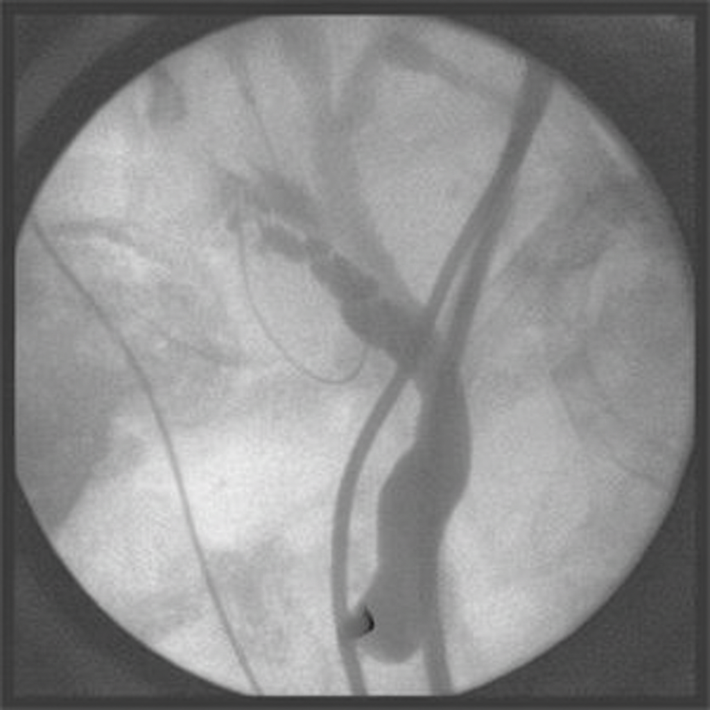
What is the most appropriate diagnostic finding in this scenario?
On barium swallow, what characteristic appearance is shown by a leiomyoma?
A 50-year-old male patient complains of facial heaviness, headache, and nasal congestion. He has a history of chronic sinusitis. A Waters view X-ray was performed. What is the typical positioning of the canthomeatal line relative to the film in this projection?
In atresia of the cardiac end of the stomach, what contrast agent is best visualized on an X-ray chest?
Practice by Chapter
Iodinated Contrast Media
Practice Questions
MRI Contrast Agents
Practice Questions
Ultrasound Contrast Agents
Practice Questions
Adverse Reactions to Contrast Media
Practice Questions
Management of Contrast Reactions
Practice Questions
Contrast-Induced Nephropathy
Practice Questions
Barium Studies
Practice Questions
Intravenous Urography
Practice Questions
Angiography Techniques
Practice Questions
Lymphangiography
Practice Questions
Contrast Administration Protocols
Practice Questions
Pretesting and Premedication
Practice Questions
Want unlimited practice?
Get full access to all questions, explanations, and performance tracking.
Scan to download app