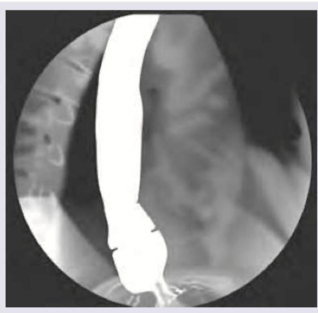
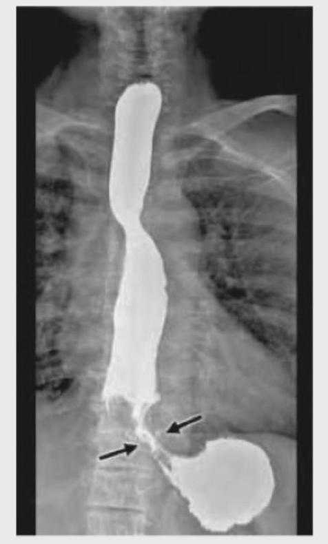
The barium swallow shows presence of:

The following barium swallow study shows which of the following?

A 45-year-old female patient complains of dysphagia for one year. UGI endoscopy was normal. Diagnosis is: (Recent NEET Pattern 2016-17)

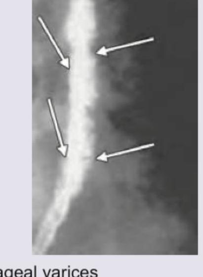
A 35-year-old woman presents with progressive dysphagia to solids. A barium swallow study is performed. Which of the following is true regarding the condition shown in the image?

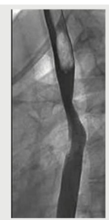
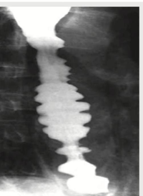
All of the following statements regarding this radiological film representing esophagus are true except:

A patient complains of episodes of dysphagia and chest pain. The barium study presentation of the patient is shown below. A radiologist will describe this condition as all except:

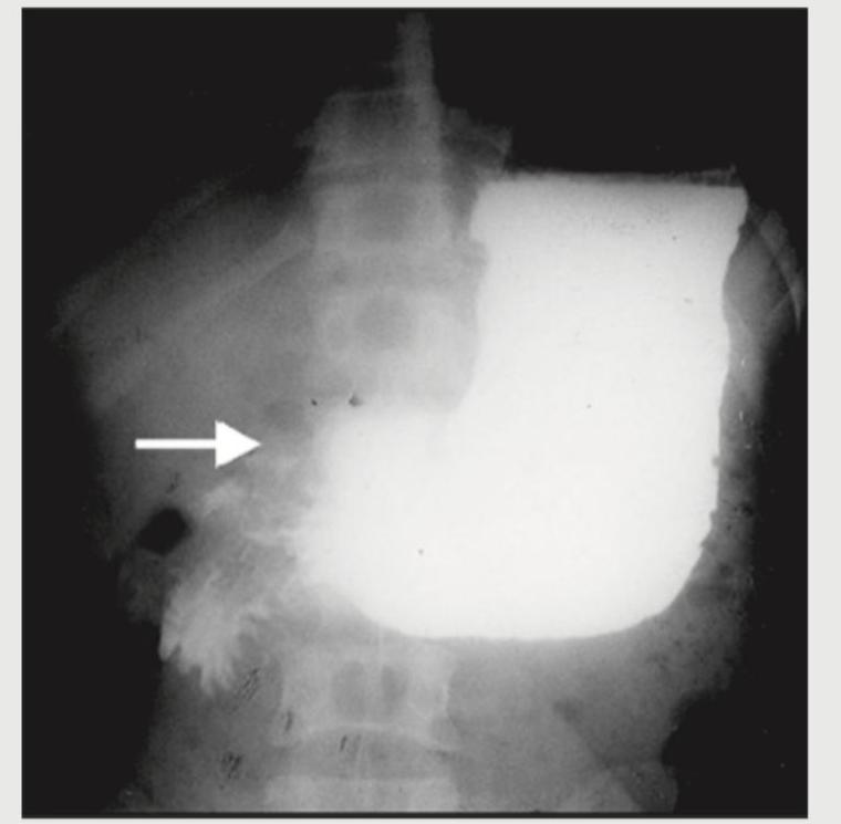
Which of the following is shown in the barium study?

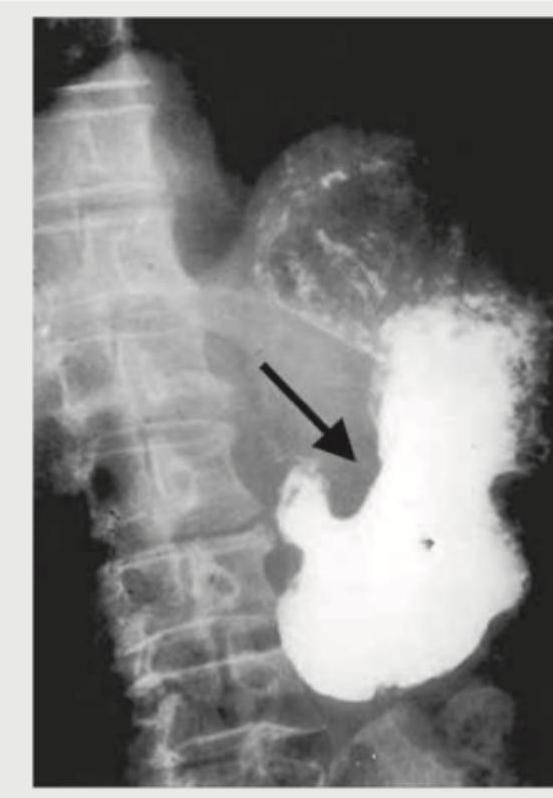
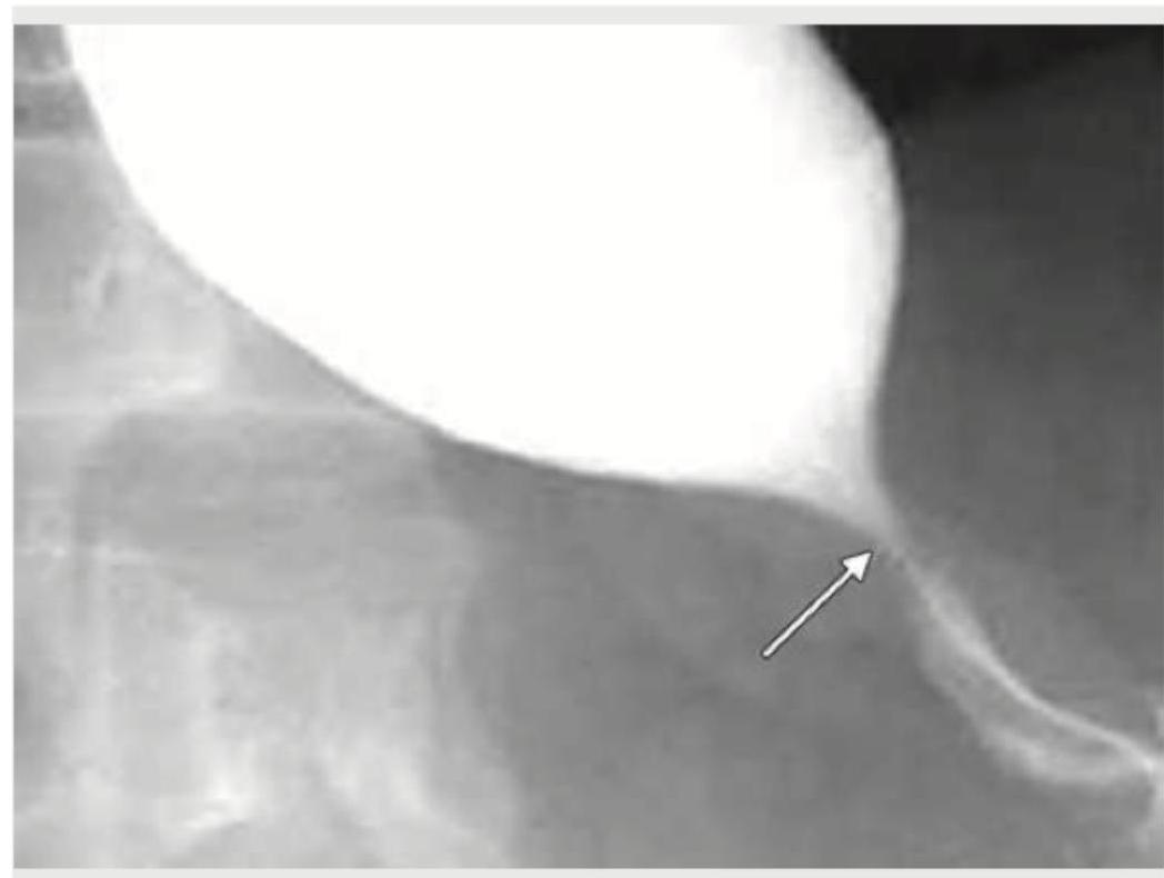
What is the diagnosis based on the barium meal study shown below?

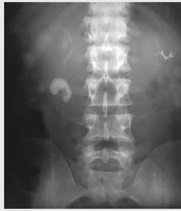
A 26-year-old construction worker with a previous history of recurrent kidney stones presents with flank pain. What is the radiological sign demonstrated in the IVP image shown below?

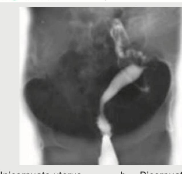
A 32-year-old lady presents with history of first trimester miscarriage and underwent HSG. The diagnosis is: (Recent NEET Pattern 2018-19)

Explanation: ***Bird beak appearance*** - The image shows a **smooth, tapered narrowing of the distal esophagus** with proximal esophageal dilation, a classic sign of **achalasia**. - This appearance resembles a **bird's beak** on a barium swallow study due to the failure of the **lower esophageal sphincter** to relax. *Apple core appearance* - The **apple core sign** is characteristic of an **annular stricture** in the colon, typically caused by a **colonic carcinoma**. - It describes irregular, circumferential narrowing of the bowel lumen with overhanging edges, which is not seen here. *Schatzki's ring* - A **Schatzki's ring** is a benign, circumferential, thin mucosal ring located at the **esophagogastric junction**. - It typically causes intermittent dysphagia for solids but does not present with the diffuse narrowing and significant proximal dilation seen in the image. *Esophageal web* - An **esophageal web** is a thin, eccentric protrusion of mucosa into the esophageal lumen, usually found in the **proximal esophagus**. - It is typically a subtle finding and does not cause the extensive, tapered narrowing and associated proximal dilation observed in this barium swallow.
Explanation: ***Esophageal varices*** - The Barium Swallow image clearly demonstrates a **serpiginous**, worm-like, or **rosary bead-like appearance** along the esophageal wall, which is characteristic of esophageal varices. - Varices are dilated submucosal veins and appear as **filling defects** on barium studies, especially when distended. *Schatzki ring* - A Schatzki ring is a **thin, symmetric mucosal ring** at the esophagogastric junction, causing solid food dysphagia. - It presents as a **smooth, circumferential narrowing** on a barium swallow, which is not seen here. *Candida esophagitis* - Candida esophagitis typically appears as **ulcerations, plaques, or nodularity** with an irregular, cobblestone pattern on barium swallow. - This image does not show the characteristic **"shaggy" or "cobblestone"** appearance associated with candidal plaques. *Reflux esophagitis* - Reflux esophagitis can manifest as **mucosal irregularity, erosions, or strictures**, but not the distinct serpiginous filling defects seen in this image. - Severe reflux can cause peptic strictures, which appear as **smooth, tapered narrowings**, different from the irregular variceal pattern.
Explanation: ***Diffuse esophageal spasm*** - The image shows a **"corkscrew" or "rosary bead" esophagus**, which is a classic radiographic finding in diffuse esophageal spasm due to uncoordinated esophageal contractions. - Dysphagia in this context, coupled with a normal UGI endoscopy (ruling out mechanical obstruction visible by scope), points to a motility disorder. *Zenker's diverticulum* - This is an **outpouching of the pharyngeal mucosa** occurring above the upper esophageal sphincter. - It would typically appear as a distinct barium-filled pouch on a barium swallow, not the diffuse spasm seen in the image. *Dysphagia lusoria* - This is dysphagia caused by **vascular compression of the esophagus**, usually an aberrant right subclavian artery. - It would manifest as a localized external compression rather than the widespread functional spasm shown. *Esophageal carcinoma* - Esophageal carcinoma would typically present as a **fixed filling defect, stricture, or ulceration** on a barium swallow, often with abnormal mucosal patterns. - The UGI endoscopy would likely show an abnormality if carcinoma were present.
Explanation: ***Associated with iron deficiency anemia*** - The image shows an **esophageal web** in the upper esophagus, characteristic of **Plummer-Vinson syndrome** (also known as **Paterson-Brown-Kelly syndrome**). - This syndrome consists of the classic triad: **iron deficiency anemia**, **dysphagia**, and **esophageal webs**. - The webs are thin membranes of tissue in the upper esophagus that cause intermittent dysphagia to solids. - Treatment involves **iron supplementation** and mechanical dilation of the web if symptomatic. - This is a **premalignant condition** with increased risk of **post-cricoid and upper esophageal squamous cell carcinoma**. *Increased tone of lower esophageal sphincter* - Increased tone of the **lower esophageal sphincter** is characteristic of **achalasia**, which presents with a **"bird's beak" appearance** on barium swallow. - Achalasia is a primary **motility disorder** caused by degeneration of the myenteric plexus, not a structural web. - The image shows a **web in the upper esophagus**, not LES pathology. *Not a premalignant condition* - Esophageal webs, particularly in **Plummer-Vinson syndrome**, are considered a **premalignant condition**. - There is increased risk of developing **squamous cell carcinoma** of the post-cricoid region and upper esophagus. - Regular **endoscopic surveillance** is recommended due to this malignant potential. *It is a normal phenomenon* - The presence of an **esophageal web** is an **abnormal anatomical structure** and pathological finding. - Esophageal webs cause **dysphagia** and are associated with underlying conditions like **iron deficiency anemia**. - Normal esophageal mucosa should be smooth without web-like strictures.
Explanation: ***Regurgitation of previous day food items is the most common and early presenting symptom*** - This symptom, along with **halitosis** and a feeling of **choking**, is more characteristic of **Zenker's diverticulum**, a pouch in the esophageal wall that retains food. - While esophageal disorders can cause regurgitation, **dysphagia (difficulty swallowing)** is typically the most common and early symptom of esophageal cancer, especially for solids. *Squamous cell usually affects the upper two-thirds* - **Squamous cell carcinoma (SCC)** of the esophagus most commonly occurs in the **middle and upper thirds** of the esophagus. - This is consistent with the distribution of **squamous epithelium** lining these parts of the esophagus. *Adenocarcinoma usually affects the lower-third* - **Adenocarcinoma** of the esophagus typically arises in the **distal esophagus**, often in the context of **Barrett's esophagus**. - Barrett's esophagus, a complication of **chronic gastroesophageal reflux disease (GERD)**, involves the metaplastic change of squamous epithelium to columnar epithelium in the lower esophagus. *Lymph node involvement is a bad prognostic factor* - The presence of **lymph node metastases** is a critical indicator of regional spread and is a well-established **poor prognostic factor** for esophageal cancer. - It significantly influences the **staging** of the cancer and guides treatment decisions, often necessitating more aggressive therapies.
Explanation: ***Cobble stone appearance*** - **Cobblestone appearance** on barium swallow studies is typically associated with **Crohn's disease** affecting the bowel, characterized by deep ulcerations intersected by edematous mucosa. - This image clearly shows severe, disorganized contractions in the esophagus, which is characteristic of a **motility disorder**, not Crohn's. *Corkscrew appearance* - The image distinctly shows a **twisted, irregular contour** of the esophageal lumen, which is well-described as a **corkscrew esophagus**. - This finding is classic for **diffuse esophageal spasm** (DES), where powerful, uncoordinated contractions occur. *Rosary bead esophagus* - This term is another descriptive phrase for the appearance of the esophagus in **diffuse esophageal spasm**. - The appearance resembles beads on a rosary due to the **simultaneous, non-peristaltic contractions** causing segmental narrowing and outpouching. *Pseudodiverticula* - While not true diverticula which are herniations of all layers, the saccular outpouchings seen between the spastic contractions can be described as **pseudodiverticula**. - These are formed by the irregular, forceful contractions transiently distorting the esophageal wall.
Explanation: ***Menetrier's disease*** - The barium study shows **markedly thickened gastric folds**, particularly in the fundus and body of the stomach, which is characteristic of Menetrier's disease. - This condition involves **hypertrophy of the gastric mucosa** with associated protein loss, which can be visualized as prominent folds on imaging. *Gastric ulcer* - A gastric ulcer would typically appear as a **focal collection of barium** (crater) that projects beyond the normal gastric lumen, often with surrounding edema. - The image does not show a discrete ulcer crater but rather diffuse thickening of the gastric folds. *Esophageal varices* - Esophageal varices would be seen as **serpiginous filling defects** within the esophagus, particularly in the distal portion. - The image focuses on the stomach and does not display the characteristic features of esophageal varices. *Mallory-Weiss syndrome* - Mallory-Weiss syndrome involves **longitudinal tears in the esophageal mucosa**, usually near the gastroesophageal junction. - These tears are best identified endoscopically and would not typically be visible as diffuse gastric fold thickening on a barium study.
Explanation: ***Peptic ulcer disease*** - The barium meal study shows a distinct **niche** (pocket of barium) within the gastric wall, indicated by the arrow, consistent with an **ulcer crater** - Additionally, there is evidence of **gastric wall edema** and possibly **converging folds** around the ulcer, which are typical radiographic findings in peptic ulcer disease - The smooth, round contour of the niche with radiating folds is characteristic of a **benign gastric ulcer** *Gastric carcinoma* - Gastric carcinoma typically appears as an **irregular, nodular filling defect** with shouldering and abrupt transitions - Malignant ulcers have **irregular margins**, do not show converging folds, and lack the smooth contour seen in this image - The benign features of the ulcer crater here make carcinoma unlikely *Esophageal varices* - Esophageal varices would appear as **serpiginous filling defects** or a "rosary bead" appearance within the esophagus - They are not typically seen in the stomach and do not create a niche or ulcer crater - This image shows findings in the stomach or duodenum, not the esophagus *Hiatal hernia* - Hiatal hernia presents as **herniation of the gastric fundus** through the diaphragmatic hiatus into the thoracic cavity - It appears as a **retrocardiac gas bubble** or mass above the diaphragm on barium studies - No evidence of herniation or diaphragmatic abnormality is seen in this image; the findings are localized to an ulcer crater
Explanation: ***Rim sign*** - The image shows a **calcified rim around a radiolucent (non-calcified) center**, which is characteristic of a **calcium oxalate monohydrate stone** on an intravenous pyelogram (IVP). - This sign is also known as the **"lucent-centered calculus"** or **"target sign"** and indicates a partially calcified stone. *Ring sign* - The **ring sign** can refer to various appearances in medical imaging, often indicating a **ring-enhancing lesion** on CT or MRI, which is not applicable to a kidney stone on IVP. - In renal imaging, a ring sign might describe a collection of contrast medium around a tumor or cyst, but not typically a stone with a radiolucent center. *Egg in cup appearance* - This sign is typically associated with **osteochondroma or enchondroma** lesions in bone imaging, where the cartilage cap creates a "cup" for the medullary bone to grow into. - It is not a recognized sign for kidney stones. *Sun burst appearance* - The **sun burst appearance** is characteristic of certain **bone tumors**, particularly **osteosarcoma**, where new bone forms perpendicular to the bone surface. - This description does not apply to the radiological features of kidney stones on IVP.
Explanation: ***Unicornuate uterus*** - A **unicornuate uterus** develops from one **Müllerian duct**, resulting in a single functional uterine horn with a characteristic **banana-shaped** or crescent-shaped cavity. - On **HSG**, it appears as a single, elongated cavity that **deviates to one side** and is typically smaller than normal, associated with increased risk of **first trimester miscarriage**. *Bicornuate uterus* - Would show **two distinct uterine horns** separated by a deep fundal cleft, with the uterus appearing **heart-shaped** externally. - HSG would demonstrate **bifurcation** of the uterine cavity into two separate horns at the fundal level with an **intercornual angle >105 degrees**. *Uterus didelphys* - Complete duplication with **two separate uterine bodies**, cervices, and often vaginas resulting from total failure of **Müllerian duct fusion**. - HSG would show **two completely separate uterine cavities** with no communication, not consistent with the single cavity shown. *Arcuate uterus* - Mildest form of **Müllerian anomaly** with only a slight **fundal indentation** and essentially normal uterine cavity size. - HSG shows a **broad, smooth, mild concave contour** of the fundus, not consistent with the elongated, deviated cavity shown.
Iodinated Contrast Media
Practice Questions
MRI Contrast Agents
Practice Questions
Ultrasound Contrast Agents
Practice Questions
Adverse Reactions to Contrast Media
Practice Questions
Management of Contrast Reactions
Practice Questions
Contrast-Induced Nephropathy
Practice Questions
Barium Studies
Practice Questions
Intravenous Urography
Practice Questions
Angiography Techniques
Practice Questions
Lymphangiography
Practice Questions
Contrast Administration Protocols
Practice Questions
Pretesting and Premedication
Practice Questions
Get full access to all questions, explanations, and performance tracking.
Start For Free