What is an X-ray artifact?
Hourglass deformity is seen in which of the following conditions?
Who is credited with inventing the orthopantomographic machine?
The following radiological image was taken to assess the urinary tract. Identify the investigation shown below.
Identify the procedure shown in the image
What is true about the investigation shown below?
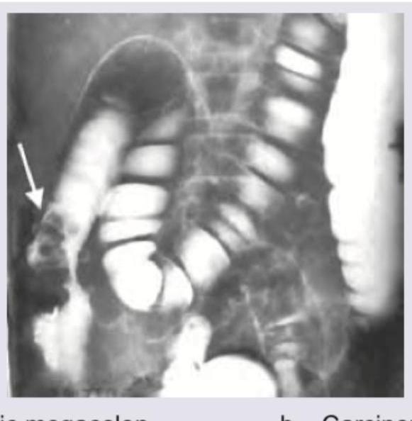
The given barium enema is diagnostic of:

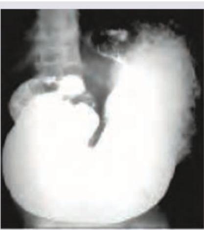
The following barium meal shows:

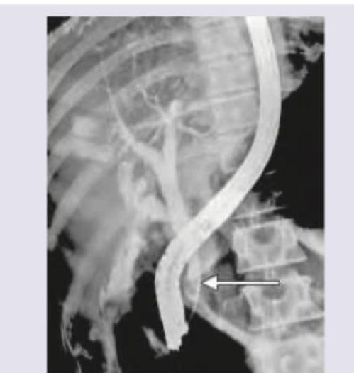
The following image shows:

The flow of Barium across the mucosal surface is highly irregular and is seen in which of the following conditions?
Explanation: ### Explanation An **artifact** in radiology is defined as any structure or feature visible on an image that does not correspond to the actual anatomy of the patient. These are undesirable optical densities or patterns that can degrade image quality and potentially lead to misdiagnosis. **Why "All of the above" is correct:** * **Option A (Radiolucent area):** Artifacts can appear as dark (radiolucent) areas. For example, a scratch on the emulsion of an X-ray film or a static electricity discharge can result in localized areas of decreased density. * **Option B (Abnormal opacity):** Artifacts frequently appear as light (radiopaque) areas. Common examples include external objects like jewelry, buttons, or medical leads, as well as internal artifacts like surgical clips or "dust spots" on a digital imaging plate. * **Option C (Patient movement):** Motion is one of the most common causes of artifacts. When a patient moves during exposure, it results in **motion blur**, which obscures anatomical detail and creates a distorted image that does not accurately represent the patient's true anatomy. Since artifacts can manifest as lucencies, opacities, or distortions due to motion, all statements are correct. ### High-Yield Clinical Pearls for NEET-PG: * **Grid Cut-off:** A common artifact in conventional radiography caused by improper alignment of the X-ray beam with the grid, resulting in a uniform loss of density (underexposure). * **Ring Artifact:** Specifically seen in **CT scans**, usually due to a miscalibrated or faulty detector element. * **Star Artifact:** Caused by high-density metallic implants (e.g., dental fillings or prosthetic joints) in CT, resulting from "beam hardening" and "photon starvation." * **Aliasing (Nyquist) Artifact:** In MRI, this occurs when the Field of View (FOV) is smaller than the body part being imaged, causing the anatomy to "wrap around" to the opposite side.
Explanation: **Explanation:** **Hourglass deformity** (also known as the "B-shaped stomach") is a classic radiological sign seen in **Peptic Ulcer Disease (PUD)**, specifically chronic gastric ulcers. It occurs due to the formation of extensive fibrosis and cicatrization (scarring) on the **lesser curvature** of the stomach. This fibrous contraction pulls the **greater curvature** inward, creating a deep indentation or "cleft" that divides the stomach into two distinct pouches connected by a narrow channel, resembling an hourglass. **Analysis of Incorrect Options:** * **Carcinoma of the stomach:** Typically presents with a "Leather bottle stomach" (**Linitis Plastica**) due to diffuse infiltration, or irregular filling defects. While malignant ulcers can cause deformity, the classic "hourglass" description is reserved for benign cicatrization. * **Duodenal atresia:** Characterized by the **"Double Bubble Sign"** on X-ray (air in the stomach and proximal duodenum), representing a complete obstruction. * **Congenital Hypertrophic Pyloric Stenosis (CHPS):** Associated with signs like the **"String sign"** (narrowed pyloric canal), **"Shoulder sign"**, and **"Mushroom sign"** on barium studies, but not an hourglass shape. **High-Yield Clinical Pearls for NEET-PG:** * **Tea-pot deformity:** Seen when chronic gastric ulcer scarring causes shortening of the lesser curvature, pulling the antrum upward. * **Cup-and-spill (Cascade) stomach:** A functional or structural variant where the fundus folds posteriorly, often confused with organic deformities. * **Leather bottle stomach:** Associated with diffuse-type gastric adenocarcinoma (Krukenberg tumor precursor).
Explanation: **Explanation:** **Correct Answer: D. Numata** The **Orthopantomogram (OPG)**, or panoramic radiograph, is a specialized tomographic technique used to produce a single image of the entire dentition and surrounding structures. The concept of panoramic radiography was first pioneered by **Hisatugu Numata** in 1933. He was the first to experiment with placing a curved film inside the mouth while the X-ray source rotated around the patient. Later, **Yrjö Paatero** (often called the "father of panoramic radiography") refined this by placing the film outside the mouth, leading to the modern OPG machines used today. **Analysis of Incorrect Options:** * **A. Hounsfield:** Sir Godfrey Hounsfield is credited with the invention of **Computed Tomography (CT)**. The "Hounsfield Unit" (HU) is the standard scale for measuring radiodensity in CT scans. * **B. Roentgen:** Wilhelm Conrad Roentgen discovered **X-rays** in 1895. He is considered the father of diagnostic radiology and was the first Nobel Prize winner in Physics. * **C. Kell:** Edmund Kells (C. Edmund Kells) was a pioneer in **dental radiography** who is credited with taking the first intraoral radiograph in the United States and advocating for the clinical use of X-rays in dentistry. **High-Yield Clinical Pearls for NEET-PG:** * **Principle of OPG:** It is based on the principle of **curved plane tomography** (slit-scan radiography). * **Indications:** Assessment of impacted teeth (especially 3rd molars), orthodontic planning, mandibular fractures, and screening for bony lesions like ameloblastoma. * **Ghost Images:** A common OPG artifact where dense objects (like earrings or the contralateral mandible) appear on the opposite side, higher up, and blurred. * **Ideal Positioning:** The patient must bite on a plastic rod to ensure the teeth are in the "focal trough" (the zone of sharpest image).
Explanation: ***Intravenous pyelogram (IVP)*** - An **IVP**, also known as intravenous urography (IVU), involves injecting a radiopaque **contrast agent** intravenously, which is then excreted by the kidneys, allowing for visualization of the entire urinary tract including the kidneys, ureters, and bladder. - This investigation provides both anatomical detail, such as identifying obstructions or masses, and functional information about the kidneys' ability to excrete the contrast. *Pelvic X-ray* - A standard **pelvic X-ray** is primarily used to evaluate the bony structures of the pelvis and would not typically involve contrast media to outline the urinary tract. - While it can reveal radiopaque **kidney stones** (calculi) or bladder stones, it does not provide detailed visualization of the kidneys or ureters as an IVP does. *Retrograde pyelogram* - In a **retrograde pyelogram**, contrast dye is injected directly into the ureters or renal pelvis via a catheter inserted through a **cystoscope** into the bladder. - This procedure visualizes the collecting system in a retrograde fashion (from bottom to top) and does not assess renal excretory function. *Micturating cystourethrogram (MCU)* - An **MCU** (also known as voiding cystourethrogram or VCUG) involves retrograde filling of the bladder with contrast through a urethral catheter, followed by fluoroscopic imaging during voiding. - This investigation is primarily used to evaluate **vesicoureteral reflux** and urethral anatomy, not for assessing the upper urinary tract (kidneys and ureters) as shown in an IVP.
Explanation: ***Intravenous Pyelogram*** - In this procedure, a contrast agent is injected **intravenously** and is then excreted by the kidneys, allowing for visualization of the entire urinary tract, including the renal calyces, pelvis, ureters, and bladder. - The image demonstrates the **nephrogram phase** (contrast in the kidney parenchyma) and the **excretory phase** (contrast in the collecting systems and ureters) simultaneously on both sides, which is characteristic of an IVP. *Retrograde pyelogram* - This procedure involves the injection of contrast material directly into the ureter via a **cystoscope** passed into the bladder, resulting in retrograde (bottom-to-top) filling of the collecting system. - A **ureteral catheter** would typically be visible on the image, and usually, only one side is opacified at a time, which is not the case here. *Combined Retrograde Pyelogram and Ureteroscopy* - This is an invasive procedure where a **ureteroscope** (a thin, flexible camera) is passed up the ureter to directly visualize and treat pathologies like stones or tumors. - The presence of the **ureteroscope** or other instruments would be evident on the radiograph, which are absent in this image. *Antegrade pyelography* - This technique involves inserting a needle or catheter through the skin of the back directly into the renal collecting system (**percutaneous access**) to inject contrast. - A **percutaneous nephrostomy tube** or needle would be visible on the image, which is not seen here.
Explanation: ***Invasive procedure*** - The image depicts a form of **Urography** (likely Intravenous Urography or IVU), which requires the **intravenous injection** of a contrast medium, making it an invasive procedure. - If the image were a Retrograde Pyelogram (RGP), it would also be invasive, requiring instrumentation via **cystoscopy** up to the ureteric orifices. *Non invasive procedure to visualize ureteropelvic junction* - This statement is incorrect because the procedure requires the introduction of contrast material into the body, either intravenously or directly into the urinary system, which classifies it as **invasive**. - While it effectively visualizes the **ureteropelvic junction (UPJ)**, non-invasive imaging like **Ultrasound** or **Non-contrast CT** does not require contrast injection. *Gold standard for bladder cancer* - The gold standard investigation for diagnosing and staging bladder cancer is **Cystoscopy with Biopsy**, not urography. - Urography is primarily used to evaluate the **upper urinary tract** (kidneys and ureters) for filling defects, strictures, or stones. *Done percutaneously* - This procedure, typically an IVU, involves **intravenous access** for contrast injection, not a percutaneous stab into the kidney or other structures. - **Antegrade pyelography** is the investigation done percutaneously, usually through a **nephrostomy tube**, but this image represents broader visualization.
Explanation: ***Toxic megacolon*** - The barium enema shows **marked colonic dilatation** and loss of **haustral folds** (the normal sacculations of the colon), which are classic features of toxic megacolon. - The dilated colon with absent haustrations indicates severe inflammation and paralysis of the colonic smooth muscle, often seen in severe inflammatory bowel disease. *Carcinoma colon* - Colorectal carcinoma typically presents as a **focal mass**, **apple-core lesion**, or a stricture with irregular borders on barium enema, not diffuse dilatation. - It would not cause the widespread loss of haustral markings seen in the image. *Angiodysplasia colon* - **Angiodysplasia** is characterized by submucosal vascular malformations and is usually diagnosed by **colonoscopy** with characteristic red spots or bleeding from the lesion. - It does not typically cause luminal changes visible on barium enema, nor diffuse colonic dilatation. *Normal study* - A normal barium enema would show consistently sized colon with **intact haustral markings** throughout. - The imaging clearly demonstrates significant abnormalities, including dilatation and loss of haustrations, ruling out a normal study.
Explanation: ***Gastric outlet obstruction*** - The barium meal shows a **markedly distended stomach** with a large amount of retained contrast material filling the stomach, indicating impaired emptying. - There is minimal or no passage of barium into the duodenum, characteristic of a **physical barrier** preventing gastric emptying. *Normal study* - A normal barium meal would show the contrast passing readily from the stomach into the **duodenum and small bowel** within a short period. - The stomach size and emptying rate would be within typical physiological limits, unlike the distension seen here. *Esophageal varices* - Esophageal varices would appear as **irregular, serpiginous filling defects** within the esophagus, which is not the primary finding in this image. - The major abnormality seen is related to gastric emptying, not esophageal morphology. *Gastric antral vascular ectasia* - Gastric antral vascular ectasia (water-melon stomach) typically appears as **longitudinal red streaks or spots** in the antrum on endoscopy. - It would not manifest as severe gastric distention and obstruction to outflow on a barium meal; radiological findings are often subtle or related to mucosal abnormalities.
Explanation: ***ERCP showing smooth filling defect*** - The image displays a **retrograde filling** of the biliary tree with contrast, characteristic of an **Endoscopic Retrograde Cholangiopancreatography (ERCP)**. - The arrow points to a smooth, crescent-shaped defect within the common bile duct, consistent with a **gallstone (choledocholithiasis)** causing a filling defect. *PTC showing smooth filling defect* - **Percutaneous Transhepatic Cholangiography (PTC)** involves an antegrade injection of contrast directly into the intrahepatic bile ducts through the skin, which is not what is seen here. - While PTC can show filling defects, the **retrograde cannulation** of the common bile duct from the duodenum is clearly visible, ruling out PTC. *Oral cholecystogram showing stricture* - An **oral cholecystogram** assesses gallbladder function and stones, not the common bile duct as clearly visualized here. - There is no evidence of a **stricture**, which would appear as a focal narrowing with upstream dilation, instead, a filling defect is central to the finding. *HIDA scan showing cystic duct agenesis* - A **HIDA scan (Hepatobiliary Iminodiacetic Acid scan)** is a nuclear medicine study that assesses gallbladder function and bile flow, not anatomical details of the biliary tree in the same way as a contrast study. - **Cystic duct agenesis** would manifest as the absence of gallbladder visualization, which cannot be determined from this image.
Explanation: ***Candida esophagitis*** - **Candida esophagitis** often presents with a characteristic imaging finding called a "**shaggy esophagus**" on barium swallow studies. - This "shaggy" appearance is due to the irregular adherence of barium to the **candidal plaques and pseudomembranes** on the esophageal mucosa, leading to an irregular flow pattern. *Esophageal cancer* - Esophageal cancer typically appears as a **filling defect**, stricture, or focal irregularity with **shouldering** or mucosal nodularity on barium studies. - The barium flow would be obstructed or narrowed, but usually not described as "highly irregular" across the entire mucosal surface in the same diffuse manner as Candida. *Esophageal varices* - Esophageal varices appear as **snake-like** or **serpiginous filling defects** that are typically longitudinal and alter with respiration, giving a "rosary bead" appearance. - While they cause irregularities, the description of "highly irregular flow across the mucosal surface" is not the primary way varices are characterized on barium studies. *Reflux esophagitis* - Reflux esophagitis can show mild mucosal irregularities, thickening of folds, or strictures, especially in chronic cases. - However, the irregular barium flow from diffuse mucosal plaque adherence characteristic of Candida is not a typical finding in reflux esophagitis.
Iodinated Contrast Media
Practice Questions
MRI Contrast Agents
Practice Questions
Ultrasound Contrast Agents
Practice Questions
Adverse Reactions to Contrast Media
Practice Questions
Management of Contrast Reactions
Practice Questions
Contrast-Induced Nephropathy
Practice Questions
Barium Studies
Practice Questions
Intravenous Urography
Practice Questions
Angiography Techniques
Practice Questions
Lymphangiography
Practice Questions
Contrast Administration Protocols
Practice Questions
Pretesting and Premedication
Practice Questions
Get full access to all questions, explanations, and performance tracking.
Start For Free