
Contrast and Radiological Procedures — MCQs

On this page
What is the primary use of T2-weighted imaging in MRI?
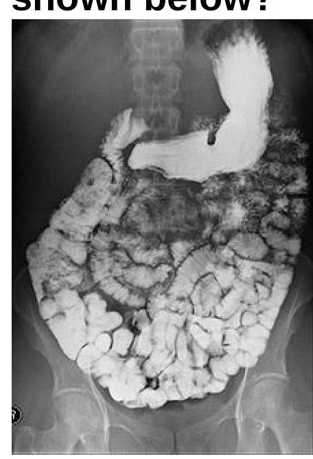
Barium meal follow through is helpful in diagnosing -
Which of the following is NOT an indication for a barium meal X-ray?
What is the primary purpose of the Caldwell view in radiology?
Identify the radiological procedure shown in the image?
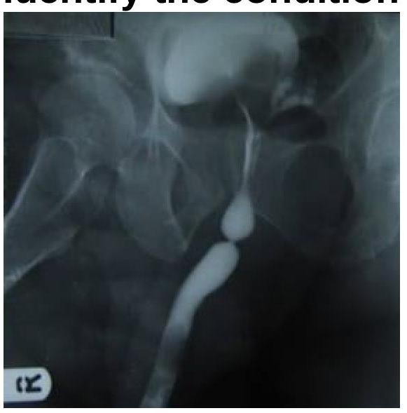
Identify the imaging modality and the location of pathology shown in the image.
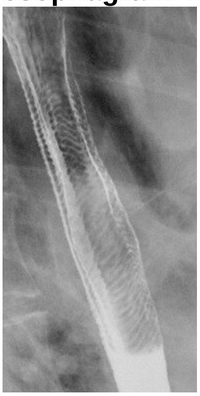
What does the following radiograph from a double contrast esophagram represent?
Which of the following is not considered a contraindication for undergoing an MRI?
Barium swallow is used for -
The posterior urethra is best visualized by:
Practice by Chapter
Iodinated Contrast Media
Practice Questions
MRI Contrast Agents
Practice Questions
Ultrasound Contrast Agents
Practice Questions
Adverse Reactions to Contrast Media
Practice Questions
Management of Contrast Reactions
Practice Questions
Contrast-Induced Nephropathy
Practice Questions
Barium Studies
Practice Questions
Intravenous Urography
Practice Questions
Angiography Techniques
Practice Questions
Lymphangiography
Practice Questions
Contrast Administration Protocols
Practice Questions
Pretesting and Premedication
Practice Questions
Want unlimited practice?
Get full access to all questions, explanations, and performance tracking.
Scan to download app