I/V contrast is not used in -
Which of the following contrast agents is PREFERRED in a patient with renal dysfunction for the prevention of contrast-induced nephropathy?
Investigation of choice for leptomeningeal carcinomatosis:
Enhancement in CT contrast is due to -
Which of the following is a non-ionic contrast agent?
Which of the following non-depolarising muscle relaxant is excreted maximally through the kidney?
Gyromagnetic property of proton is seen in -
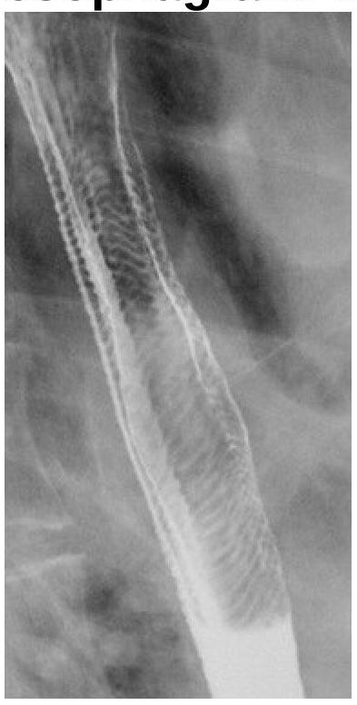
What does the following radiograph from a double contrast esophagram represent?

Even conventional radiological procedures are contraindicated in which neurological disease?
The technique involving injection of contrast material for evaluation of salivary glands is called:
Explanation: ***Myelography*** - Myelography involves injecting contrast material directly into the **subarachnoid space** of the spinal canal to visualize nerve roots and the spinal cord, and therefore does not use intravenous contrast. - The contrast in myelography is typically **iodinated non-ionic contrast** injected intrathecally, not intravenously. *IVP* - **Intravenous Pyelogram (IVP)** is a radiological procedure that specifically uses **intravenous iodinated contrast** to visualize the kidneys, ureters, and bladder. - The contrast is excreted by the kidneys, highlighting the urinary tract structures on X-ray images. *MRI* - While many MRI scans do not require contrast, **intravenous gadolinium-based contrast agents** are commonly used to enhance visualization of certain pathologies like tumors, inflammation, or vascular anomalies. - The contrast is administered intravenously to accumulate in areas with increased vascularity or disrupted blood-brain barrier. *CT scan* - **CT scans** frequently utilize **intravenous iodinated contrast** to improve the visibility of blood vessels, organs, and various lesions like tumors or inflammatory processes. - The contrast enhances density differences between tissues, making pathologies more conspicuous.
Explanation: ***Iso-osmolar contrast*** - **Iso-osmolar contrast agents** (e.g., iodixanol) have an osmolality of ~290 mOsm/kg, which is identical to that of plasma. - **This is the PREFERRED choice** in patients with renal dysfunction as multiple studies demonstrate the lowest risk of contrast-induced nephropathy (CIN). - The iso-osmolar formulation minimizes osmotic stress on renal tubules and reduces the risk of acute kidney injury. - **Current guidelines recommend iso-osmolar agents as first-line** in high-risk patients with pre-existing renal impairment. *Low osmolar contrast* - **Low osmolar contrast agents** have osmolality of 600-900 mOsm/kg, which is significantly lower than high osmolar agents but still 2-3 times higher than plasma. - While **acceptable and safer than high osmolar agents**, they are not as optimal as iso-osmolar contrast for patients with renal dysfunction. - These agents are widely used and represent a reasonable alternative when iso-osmolar agents are not available. *High osmolar contrast* - **High osmolar contrast agents** have osmolality >1400 mOsm/kg (about 5 times that of plasma). - They carry the **highest risk of contrast-induced nephropathy** due to severe osmotic load and direct tubular toxicity. - **Contraindicated or strongly avoided** in patients with pre-existing renal dysfunction. *Ionic contrast* - **Ionic contrast** refers to the chemical structure (dissociates into ions) rather than osmolality. - Can be either high or low osmolar—the ionic nature alone does not determine renal safety. - The critical factor for nephrotoxicity prevention is osmolality, not ionic charge.
Explanation: ***Gd enhanced MRI*** - **Gadolinium-enhanced MRI** is the investigation of choice for **leptomeningeal carcinomatosis** as it can visualize the subtle nodular or linear enhancement along the leptomeninges, indicating tumor dissemination. - It offers superior **soft tissue contrast** and spatial resolution compared to CT, enabling detection of small lesions and accurate mapping of disease extent. *CT scan* - A **CT scan** has limited sensitivity for detecting leptomeningeal involvement due to poor contrast resolution of soft tissues and the dura/arachnoid spaces. - It might show hydrocephalus or large tumor deposits, but subtle leptomeningeal enhancement is often missed. *SPECT* - **Single photon emission computed tomography (SPECT)** is primarily used for functional imaging and is not the investigation of choice for anatomical visualization of leptomeningeal carcinomatosis. - Its resolution is too low to detect the fine structural changes associated with leptomeningeal spread. *PET* - **Positron emission tomography (PET)**, often combined with CT, identifies metabolically active tumor cells and can detect diffuse metastatic disease. - While useful for overall cancer staging and identifying primary lesions, it is less effective than gadolinium-enhanced MRI for directly visualizing the morphology and enhancement patterns of leptomeningeal carcinomatosis due to limited spatial resolution in the CSF spaces.
Explanation: ***Iodine*** - **Iodine-based contrast agents** are commonly used in CT scans to enhance the visualization of blood vessels, organs, and certain lesions due to their **high atomic number** and ability to absorb X-rays. - The degree of enhancement observed on a CT image is directly proportional to the concentration of **iodine** in the tissue or blood. *Gadolinium* - **Gadolinium-based contrast agents** are predominantly used in **Magnetic Resonance Imaging (MRI)**, not CT scans. - Gadolinium works by altering the **magnetic properties** of water molecules in tissues, thereby improving MRI signal intensity. *Mercury* - **Mercury** is a highly toxic heavy metal and is **not used as a contrast agent** in any imaging modality due to its severe health risks. - While historically used in some medical applications, it has been replaced by safer alternatives. *Silver* - **Silver** is not used as a contrast agent in medical imaging; it has no suitable properties for enhancing images in CT or other common modalities. - It is known for its **antimicrobial properties** and is sometimes used in wound dressings.
Explanation: ***Iohexol*** - **Iohexol** is a well-known example of a **non-ionic, low osmolar contrast agent**. It's widely used due to its lower incidence of adverse reactions compared to ionic agents. - Non-ionic contrast agents remain as **intact molecules** in solution and do not dissociate into charged ions, contributing to their lower osmolality and better tolerability. *Amidotrizoate* - **Amidotrizoate** (also known as diatrizoate) is an **ionic, high osmolar contrast agent**. It dissociates into two ions in solution. - Due to its high osmolality, it is associated with a higher risk of adverse effects, such as **nausea**, **vomiting**, and **nephrotoxicity**. *Iothalamate* - **Iothalamate** is another example of an **ionic, high osmolar contrast agent**. It also dissociates into charged ions when dissolved. - Its use has decreased significantly with the development of safer non-ionic alternatives due to its higher potential for **adverse drug reactions**. *Ioxoglate* - **Ioxoglate** is a **dimeric, ionic contrast agent**. Although it's ionic, it has a lower osmolality than monomeric ionic agents due to its dimeric structure. - Despite being dimeric, it still dissociates into ions, distinguishing it from truly non-ionic compounds like iohexol.
Explanation: ***Gallamine*** - **Gallamine** is predominantly cleared by the kidneys, with **80-100% of the unchanged drug** excreted via **renal elimination**. - Its use can be problematic in patients with renal impairment due to the risk of **prolonged paralysis** and **tachycardia**. *Rocuronium* - Rocuronium is primarily eliminated through the **bile** and **liver**, with a small fraction excreted renally. - While some renal excretion occurs, it is not the main pathway, making it a safer option than gallamine in patients with renal dysfunction. *Vecuronium* - Vecuronium undergoes significant **hepatic metabolism** to inactive metabolites, with subsequent biliary and renal excretion. - Its elimination half-life can be extended in patients with **liver disease**, but **renal excretion is minimal** for the parent drug. *Pancuronium* - **Pancuronium** is mainly eliminated by a combination of **hepatic metabolism** and **renal excretion**. - Approximately **40-60%** of the drug is excreted unchanged in the urine, but a substantial portion is metabolized by the liver.
Explanation: ***MRI*** - Magnetic Resonance Imaging (MRI) relies on the **gyromagnetic properties of protons**, primarily hydrogen nuclei in water and fat. - These protons align with a strong magnetic field and, when pulsed with radiofrequency waves, emit detectable signals that form the image. *CT* - Computed Tomography (CT) utilizes **X-rays** and their differential absorption by various tissues to create cross-sectional images. - It does not involve the gyromagnetic properties of protons. *PET scan* - Positron Emission Tomography (PET) scans detect **gamma rays** emitted from radiotracers, typically radionuclides like Fluorine-18, that accumulate in metabolically active tissues. - This imaging modality is based on radioactive decay, not proton spin. *USG* - Ultrasonography (USG) generates images by sending **high-frequency sound waves** into the body and detecting the echoes that bounce back from various tissues. - It relies on acoustic properties and tissue interfaces, not magnetic properties of protons.
Explanation: **Feline esophagus (Correct)** - The image displays a characteristic transverse striation pattern along the esophageal mucosa, resembling the rings seen in the esophagus of a cat (hence "feline esophagus") - This finding is often associated with gastroesophageal reflux disease (GERD) or eosinophilic esophagitis, representing mucosal edema and inflammation - Also known as "ringed esophagus" or "corrugated esophagus" *Esophageal atresia (Incorrect)* - This is a congenital condition where the esophagus ends in a blind pouch, making it impossible for food to reach the stomach - The radiograph clearly shows a patent esophagus with contrast flowing through it, which rules out atresia *Esophageal stenosis (Incorrect)* - Esophageal stenosis refers to a narrowing of the esophagus, which would appear as a constricted segment on a barium swallow - While there is some irregularity, the primary pattern seen is transverse rings, not a focal, sustained narrowing typical of stenosis *Tracheoesophageal fistula (Incorrect)* - A tracheoesophageal fistula is an abnormal connection between the esophagus and the trachea - This would typically present with aspiration of contrast into the trachea or lungs, which is not evident in the provided image
Explanation: ***Ataxia telangiectasia*** - Patients with **ataxia telangiectasia** have a defect in the **ATM gene**, leading to extreme sensitivity to **ionizing radiation**, making conventional radiological procedures unsafe. - This increased radiosensitivity can result in severe adverse reactions, including increased risk of **malignancy** and neurological damage if exposed to routine diagnostic radiation. *Cockayne Syndrome* - **Cockayne Syndrome** is characterized by a defect in **DNA repair**, specifically **transcription-coupled repair**, leading to pronounced sun sensitivity and premature aging. - While these patients are sensitive to UV radiation, they do not have the profound hypersensitivity to **ionizing radiation** that contraindicates conventional X-ray imaging, distinguishing them from ataxia telangiectasia. *Gorlin Syndrome* - **Gorlin Syndrome** (Nevoid Basal Cell Carcinoma Syndrome) is associated with an increased risk of developing various cancers, including **basal cell carcinomas**, and is linked to the **PTCH1 gene**. - Although individuals with Gorlin Syndrome have an increased lifetime risk of developing tumors with **ionizing radiation exposure**, it does not typically contraindicate conventional diagnostic imaging, unlike the extreme radiosensitivity seen in ataxia telangiectasia. *All of the options* - This option is incorrect because while Cockayne Syndrome and Gorlin Syndrome involve heightened cancer risks or sensitivities, only **ataxia telangiectasia** presents a direct and severe contraindication to conventional radiological procedures due to extreme **radiosensitivity**.
Explanation: **Sialography** - This technique specifically involves the injection of **radiopaque contrast material** into the salivary ducts (typically Stensen's or Wharton's ducts). - It is used to visualize the **ductal system and parenchyma** of the major salivary glands, aiding in the diagnosis of conditions like **sialolithiasis**, strictures, or tumors. *Angiogram* - An angiogram is a medical imaging technique used to visualize the **blood vessels**, including arteries and veins, using contrast material. - It is primarily for evaluating **vascular pathology** and not the ductal system of salivary glands. *Tomography* - Tomography is a general term for imaging techniques (like CT or MRI) that produce **cross-sectional images** of the body. - While it can visualize salivary glands, it does not involve the direct injection of contrast into the salivary ducts for ductal evaluation unless specifically combined with sialography. *Ultrasound* - Ultrasound uses **sound waves** to create images of internal body structures. - It is a useful non-invasive tool for evaluating salivary glands for masses or inflammation but does not involve **contrast injection into the ducts** to map the ductal system.
Get full access to all questions, explanations, and performance tracking.
Start For Free