Which of the following contrast agents is PREFERRED in a patient with renal dysfunction for the prevention of contrast-induced nephropathy?
The most sensitive imaging modality to detect early renal tuberculosis is:
What is the investigation of choice in a patient with blunt abdominal trauma with hematuria?
A patient presents with acute renal failure (ARF) and complete anuria, with a normal ultrasound of the kidneys. Which investigation will provide the best initial information regarding renal function?
A dense nephrogram is obtained by
Which of the following statements about contrast in radiography is true:
A one-year-old male child presented with a poor urinary stream since birth. The initial investigation of choice for evaluation is:
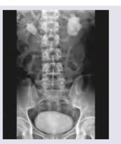
The following IVU shows:

What is the investigation of choice in a patient with blunt abdominal trauma with hematuria?
A dense persistent nephrogram may be seen in all of the following except:
Explanation: ***Iso-osmolar contrast*** - **Iso-osmolar contrast agents** (e.g., iodixanol) have an osmolality of ~290 mOsm/kg, which is identical to that of plasma. - **This is the PREFERRED choice** in patients with renal dysfunction as multiple studies demonstrate the lowest risk of contrast-induced nephropathy (CIN). - The iso-osmolar formulation minimizes osmotic stress on renal tubules and reduces the risk of acute kidney injury. - **Current guidelines recommend iso-osmolar agents as first-line** in high-risk patients with pre-existing renal impairment. *Low osmolar contrast* - **Low osmolar contrast agents** have osmolality of 600-900 mOsm/kg, which is significantly lower than high osmolar agents but still 2-3 times higher than plasma. - While **acceptable and safer than high osmolar agents**, they are not as optimal as iso-osmolar contrast for patients with renal dysfunction. - These agents are widely used and represent a reasonable alternative when iso-osmolar agents are not available. *High osmolar contrast* - **High osmolar contrast agents** have osmolality >1400 mOsm/kg (about 5 times that of plasma). - They carry the **highest risk of contrast-induced nephropathy** due to severe osmotic load and direct tubular toxicity. - **Contraindicated or strongly avoided** in patients with pre-existing renal dysfunction. *Ionic contrast* - **Ionic contrast** refers to the chemical structure (dissociates into ions) rather than osmolality. - Can be either high or low osmolar—the ionic nature alone does not determine renal safety. - The critical factor for nephrotoxicity prevention is osmolality, not ionic charge.
Explanation: ***Magnetic resonance imaging*** - **MRI** is the most sensitive imaging modality for detecting **early renal tuberculosis** due to its superior soft tissue contrast resolution. - It can identify subtle **parenchymal inflammatory changes**, early granulomas, and edema before calcification or cavitation occurs. - MRI is particularly useful for detecting **early renal cortical involvement** and small lesions that may be missed by other modalities. - Provides excellent anatomical detail without ionizing radiation. *Computed tomography* - **CT** is excellent for detecting **established renal tuberculosis** with calcifications, cavitations, and collecting system abnormalities. - However, calcifications and cavitations represent **intermediate to late findings**, not early disease. - CT is less sensitive than MRI for detecting subtle parenchymal inflammatory changes in early stages. - More useful once the disease has progressed to structural changes. *Ultrasound* - **Ultrasound** is useful for screening and follow-up but has significant limitations for early disease detection. - May miss small lesions, subtle parenchymal changes, and early calcifications. - Operator-dependent and limited by **acoustic windows** and patient body habitus. *Intravenous urography* - **IVU** primarily detects changes in the **collecting system** such as strictures, calyceal clubbing, and ureteral abnormalities. - These represent **late manifestations** of renal tuberculosis, not early disease. - Has largely been replaced by cross-sectional imaging (CT/MRI) for evaluating renal pathology. - Less sensitive for parenchymal involvement compared to CT or MRI.
Explanation: ***Correct Answer: CECT*** - **Contrast-enhanced computed tomography (CECT)** is the investigation of choice for evaluating blunt abdominal trauma with hematuria as it accurately assesses the extent of injury to the **kidneys, ureters, bladder**, and surrounding structures. - It provides detailed images for detecting **renal lacerations, hematomas, urine extravasation**, and other abdominal organ injuries. - **Gold standard** in trauma protocols for comprehensive evaluation of renal and abdominal injuries. *Incorrect: USG of the abdomen* - **Ultrasound** can identify gross abnormalities like large hematomas or free fluid but is less sensitive than CECT for subtle renal injuries or collecting system disruptions. - It is often used as an initial screening tool (FAST exam) but not the definitive investigation of choice in this context. *Incorrect: Retrograde urogram* - A **retrograde urogram** primarily evaluates the **lower urinary tract** (ureters and bladder) by injecting contrast directly into the urethra. - It is not suitable for assessing the extent of renal parenchymal injury or other abdominal organ damage in blunt trauma. *Incorrect: IVP* - **Intravenous pyelogram (IVP)** uses intravenous contrast to visualize the kidneys, ureters, and bladder, but it has largely been replaced by CECT due to its lower sensitivity and specificity for traumatic injuries. - It provides less detailed anatomical information about surrounding soft tissues and can miss subtle parenchymal or vascular injuries.
Explanation: DTPA Scan (Radiorenogram) - A DTPA scan can assess renal perfusion and glomerular filtration rate (GFR), providing crucial information about the functional status of the kidneys, particularly in cases of anuria [1]. - In the context of anuria with a normal ultrasound, it helps differentiate between pre-renal (poor perfusion), post-renal (if obstruction was missed), or intrinsic renal failure by evaluating blood flow and tracer uptake [1]. Serum Creatinine - While serum creatinine is a marker of renal function, it reflects the current state and doesn't provide real-time or dynamic information about renal blood flow or GFR at the onset of anuria [2]. - In acute renal failure, particularly with anuria, creatinine levels can rise rapidly, but assessing the cause and degree of functional impairment beyond simple filtration requires more advanced imaging. Urine Output Assessment - The patient is already presenting with complete anuria, meaning there is no urine output, rendering this assessment uninformative for further diagnostic steps. - While typically a crucial initial indicator, in this specific scenario, it only confirms the clinical presentation and does not provide insight into the underlying etiology of the anuria. Renal Biopsy - A renal biopsy is an invasive procedure primarily used for definitive diagnosis of intrinsic renal diseases and is not a first-line investigation for immediate assessment of renal function in acute anuria [2]. - It would typically be considered after non-invasive tests have failed to provide a diagnosis or if a specific intrinsic renal disease is strongly suspected and requires histological confirmation [2].
Explanation: ***Rapid (Bolus) injection of dye*** - A **rapid bolus injection** of contrast material ensures a high concentration reaches the kidneys simultaneously, leading to optimal opacification and a **dense nephrogram**. - This method allows for the collection of a **large bolus of undiluted contrast** in the renal vessels and parenchyma, improving visualization of the renal parenchyma during the nephrographic phase. - The dense nephrogram phase occurs when contrast is within the renal tubules and interstitium, producing uniform opacification. *Dehydrating the patient* - **Dehydration** would concentrate the urine in the collecting system, but it does not directly contribute to the **dense nephrogram** appearance of the renal parenchyma. - While dehydration may improve visualization of the pelvicalyceal system on delayed images, it can increase the risk of **contrast-induced nephropathy**. *Using non ionic media* - **Non-ionic contrast media** are associated with fewer adverse reactions and greater patient safety compared to ionic media due to their lower osmolality. - However, the type of contrast media (ionic vs. non-ionic) does not primarily determine the **density of the nephrogram** itself, but rather patient tolerability and safety profile. *Increasing the dose of contrast media* - While increasing the dose might provide more contrast overall, it does not guarantee a **dense nephrogram**, which requires a high concentration of contrast to be present acutely in the renal parenchyma. - A dense nephrogram is better achieved by **rapid bolus injection technique** rather than simply increasing the total dose. - Excessive contrast increases the risk of **adverse reactions** and contrast-induced nephropathy without necessarily improving nephrographic density proportionally.
Explanation: ***Ionic monomers have three iodine atoms per two particles in solution*** - **Ionic monomeric contrast agents** (e.g., diatrizoate, iothalamate) dissociate in solution, producing **two particles** (one cation and one anion containing three iodine atoms) per molecule. - This dissociation results in a **high osmolality** compared to non-ionic agents, as osmolality is determined by the number of particles in solution. *Gadolinium cannot cross an intact blood brain barrier* - This statement is **FALSE** - **Gadolinium-based contrast agents CAN cross the blood-brain barrier when it is compromised**. - They are used in MRI precisely because they extravasate into tissues where the **blood-brain barrier is disrupted**, such as in tumors, inflammation, or infections. - However, they do **not cross an intact BBB** due to their size and hydrophilicity. *Iohexol is a high osmolar contrast media* - **Iohexol** is a **non-ionic, low osmolality contrast medium** (LOCM). - Its non-ionic nature means it does not dissociate in solution, leading to a significantly lower osmolality compared to older ionic agents. *Non-ionic contrast agents are always high osmolar* - **Non-ionic contrast agents** are characterized by their molecular structure which **does not dissociate into ions** in solution. - This property makes them **low osmolar** or **iso-osmolar**, meaning they have fewer particles in solution compared to ionic agents, thereby reducing osmolality.
Explanation: ***Voiding cystourethrography (VCUG)*** - A **one-year-old male child with poor urinary stream since birth** is highly suggestive of **posterior urethral valves (PUV)**, the most common cause of bladder outlet obstruction in male infants. - **VCUG is the investigation of choice** for diagnosing PUV as it directly visualizes the posterior urethra during voiding and can demonstrate the characteristic findings: dilated posterior urethra, valve leaflets, bladder trabeculation, and vesicoureteral reflux. - While it involves catheterization and radiation, in this classic presentation, VCUG provides definitive diagnosis and is essential for surgical planning. *USG bladder* - Ultrasound is a useful **non-invasive screening tool** that can detect secondary findings such as hydronephrosis, bladder wall thickening, and increased post-void residual. - However, **USG cannot visualize the urethral valves** themselves and cannot definitively diagnose PUV. - In practice, many centers may perform ultrasound first, but it must be followed by VCUG for definitive diagnosis in this clinical scenario. *Uroflowmetry* - This test measures the **rate of urine flow** and requires patient cooperation with voiding. - A **one-year-old child cannot reliably follow instructions** to perform uroflowmetry. - It is more useful in older, cooperative children and adults. *Intravenous urography* - **Intravenous urography (IVU)** involves contrast administration and multiple X-rays to visualize the urinary tract. - It has been largely **replaced by ultrasound and CT urography** due to better imaging quality and safety profile. - IVU does not adequately visualize the urethra or diagnose urethral pathology like PUV.
Explanation: ***Horseshoe kidney*** - The image shows both kidneys are **fused at their lower poles** across the midline, forming a "horseshoe" shape, which is a classic radiographic finding for this condition. - The **calyces and renal pelves are seen medially oriented**, supporting the diagnosis of a horseshoe kidney. *Hydronephrosis* - **Hydronephrosis** would appear as a dilation of the renal pelvis and calyces due to obstruction, which is not the primary finding here. - While a horseshoe kidney can be associated with hydronephrosis due to an abnormal ureteral course, the image clearly depicts the **fused renal parenchyma** rather than just dilation. *Polycystic kidney* - **Polycystic kidneys** are characterized by numerous cysts of varying sizes replacing normal renal parenchyma, which would present as enlarged, multi-cystic kidneys on imaging. - The image does not show multiple cysts replacing the renal tissue but rather a **fused, single-mass structure** in the lower abdomen. *Duplication of collecting system* - **Duplication of the collecting system** involves two ureters draining a single kidney, or a bifid renal pelvis, which would appear as double ureters or collecting systems on an IVU. - This condition does not present with the characteristic **fusion of the renal poles** across the midline as seen in the image.
Explanation: ***Contrast enhanced computed tomography (CECT)*** - **CECT** is the **investigation of choice** for evaluating solid organ injuries, including renal trauma, in hemodynamically stable patients with blunt abdominal trauma and hematuria. - It provides detailed imaging of the kidneys, urinary tract, and surrounding structures, allowing for the classification of injury severity and identification of associated injuries. *Retrograde urogram* - A retrograde urogram is primarily used to evaluate the **lower urinary tract** (urethra and bladder) for strictures or injuries, specifically when there is a suspicion of urethral injury. - It is not the primary imaging modality for assessing renal parenchymal or collecting system injuries from blunt trauma. *Intravenous urogram (IVU)* - While an IVU can assess the upper urinary tract, it has largely been replaced by **CECT** in the acute trauma setting due to CECT's superior resolution and ability to evaluate renal parenchyma and other abdominal organs. - IVU exposes the patient to radiation and requires contrast administration, and it may not adequately visualize subtle renal injuries or hematomas as effectively as CECT. *Ultrasonography of abdomen* - **Ultrasound** is useful for rapidly detecting free fluid (suggesting hemorrhage) or gross hydronephrosis in trauma, but it has limited sensitivity for diagnosing specific renal parenchymal injuries or urinary extravasation. - Its role in blunt abdominal trauma with hematuria is often as an initial screening tool, but it is not sufficient for definitive diagnosis or grading of renal injuries.
Explanation: ***Systemic hypertension*** - **Systemic hypertension** is not typically associated with a dense, persistent nephrogram on imaging. While chronic hypertension can cause renal damage, it does not directly lead to the characteristic prolonged parenchymal enhancement. - A dense, persistent nephrogram suggests impaired contrast excretion or increased reabsorption, neither of which is a primary manifestation of systemic hypertension itself. *Severe hydronephrosis* - **Severe hydronephrosis** leads to impaired urine flow and delayed transit of contrast medium through the renal tubules, resulting in a persistent nephrogram. - The dilated collecting system and compressed parenchyma can retain contrast for an extended period due to reduced glomerular filtration rate (GFR) in the affected kidney. *Dehydration* - In cases of **dehydration**, the kidneys attempt to conserve water, leading to increased reabsorption of water from the renal tubules. - This process can concentrate the contrast medium within the tubules, resulting in a denser and more persistent nephrogram as it slowly transits through the kidney. *Acute ureteral obstruction* - **Acute ureteral obstruction** causes a build-up of pressure within the renal collecting system, impairing glomerular filtration and slowing the passage of contrast. - The contrast medium remains within the renal parenchyma for a prolonged period due to the blockage, leading to a dense and persistent nephrogram and delayed excretion.
Get full access to all questions, explanations, and performance tracking.
Start For Free