All of the following are indications for hemodialysis in acute kidney injury, EXCEPT:
Which of the following is Iso-osmolar agent?
A dense persistent nephrogram may be seen in all of the following except:
Which part of the kidney is first affected by ischemia in the context of acute kidney injury?
Immediately after kidney donation what happens to the creatinine level in the donors?
Which of the following drugs is associated with untoward side effect of renal tubular damage?
Following surgery, a patient develops oliguria. You believe the patient is hypovolemic, but you seek corroborative data before increasing intravenous fluids. The best data is?
Uremic complications typically arise during which of the following phases of renal failure?
Excretory urography should be cautiously performed in
Even conventional radiological procedures are contraindicated in which neurological disease?
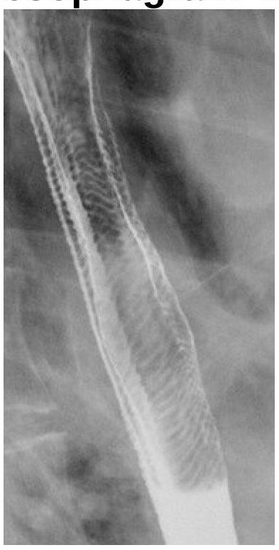
Explanation: ***Hypertension*** - While hypertension can be a complication of **acute kidney injury (AKI)**, it is generally managed with **antihypertensive medications** and **fluid removal**, and does not by itself necessitate urgent hemodialysis unless it is severe and refractory, alongside other uremic symptoms. - Hemodialysis primarily addresses life-threatening electrolyte imbalances, fluid overload, and uremic symptoms. [2] *Severe metabolic acidosis* - **Severe metabolic acidosis (pH < 7.1)** is a critical indication for hemodialysis in AKI because the kidneys are unable to excrete acid or regenerate bicarbonate. - Hemodialysis can rapidly remove acids and correct the pH imbalance, preventing further organ dysfunction. *Hyperkalemia* - **Life-threatening hyperkalemia (potassium > 6.5 mEq/L)**, especially when refractory to medical management (e.g., insulin, glucose, calcium gluconate), is a major indication for hemodialysis. [1] - Hemodialysis is highly effective at rapidly lowering potassium levels, which is crucial to prevent cardiac arrhythmias. [1] *Pulmonary edema* - **Severe fluid overload** leading to **pulmonary edema** that is refractory to diuretic therapy is a strong indication for hemodialysis in AKI. [2] - Hemodialysis can efficiently remove excess fluid, thereby alleviating respiratory distress and improving oxygenation.
Explanation: ***Non-ionic Dimer contrast media*** - **Iodixanol** is the only available non-ionic dimer contrast agent, and it is **iso-osmolar** with blood plasma (290 mOsm/kg). - Its iso-osmolality contributes to a lower incidence of adverse reactions, particularly in patients at high risk. *Ionic Monomer - High osmolality contrast media* - These agents have an osmolality significantly higher than that of blood plasma, often 6-8 times greater. - High osmolality leads to a higher incidence of adverse effects due to cellular fluid shifts and direct endothelial damage. *Non-ionic Monomer - Low osmolality contrast media* - These agents have an osmolality lower than ionic monomers but are still hyperosmolar compared to blood plasma (typically 2-3 times higher). - While generally safer than high-osmolality agents, they can still cause discomfort and adverse reactions due to their hyperosmolality. *Ionic Dimer - Low osmolality contrast media* - Ionic dimers, such as **ioxaglate**, are considered low-osmolality agents but are still hyperosmolar relative to plasma. - They feature two benzene rings with iodine atoms and are salts, contributing to their osmolality.
Explanation: ***Systemic hypertension*** - **Systemic hypertension** is not typically associated with a dense, persistent nephrogram on imaging. While chronic hypertension can cause renal damage, it does not directly lead to the characteristic prolonged parenchymal enhancement. - A dense, persistent nephrogram suggests impaired contrast excretion or increased reabsorption, neither of which is a primary manifestation of systemic hypertension itself. *Severe hydronephrosis* - **Severe hydronephrosis** leads to impaired urine flow and delayed transit of contrast medium through the renal tubules, resulting in a persistent nephrogram. - The dilated collecting system and compressed parenchyma can retain contrast for an extended period due to reduced glomerular filtration rate (GFR) in the affected kidney. *Dehydration* - In cases of **dehydration**, the kidneys attempt to conserve water, leading to increased reabsorption of water from the renal tubules. - This process can concentrate the contrast medium within the tubules, resulting in a denser and more persistent nephrogram as it slowly transits through the kidney. *Acute ureteral obstruction* - **Acute ureteral obstruction** causes a build-up of pressure within the renal collecting system, impairing glomerular filtration and slowing the passage of contrast. - The contrast medium remains within the renal parenchyma for a prolonged period due to the blockage, leading to a dense and persistent nephrogram and delayed excretion.
Explanation: ***Outer medulla*** - The **outer medulla** is particularly vulnerable to ischemia due to its high metabolic demand and limited blood supply. - Ischemic damage typically begins here as it receives blood supply from the **vasa recta**, which are more susceptible to drops in perfusion pressure. *Glumerulus* - The **glomerulus** is primarily affected in conditions like **glomerulonephritis**, not in acute ischemic injury where tubular structures are first impacted [1]. - It is well-perfused under normal conditions, making it less likely to be the first area affected during acute kidney injury. *Cortex* - The **cortex** is indeed involved in acute kidney damage but is not the first area affected by ischemia. - The cortical region can withstand lower perfusion volumes for a shorter time compared to the outer medulla. *Inner medulla* - The **inner medulla** is the last area to suffer from ischemic damage as it is more tolerant to **hypoxic conditions**. - It primarily encounters ischemia after the outer medulla has already been compromised, thus not the first area affected. **References:** [1] Kumar V, Abbas AK, et al.. Robbins and Cotran Pathologic Basis of Disease. 9th ed. The Kidney, p. 933.
Explanation: ***Increases*** - Following the donation of one kidney, the remaining kidney experiences a temporary **reduction in overall renal mass** and a subsequent **transient decrease in glomerular filtration rate (GFR)**. - This immediate post-operative decrease in GFR leads to a **temporary rise in serum creatinine** as the body adjusts to the function of a single kidney. *Level is independent of the donation* - This statement is incorrect because the GFR is directly related to the total functional renal mass, which changes significantly after **nephrectomy**. - Renal function, as measured by creatinine, is undeniably affected by the **loss of a kidney**. *Decreases* - Creatinine levels would decrease if the **GFR of the remaining kidney improved significantly** or if there was an underlying condition causing an already elevated creatinine to normalize post-donation, neither of which is the immediate physiological response. - A decrease in creatinine after donation would imply improved kidney function or reduced burden, which is not what occurs acutely. *Remains Same* - This is unlikely because the removal of one kidney immediately **reduces the total filtration capacity** of the body by approximately half, even if there's rapid compensatory hypertrophy. - While the remaining kidney will undergo **compensatory hypertrophy and hyperfiltration** in the long term, the immediate effect is a reduction in overall GFR.
Explanation: ***Cisplatin*** - **Cisplatin** is a platinum-based chemotherapy drug well-known for its dose-limiting nephrotoxicity, primarily causing **renal tubular damage**. - Its mechanism involves direct DNA damage within renal tubular cells, leading to **acute tubular necrosis** if not managed with aggressive hydration and other protective measures. *Streptozotocin* - **Streptozotocin** is an alkylating agent primarily used in treating **pancreatic neuroendocrine tumors**; its main toxicity is typically to pancreatic beta cells (leading to hypoglycemia) and to the liver. - While it can be nephrotoxic, its predominant and most recognized untoward side effect is not renal tubular damage, but rather **pancreatic beta-cell destruction**. *Methysergide* - **Methysergide** is an ergot alkaloid used for **migraine prophylaxis** but is largely discontinued due to severe side effects like **retroperitoneal fibrosis**. - Renal damage in the context of methysergide is typically due to this fibrosis compressing the ureters, rather than direct tubular toxicity. *Cyclophosphamide* - **Cyclophosphamide** is an alkylating agent known for its immunosuppressive and chemotherapeutic effects; its major side effects include **hemorrhagic cystitis** and myelosuppression. - While high doses can cause nephrotoxicity, its primary and most feared renal-related toxicity is hemorrhagic cystitis, not direct tubular damage as seen with cisplatin.
Explanation: ***Fractional excretion of sodium less than 1*** - A **fractional excretion of sodium (FENa) less than 1%** is a classic indicator of **prerenal azotemia** or hypovolemia, as the kidneys are avidly reabsorbing sodium and water to preserve circulating volume. - This indicates the kidneys are functioning appropriately in response to perceived hypoperfusion, attempting to conserve sodium and thus water. *Urine chloride of 15 meq/L* - While a **low urine chloride** can sometimes be seen in volume depletion, it is not as specific or reliable an indicator of hypovolemia as FENa. - Urine chloride is more helpful in differentiating causes of **metabolic alkalosis**, particularly saline-responsive versus saline-unresponsive. *Urine sodium of 28 meq/L* - A urine sodium concentration of **less than 20 mEq/L** is a more classic cutoff for prerenal azotemia/hypovolemia, indicating aggressive sodium reabsorption. - A value of 28 mEq/L, although relatively low, is less definitive than a low FENa in strongly supporting hypovolemia. *Urine/Serum creatinine ratio of 20* - A **urine/serum creatinine ratio greater than 20:1** is indicative of prerenal azotemia, suggesting the kidneys are concentrating urine in response to hypovolemia. - While supportive, FENa is often considered a more precise and widely accepted marker, especially in the absence of diuretic use or chronic kidney disease.
Explanation: ***Maintenance*** - During the **maintenance phase**, renal function is severely impaired, leading to the accumulation of **uremic toxins** and metabolic waste products. - This prolonged period of reduced kidney function is when **uremic complications** such as pericarditis, encephalopathy, and coagulopathy typically manifest. *Initiation* - The **initiation phase** is characterized by the initial insult to the kidneys and the onset of reduced glomerular filtration, but significant uremic complications are usually not yet apparent. - It is a period of evolving injury, and the body's compensatory mechanisms may still be able to mitigate acute toxicity. *Diuretic Phase* - The **diuretic phase** is a period of gradual improvement from renal failure, where urine output increases, but the kidneys may still have impaired ability to concentrate urine or fully excrete waste. - While electrolyte imbalances can occur, severe uremic complications are less common as renal function starts to recover. *Recovery Phase* - In the **recovery phase**, renal function gradually normalizes, and the kidneys regain their ability to excrete waste products effectively. - Uremic complications would typically be resolving, not arising, during this phase as **renal repair** takes place.
Explanation: ***Multiple myeloma*** - Excretory urography (intravenous pyelography or IVP) involves the administration of **iodinated contrast media**, which can precipitate **Bence Jones proteins** in the renal tubules, leading to or worsening **acute kidney injury** in patients with multiple myeloma. - Patients with multiple myeloma often have **pre-existing renal dysfunction** (myeloma kidney) due to light chain deposition, making them highly susceptible to contrast-induced nephropathy. *Bone metastases* - While bone metastases can be painful and may require imaging, they do not directly contraindicate excretory urography; the primary concern with IVP is renal function. - The presence of bone lesions itself does not increase the risk of **contrast-induced nephropathy** in the same way that proteinuria from multiple myeloma does. *Neuroblastoma* - Neuroblastoma is a **childhood cancer** affecting the adrenal glands or sympathetic nervous system, and it is not typically associated with a specific risk for contrast-induced nephropathy from excretory urography. - The primary diagnostic imaging for neuroblastoma often involves ultrasound, CT, or MRI, and while contrast may be used, the specific renal risk seen in multiple myeloma is not present. *Leukemia* - While some forms of leukemia can affect the kidneys, particularly through infiltration, it does not typically pose the same specific risk for **contrast-induced nephropathy** as multiple myeloma. - The renal manifestations in leukemia are generally different from the **light chain proteinuria** seen in multiple myeloma, which directly interacts with iodinated contrast.
Explanation: ***Ataxia telangiectasia*** - Patients with **ataxia telangiectasia** have a defect in the **ATM gene**, leading to extreme sensitivity to **ionizing radiation**, making conventional radiological procedures unsafe. - This increased radiosensitivity can result in severe adverse reactions, including increased risk of **malignancy** and neurological damage if exposed to routine diagnostic radiation. *Cockayne Syndrome* - **Cockayne Syndrome** is characterized by a defect in **DNA repair**, specifically **transcription-coupled repair**, leading to pronounced sun sensitivity and premature aging. - While these patients are sensitive to UV radiation, they do not have the profound hypersensitivity to **ionizing radiation** that contraindicates conventional X-ray imaging, distinguishing them from ataxia telangiectasia. *Gorlin Syndrome* - **Gorlin Syndrome** (Nevoid Basal Cell Carcinoma Syndrome) is associated with an increased risk of developing various cancers, including **basal cell carcinomas**, and is linked to the **PTCH1 gene**. - Although individuals with Gorlin Syndrome have an increased lifetime risk of developing tumors with **ionizing radiation exposure**, it does not typically contraindicate conventional diagnostic imaging, unlike the extreme radiosensitivity seen in ataxia telangiectasia. *All of the options* - This option is incorrect because while Cockayne Syndrome and Gorlin Syndrome involve heightened cancer risks or sensitivities, only **ataxia telangiectasia** presents a direct and severe contraindication to conventional radiological procedures due to extreme **radiosensitivity**.
Get full access to all questions, explanations, and performance tracking.
Start For Free