What is the most common view used for a chest X-ray?
The 'Floating Water Lily' sign is associated with which of the following conditions?
What is the investigation of choice for a Pancoast tumor?
Round pneumonia is seen with
Finger-in-glove sign is seen in
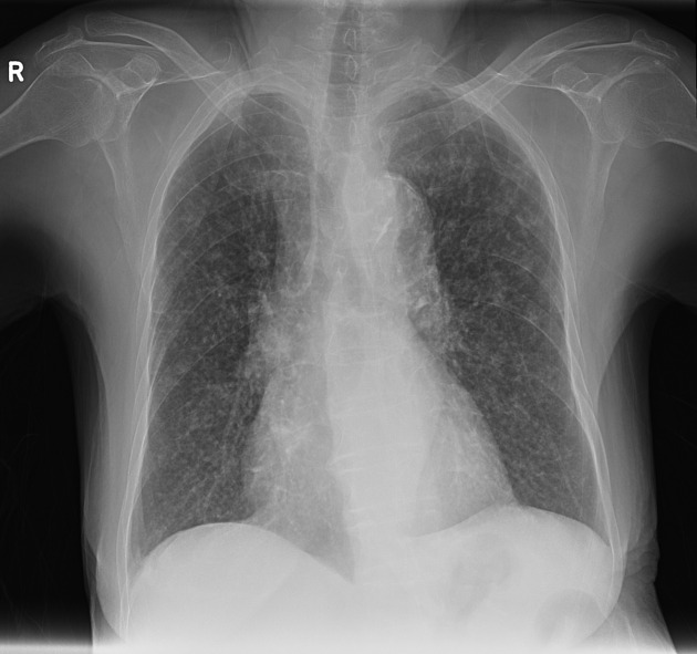
Based on the provided chest X-ray image of a patient presenting with low-grade fever, which infection is most likely?

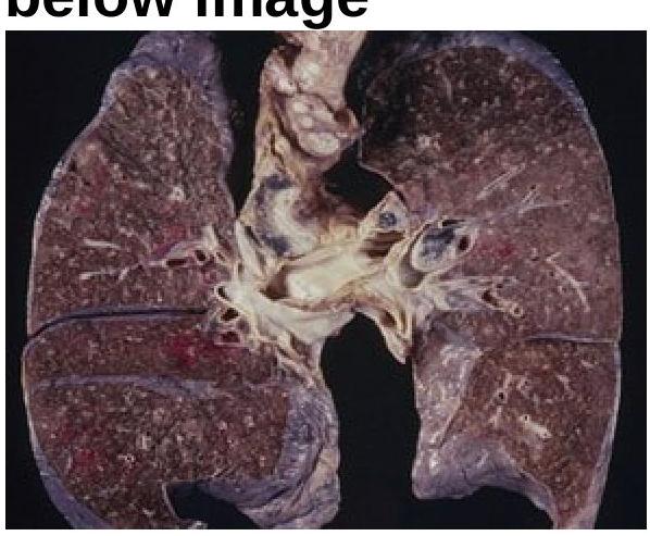
Identify the condition represented in the image.

A 55-year-old hypertensive patient presents with severe chest pain radiating to the back. What does a CT scan of the thorax typically reveal?
On CT chest, the 'halo sign' is particularly associated with which condition in immunocompromised patients?
Which condition is characterized by a specific appearance on CT scans that resembles small centrilobular nodules with branching linear structures?
Explanation: **PA view** - The **posteroanterior (PA) view** is the most common and preferred projection for routine chest X-rays. - This view minimizes **cardiac magnification** and provides a clearer demonstration of lung fields and pulmonary vasculature. *AP view* - The **anteroposterior (AP) view** is typically used for patients who are unable to stand, such as those who are bedridden or critically ill. - It causes **magnification of the heart shadow** and often results in poorer image quality due to the shorter source-to-image receptor distance. *Lateral view* - A **lateral view** is commonly performed in addition to the PA view to provide a three-dimensional perspective and help localize abnormalities. - It is not usually taken as the sole or primary projection for a general chest X-ray. *Oblique view* - **Oblique views** are specialized projections used to visualize specific areas of the chest more clearly, often to differentiate structures or investigate findings seen on standard PA or lateral views. - They are not considered a standard or routine view for initial chest imaging.
Explanation: ***Ruptured Hydatid Cyst*** - The **'Floating Water Lily' sign**, also known as the **'Water Lily sign'** or **'Camelot sign'**, is pathognomonic for a **ruptured pulmonary hydatid cyst**. - It occurs when the endocyst membrane ruptures and detaches from the pericyst, flaring up within the existing cyst fluid, creating a characteristic appearance on imaging. *Pulmonary Aspergillosis* - While pulmonary aspergillosis can form **fungus balls (aspergillomas)** within pre-existing lung cavities, the characteristic sign is the **'air crescent sign'**, not the 'floating water lily' sign. - Aspergillomas typically involve the fungus growing in a cavity, often with an air interface, but do not feature a detached, floating membrane. *Pulmonary Hamartoma* - A pulmonary hamartoma is a **benign tumor** of the lung, typically appearing as a solitary, well-defined nodule. - It does not rupture or form fluid-filled structures that would give rise to the 'floating water lily' sign. *Cavitating Lung Metastasis* - Cavitating lung metastases are malignant lesions that undergo **central necrosis**, leading to cavity formation. - Although they form cavities, they do not involve the detachment of parasitic membranes, and therefore, the 'floating water lily' sign is not observed.
Explanation: ***MRI*** - **Magnetic Resonance Imaging (MRI)** is the investigation of choice for a Pancoast tumor due to its superior ability to visualize **soft tissue invasion** of structures in the **thoracic inlet**, such as the brachial plexus, subclavian vessels, and vertebral bodies. - This detailed assessment of local invasion is crucial for **surgical planning** and determining resectability. *HRCT* - **High-Resolution Computed Tomography (HRCT)** is primarily used for evaluating **diffuse parenchymal lung diseases** and provides detailed imaging of lung architecture, not for tumor staging. - It is not optimal for assessing **soft tissue extension** into the surrounding structures of the thoracic inlet, which is characteristic of a Pancoast tumor. *CECT* - **Contrast-Enhanced Computed Tomography (CECT)** is excellent for identifying the presence of a lung mass and evaluating its relationship to adjacent structures, as well as for **mediastinal lymph node staging** and detecting distant metastases. - While useful, its ability to precisely delineate **neural and vascular invasion** in the thoracic inlet is inferior to MRI. *Bronchography* - **Bronchography** is an outdated and invasive procedure that involves injecting contrast into the tracheobronchial tree to visualize the airways. - It has been largely replaced by CT and bronchoscopy and is not used for the diagnosis or staging of lung tumors like a Pancoast tumor.
Explanation: ***Streptococcus pneumoniae*** - **Round pneumonia**, a well-defined spherical opacification on chest radiography, is a characteristic presentation of pneumonia caused by ***Streptococcus pneumoniae***, particularly in children. - The organism's ability to incite a rapid, localized inflammatory response can lead to the formation of this focal, rounded consolidation. *Kerosene oil aspiration pneumonia* - Kerosene oil aspiration typically causes a **diffuse, bilateral pneumonitis** with chemical irritation, not a well-circumscribed round lesion. - The radiographic pattern is usually one of prominent **ground-glass opacities** and consolidations, often in the lower lobes. *Mendelson syndrome pneumonia* - Mendelson syndrome, or **aspiration pneumonitis**, involves chemical lung injury from aspirating gastric contents, leading to **diffuse inflammation** and edema. - It typically presents as **patchy or diffuse infiltrates**, often bilateral and basilar, rather than a solitary round infiltrate. *Lung cancer* - While lung cancer can present as a **round mass** or nodule, it is a neoplastic process, not an infectious pneumonia. - A distinguishing feature is that lung cancer masses typically show **growth over time** and may have irregular margins or spiculation, unlike the acute inflammatory nature of round pneumonia.
Explanation: ***Bronchocele*** - The **finger-in-glove sign** is a characteristic radiological finding that represents **mucoid impaction** – a **bronchus distended with mucus**, appearing as branching, finger-like opacities. - **Bronchocele** refers to this mucus-filled, dilated bronchus caused by bronchial obstruction. - The **most common cause** of the finger-in-glove sign is **allergic bronchopulmonary aspergillosis (ABPA)**, though it can also be seen in bronchial atresia, asthma with mucus plugging, and other causes of bronchial obstruction with mucostasis. *Pulmonary alveolar proteinosis* - This condition is characterized by the accumulation of **lipoproteinaceous material within the alveoli**, not the bronchi. - Radiologically, it often presents as **diffuse ground-glass opacities** and **interlobular septal thickening**, known as a "crazy-paving" pattern, which is distinct from the finger-in-glove sign. *Pneumocystis jirovecii* - **Pneumocystis pneumonia (PCP)** typically affects immunocompromised individuals and is characterized by a diffuse **interstitial pneumonia**. - Radiological features include diffuse **ground-glass opacities**, reticular patterns, and sometimes cysts, but not the mucinous impaction seen in the finger-in-glove sign. *Tuberculosis* - Tuberculosis can manifest in various forms, including **cavitary lesions**, **nodules**, consolidation, and **pleural effusions**. - While it can cause bronchial narrowing, it does not typically lead to the distinct finger-in-glove appearance caused by mucoid impaction within dilated bronchi.
Explanation: ***Miliary Tuberculosis*** - The chest X-ray shows diffuse, fine, granular infiltrates, often described as a **"millet seed" pattern**, which is highly characteristic of **miliary tuberculosis**. - This pattern represents widespread hematogenous dissemination of *Mycobacterium tuberculosis* and is consistent with systemic symptoms like **low-grade fever**. - The **uniform distribution** of 1-3 mm nodules throughout both lung fields is pathognomonic for miliary TB. *Lobar Pneumonia* - **Lobar pneumonia** appears as a homogenous opacification of a lung segment or entire lobe, often with obscuration of vascular markings and sometimes with **air bronchograms**. - The image does not show a dense, localized area of consolidation, but rather a diffuse, fine nodular pattern throughout both lung fields. - Lobar pneumonia typically presents with high-grade fever and acute symptoms, not the low-grade fever described. *Interstitial Lung Disease* - While interstitial lung disease can present with widespread infiltrates, these are typically more varied, showing patterns like **reticular nodularities**, **honeycombing**, or **ground-glass opacities**. - The uniform, fine nodularity seen in the image is not typical for most forms of interstitial lung disease. - ILD is usually a chronic process, whereas miliary TB can present acutely or subacutely. *Bronchopneumonia* - **Bronchopneumonia** commonly presents as patchy, multifocal areas of consolidation or opacities that are often centered around bronchi in a **bronchocentric distribution**. - The widespread, uniform, and fine nodular pattern seen on this X-ray is distinct from the typical patchy, asymmetric appearance of bronchopneumonia. - Bronchopneumonia shows more confluent opacities rather than discrete miliary nodules.
Explanation: ***Miliary tuberculosis*** - The image displays lungs riddled with numerous small (1-5 mm), uniform **granulomas**, resembling millet seeds, which is characteristic of **miliary tuberculosis**. - This pattern results from the **hematogenous dissemination** of **Mycobacterium tuberculosis** throughout the body, including the lungs. *Bronchiectasis* - Bronchiectasis is characterized by **irreversible dilation of bronchi** with chronic inflammation and infection, which would appear as dilated airways and associated scarring on gross pathology, not diffuse small nodules. - While it can be a complication of tuberculosis, the primary gross feature in the image is not dilated bronchi. *COPD* - Chronic Obstructive Pulmonary Disease (COPD) typically manifests as **emphysema** (enlarged airspaces with destruction of alveolar walls) or **chronic bronchitis** (mucus gland hypertrophy and inflammation of airways). - These conditions would present with large areas of destroyed lung tissue, mucous plugging, or thickened bronchial walls, which are distinctly different from the numerous small nodules seen in the image. *Lung cancer* - Lung cancer usually presents as a **mass or nodule**, which can be solitary or multiple, but typically larger and more irregular than the uniform small nodules in this image. - While metastatic lung cancer can present with multiple nodules, the uniform size and diffuse distribution seen here are far more typical of miliary spread of an infection.
Explanation: ***Type B Aortic dissection*** - A CT scan of the thorax would show a **tear in the descending aorta**, typically distal to the left subclavian artery, consistent with a type B dissection. - The combination of **hypertension** and **severe chest pain radiating to the back** is highly suggestive of an aortic dissection, and involvement of the descending aorta (Type B) often presents with radiating back pain. *Type A Aortic dissection* - A type A dissection involves the **ascending aorta**, which would typically present with chest pain radiating to the neck or jaw, or acute onset of symptoms suggestive of a stroke or myocardial infarction. - While it's also an aortic dissection, its location is different, and the pain typically radiates to different areas. *Pulmonary Embolism* - A **pulmonary embolism** would appear as a filling defect in the pulmonary arteries on a CT angiogram, not a tear in the aorta. - Though it can cause chest pain, it's typically pleuritic and not usually described as severe and radiating to the back in the same manner as an aortic dissection. *Thoracic Aortic Aneurysm* - A **thoracic aortic aneurysm** is a localized dilation of the aorta, not a tear, and it typically presents with chronic or less acute symptoms unless it ruptures or causes compression. - While an aneurysm can be present, the acute and severe nature of the pain described points more strongly to an acute event like dissection rather than a stable aneurysm.
Explanation: ***Invasive pulmonary aspergillosis*** - The **halo sign** on CT chest, characterized by a ground-glass opacity surrounding a nodule, is a classic radiographic finding in **invasive pulmonary aspergillosis**, especially in immunocompromised patients. - This sign represents hemorrhage around the fungal nodule and indicates active tissue invasion by *Aspergillus* species. *Pulmonary hydatid cyst* - Hydatid cysts are typically well-defined, thin-walled cystic lesions, often displaying the **water lily sign** if complicated by rupture, which is different from the halo sign. - These cysts are caused by the larval stage of *Echinococcus granulosus* and are not associated with a peripheral ground-glass opacity. *Round pneumonia* - Round pneumonia is a localized, **spherical consolidation** often seen in children, which does not typically exhibit the perilesional ground-glass opacity characteristic of the halo sign. - It usually represents bacterial infection and resolves with antibiotics, unlike the invasive fungal disease suggested by the halo sign. *Bronchiectasis* - Bronchiectasis is characterized by **irreversible dilation of the bronchi**, often appearing as "tram-track" opacities or "signet ring" signs on CT. - It is a chronic condition related to airway damage and mucus retention, and not associated with acute nodular lesions or the halo sign.
Explanation: ***Pulmonary tuberculosis*** - This description ("small centrilobular nodules with **branching linear structures**") is characteristic of the **tree-in-bud pattern** seen on CT scans, which is a hallmark finding in active **endobronchial spread of tuberculosis**. - The tree-in-bud pattern results from the impaction of tuberculous **granulomas** and caseous material in the terminal and respiratory bronchioles. *Silicosis* - Characterized by multiple small, well-defined **nodules** (often in the upper lobes) that tend to calcify, but typically lacks the fine **branching linear structures**. - It’s associated with occupational exposure to **silica dust** and may progress to **massive progressive fibrosis**. *Pulmonary hydatid cyst* - Presents as a well-defined, usually **single, large cystic lesion** on CT, often with internal membranes if ruptured (water lily sign or crumpled membrane sign). - It does not typically manifest with small centrilobular nodules or branching linear structures. *Small cell carcinoma* - Usually appears as a **large central mass**, often with mediastinal lymphadenopathy, and sometimes associated with obstructive pneumonitis. - It does not typically present as diffuse small centrilobular nodules with branching patterns.
Normal Chest Radiographic Anatomy
Practice Questions
Radiographic Signs in Chest Imaging
Practice Questions
Pulmonary Infections
Practice Questions
Chronic Obstructive Pulmonary Disease
Practice Questions
Interstitial Lung Diseases
Practice Questions
Pulmonary Neoplasms
Practice Questions
Pleural Diseases
Practice Questions
Mediastinal Pathology
Practice Questions
Congenital and Developmental Chest Anomalies
Practice Questions
Pulmonary Vascular Diseases
Practice Questions
Chest Trauma Imaging
Practice Questions
Post-Surgical Chest Imaging
Practice Questions
Get full access to all questions, explanations, and performance tracking.
Start For Free