A chest X-ray shows a 'silhouette sign' with opacity obscuring the right heart border. Which lobe of the lung is most likely affected?
Which is NOT a feature of pleural effusion?
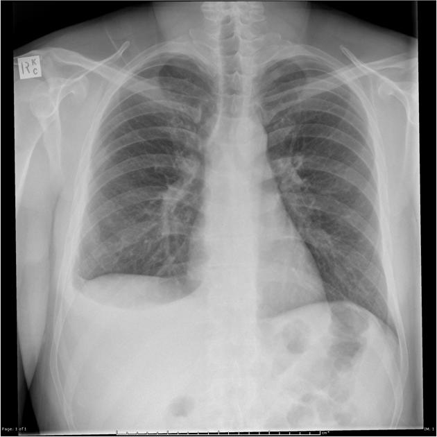
PA view of chest X-ray is given here. What is the diagnosis?

A patient presented with complaints of dyspnoea. The shown X-ray is suggestive of:

Mark the false statement regarding testing of COVID-19.
In a child with coarctation of aorta, all the following are seen in plain chest radiograph except:
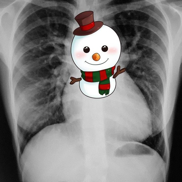
Snowman appearance on x-ray is seen in which cardiac pathology -

A female patient with clinical symptoms of systemic sclerosis presents with shortness of breath and bilateral basal rales. Her chest X-ray showed reticular opacities in bilateral basal fields. What is the next best step?
A patient of Scleroderma presents with acute respiratory distress. Chest X-ray shows B/L reticular basilar shadows. What is the next line of investigation in this patient?
Chest CT shows bilateral ground-glass opacities with crazy paving pattern and preserved bronchial markings. Likely diagnosis?
Explanation: ***Right middle lobe*** - The **silhouette sign** occurs when two objects of similar radiographic density are in direct contact, obscuring their common border. - The **right middle lobe** is adjacent to the right heart border, so an opacity in this lobe will typically obscure the border. *Right upper lobe* - The right upper lobe is positioned superiorly and medially, meaning opacification would more likely obscure the **right paratracheal stripe** or the superior mediastinal borders. - It does not directly border the right heart, thus it would not produce a silhouette sign with the cardiac outline. *Right lower lobe* - The right lower lobe is primarily associated with obscuring the **right hemidiaphragm** when it collapses or becomes consolidated. - Although it is somewhat posterior to the heart, it usually does not directly obscure the anterior right heart border. *Left lower lobe* - The left lower lobe is on the opposite side of the chest and opacification would not affect the **right heart border**. - Consolidation here would more likely obscure the **left hemidiaphragm** or the medial part of the left cardiac silhouette in certain views.
Explanation: ***Muffled heart sound*** - This is **NOT a feature of pleural effusion** and is the correct answer to this negation question. - Muffled heart sounds are characteristic of **pericardial effusion** or **cardiac tamponade**, where fluid accumulates in the pericardial sac around the heart itself. - Pleural effusion involves fluid in the pleural space surrounding the lungs, not the heart. - While massive pleural effusions can displace mediastinal structures, they do not typically cause muffled heart sounds. *Horizontal fluid level* - This **IS a feature** when air is also present in the pleural space (**hydropneumothorax**). - In **simple pleural effusion** (fluid only), the fluid typically forms a **meniscus-shaped curve** with blunting of the costophrenic angles on upright chest X-ray, not a horizontal level. - However, when both air and fluid are present, a distinct horizontal air-fluid level is visible on upright imaging. - Since the question asks about pleural effusion broadly, and effusions can be associated with air (empyema with gas-forming organisms, post-thoracentesis), this can be considered a radiological feature in certain contexts. *Low lung volume* - This **IS a feature** of pleural effusion. - The accumulating pleural fluid causes **compression atelectasis** of the adjacent lung parenchyma. - This results in **reduced functional lung volume** on the affected side, visible on chest imaging. *Decreased chest movements* - This **IS a feature** of pleural effusion. - Fluid in the pleural space restricts normal lung expansion and chest wall movement. - On physical examination, there are **diminished respiratory excursions** on the affected side. - This is one of the classic clinical signs of pleural effusion.
Explanation: ***Right Pneumothorax with left tracheal shift*** - The image shows a large **radiolucency (black area) on the right side** of the chest, indicative of **air in the pleural space**, consistent with a **right-sided pneumothorax**. - The **trachea is shifted towards the left** (away from the pneumothorax), which is the **expected finding** in pneumothorax due to increased pressure in the right pleural space pushing mediastinal structures to the contralateral side. - In pneumothorax, the trachea and mediastinum shift **away from** the affected side due to the pressure effect of air accumulation in the pleural cavity. - This **contralateral tracheal deviation** is a classic radiological sign of pneumothorax and helps confirm the diagnosis. *Right Pneumothorax with right tracheal shift* - While the **right pneumothorax** is correctly identified, the tracheal shift direction is incorrect. - In pneumothorax, the trachea shifts **away from** the affected side (contralateral), not toward it (ipsilateral). - **Ipsilateral tracheal shift** would suggest volume loss (atelectasis) or lung collapse, not pneumothorax alone. *Left Pneumothorax with right tracheal shift* - The pneumothorax is clearly on the **right side**, not the left. - The radiolucency and absent lung markings are visible on the right hemithorax. - A left pneumothorax would show these findings on the left side. *Left Pneumothorax with left tracheal shift* - There is **no pneumothorax on the left side** of the chest. - The left lung shows normal vascular markings and no evidence of pleural air. - This combination would be medically implausible as it suggests pneumothorax with ipsilateral shift.
Explanation: ***Pleural effusion*** - The X-ray shows a significant **right-sided pleural effusion** with blunting of the costophrenic angle and a meniscus sign characteristic of fluid accumulation in the pleural space. - Key radiological features include: **homogeneous opacity** in the lower zone, **obliteration of the costophrenic angle**, and the typical **concave upper border (meniscus sign)** of fluid layering. - The presence of dyspnea with these radiological findings is consistent with pleural effusion. **Note:** While the X-ray confirms pleural effusion, **differentiating between exudative and transudative effusion requires pleural fluid analysis** (Light's criteria), not imaging alone. *Pneumothorax* - A pneumothorax would appear as a **dark, air-filled space** with a visible **visceral pleural line** where the lung has collapsed away from the chest wall. - The image clearly shows **fluid opacity** (white/grey) in the right hemithorax, not air (black). *Hydropneumothorax* - This condition involves both **fluid and air** in the pleural space, typically presenting with a **straight horizontal air-fluid level** on an erect chest X-ray. - The X-ray here shows a **curved meniscus** rather than a straight air-fluid level, indicating pure fluid without air. *Consolidation* - Consolidation (as seen in pneumonia) appears as a **homogenous opacity within the lung parenchyma**, often with **air bronchograms**. - The image shows fluid in the **pleural space** (outside the lung), **displacing the lung medially**, rather than an opacity within the lung tissue itself.
Explanation: ***First line screening assay: N gene assay.*** * While the **nucleocapsid (N) gene** is a common target for COVID-19 PCR assays, the statement that it is the *first-line screening assay* is often a simplification or outdated, as many assays target multiple genes (e.g., N, E, RdRp) for increased sensitivity and specificity from the outset. * Different health organizations and diagnostic kits have varied recommendations for initial screening targets, but there isn't a universal consensus that the N gene alone is the specific 'first-line screening assay' in all contexts when considering the breadth of available PCR tests. *Confirmatory assay: RdRp gene assay.* * The **RdRp (RNA-dependent RNA polymerase) gene** is a highly specific and conserved target for SARS-CoV-2 detection and is often used in **confirmatory PCR assays**. * Detection of the RdRp gene, sometimes alongside other targets like the E (envelope) gene, helps in confirming the presence of the virus. *Peripheral ground glass opacities on CT is the hallmark feature.* * **Peripheral ground glass opacities (GGOs)** are indeed a **hallmark radiological finding** in COVID-19 pneumonia, seen on CT scans. * These opacities reflect alveolar inflammation and fluid accumulation, especially in the early and moderate stages of the disease. *Most predominant method of diagnosis of COVID-19 is PCR.* * **Reverse transcription-polymerase chain reaction (RT-PCR)** remains the **gold standard and most predominant method** for diagnosing active COVID-19 infection. * PCR tests directly detect viral genetic material, offering high sensitivity and specificity in symptomatic and asymptomatic individuals.
Explanation: ***'E' Sign or 'Reverse Three sign' (Reverse ε sign)*** - The **'E' sign** or **'reverse three sign'** is seen on **barium esophagram** (lateral view), NOT on a plain chest X-ray - On barium swallow, the esophagus shows indentation creating a reverse '3' or 'ε' shape due to impression from the dilated pre-stenotic aorta, the coarctation site, and the dilated post-stenotic aorta - **This is the correct answer** as the question asks specifically about plain chest radiograph findings - This sign requires contrast study and cannot be visualized on plain radiography *Three sign ('3' sign)* - The **'three sign'** is a **classic finding** in coarctation on plain chest X-ray (PA view) - Seen on the **left heart border** representing: (1) dilated left subclavian artery, (2) indentation at coarctation site, (3) post-stenotic dilation of descending aorta - Creates the shape of the numeral '3' along the aortic knuckle region - This is directly visible on plain radiograph *Prominent ascending aorta* - **Commonly seen** in coarctation due to increased afterload on the left ventricle - Results in **left ventricular hypertrophy** and dilation of the ascending aorta - Part of the cardiovascular remodeling in response to chronic pressure overload - Visible as widening of the superior mediastinum on plain chest X-ray *Rib notching* - **Classic finding** in long-standing coarctation of the aorta (usually after 5-6 years of age) - Due to **collateral circulation** through dilated intercostal arteries that erode the inferior rib margins - Typically affects **ribs 3-9** bilaterally - Represents chronic compensatory mechanism to bypass the obstruction
Explanation: ***TAPVC*** - The **snowman sign**, or **figure-of-8 heart**, on a chest X-ray is characteristic of supracardiac **Total Anomalous Pulmonary Venous Connection (TAPVC)**, where pulmonary veins drain into the superior vena cava via a vertical vein and an innominate vein, causing the dilated superior vena cava and innominate vein to form the "head" of the snowman and the cardiac silhouette the "body". - This appearance is due to the **dilated superior vena cava** and the **vertical vein**, which return all pulmonary venous blood to the systemic circulation, leading to right heart enlargement and increased pulmonary vascularity. *TGA* - **Transposition of the Great Arteries (TGA)** typically presents with an **egg-on-a-string** appearance on chest X-ray, characterized by a narrow vascular pedicle and an enlarged cardiac silhouette. - This is due to the transposed great arteries, where the aorta arises from the right ventricle and the pulmonary artery from the left ventricle. *Ebstein's anomaly* - **Ebstein's anomaly** usually shows a **massive cardiomegaly**, often described as a **box-shaped heart**, due to severe right atrial and right ventricular enlargement. - The characteristic finding is the apical displacement of the tricuspid valve leaflets into the right ventricle. *Fallots tetralogy* - **Tetralogy of Fallot** classic X-ray finding is a **boot-shaped heart** (coeur en sabot), caused by right ventricular hypertrophy and a concave pulmonary artery segment. - Pulmonary oligaemia is also common due to the right ventricular outflow tract obstruction.
Explanation: ***Do HRCT*** - **High-resolution computed tomography (HRCT)** is the gold standard for evaluating **interstitial lung disease (ILD)**, a common and serious complication of systemic sclerosis, characterized by **reticular opacities** seen on chest X-ray. - HRCT provides detailed images of the lung parenchyma, allowing for accurate characterization of ILD patterns (e.g., usual interstitial pneumonia and non-specific interstitial pneumonia) and assessment of disease extent and severity, which is crucial for determining prognosis and guiding treatment. *2D echocardiography* - This test is primarily used to assess **cardiac function** and evaluate for conditions like **pulmonary hypertension** or **congestive heart failure**, which can cause shortness of breath. - While pulmonary hypertension can be associated with systemic sclerosis, the **reticular opacities** and **basal rales** on chest X-ray strongly point towards a primary lung parenchymal pathology, making HRCT a more direct and immediate diagnostic step for the observed lung findings. *Do Pulmonary Function Test* - **Pulmonary function tests (PFTs)** measure lung volumes, airflow, and gas exchange and are essential for quantifying the extent of lung impairment in conditions like ILD. - While PFTs are crucial for monitoring disease progression and response to therapy, they do not provide the detailed anatomical information needed for the initial diagnosis and characterization of the **interstitial lung changes** suggested by the chest X-ray, which is better served by HRCT. *Do CECT* - **Contrast-enhanced computed tomography (CECT)** is primarily used to evaluate for **vascular abnormalities**, **masses**, or **lymphadenopathy** within the chest. - While it can provide some information about lung parenchyma, **contrast** is not typically necessary or beneficial for the initial assessment of **interstitial lung disease (ILD)** and may even pose risks if the patient has renal impairment, making HRCT a more appropriate choice for this specific clinical presentation.
Explanation: ***High resolution CT*** - A **high-resolution CT (HRCT) scan** is the gold standard for evaluating **interstitial lung disease (ILD)**, which is commonly seen in **scleroderma** and presents with basilar reticular shadows on chest X-ray. - HRCT provides detailed images of the lung parenchyma, allowing for accurate characterization of the **fibrotic changes** and extent of ILD. *Pulmonary function tests to assess lung function.* - **Pulmonary function tests (PFTs)** provide functional information about lung capacity and gas exchange but do not offer detailed anatomical imaging of the lung parenchyma. - While essential for monitoring disease progression and severity, PFTs are not the primary diagnostic tool to further characterize the **reticular basilar shadows** seen on X-ray in an acute setting. *Contrast-enhanced CT scan for vascular assessment.* - A **contrast-enhanced CT scan** is primarily used to assess **vascular structures** or rule out conditions like **pulmonary embolism**, which is not directly indicated by the description of bilateral reticular basilar shadows. - The primary concern here is **interstitial lung disease**, which is best evaluated by **HRCT** without contrast. *Echocardiography to evaluate cardiac complications.* - **Echocardiography** is used to assess cardiac function and look for complications like **pulmonary hypertension** or **myocardial fibrosis**, which can occur in scleroderma. - However, it does not directly evaluate the **lung parenchyma** or the cause of the reticular basilar shadows.
Explanation: ***Pulmonary alveolar proteinosis*** - **Bilateral ground-glass opacities** with a **"crazy paving" pattern** (interlobular septal thickening superimposed on ground-glass opacities) are highly characteristic imaging findings for pulmonary alveolar proteinosis. - The **preserved bronchial markings** further support this diagnosis, as it indicates that the airspaces are filled with proteinaceous material without significant destruction of the bronchial tree. *Edema* - While pulmonary edema can cause **ground-glass opacities**, it typically presents with additional features such as **peribronchial cuffing**, **interlobular septal thickening** uniformly without "crazy paving," and often **cardiomegaly** or **pleural effusions**. - The "crazy paving" pattern is uncommon in pure pulmonary edema. *ARDS* - **Acute Respiratory Distress Syndrome (ARDS)** is characterized by widespread **ground-glass opacities** and **consolidation**, but it usually involves significant **loss of lung volume**, **air bronchograms**, and often **bronchial distortion**, rather than perfectly preserved bronchial markings. - The "crazy paving" pattern is not a primary or characteristic finding in ARDS. *Pneumonia* - **Pneumonia** often presents with **focal consolidation**, **ground-glass opacities**, or **lobar infiltrates**. - While some forms of atypical pneumonia might have ground-glass opacities, the distinct **"crazy paving" pattern** and **preserved bronchial markings** are not typical features.
Normal Chest Radiographic Anatomy
Practice Questions
Radiographic Signs in Chest Imaging
Practice Questions
Pulmonary Infections
Practice Questions
Chronic Obstructive Pulmonary Disease
Practice Questions
Interstitial Lung Diseases
Practice Questions
Pulmonary Neoplasms
Practice Questions
Pleural Diseases
Practice Questions
Mediastinal Pathology
Practice Questions
Congenital and Developmental Chest Anomalies
Practice Questions
Pulmonary Vascular Diseases
Practice Questions
Chest Trauma Imaging
Practice Questions
Post-Surgical Chest Imaging
Practice Questions
Get full access to all questions, explanations, and performance tracking.
Start For Free