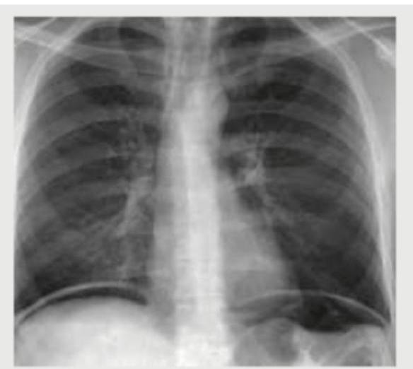
CXR was done for a 15-year-old boy with low cardiac output. Which is incorrect about the patient?

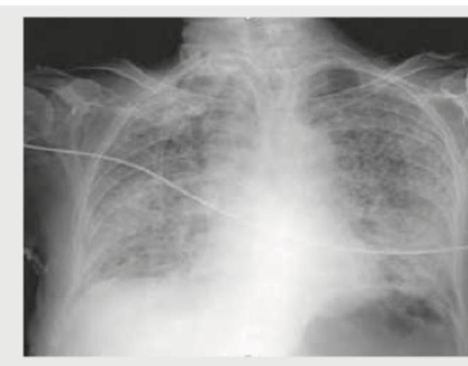
An adult undergoes multiple FFP transfusions for excessive bleeding after cardiac surgery and develops respiratory distress. CXR done is shown below. What does it indicate?

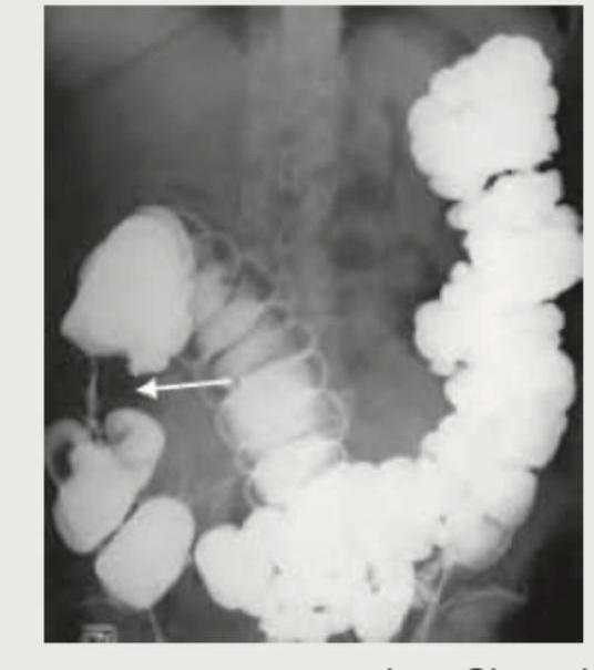
What is the diagnosis based on the CXR of a patient of cystic fibrosis shown below?

A chest X-ray shows the following appearance. Identify the pathology:

A 40-year-old male presents with sudden onset right-sided chest pain and breathlessness following a road traffic accident. On examination, breath sounds are diminished on the right side. The chest X-ray is shown below. What is the most likely diagnosis?

Which is not a finding of the Chest X-ray shown below? (AIIMS May 2017)

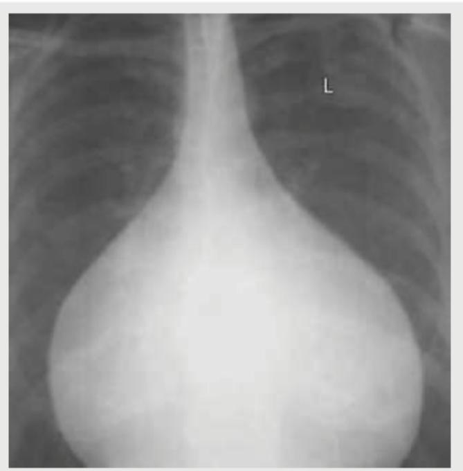
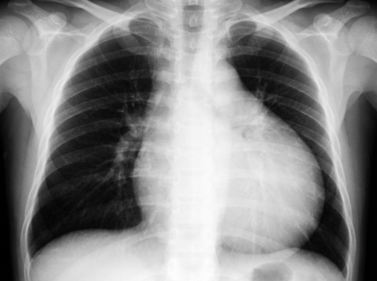
A previously healthy patient, presents with dyspnea and low grade fever since 4 months. His lungs are clear. JVP is normal. ECG showed low voltage complexes. What is the possible diagnosis?
Comment on the diagnosis of the image shown below.

On a chest radiograph, which of the following occupational diseases is most likely to be mistaken as a case of tuberculosis of lungs?
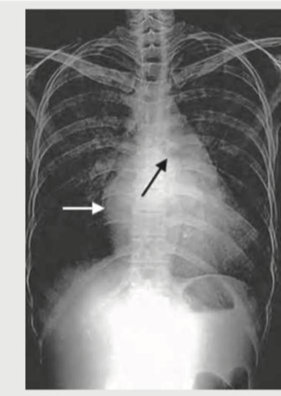
Which of the following are included in common causes of mediastinal masses in superior and anterior mediastinum? I. Goitre II. Thymic tumour III. Neurogenic tumour Select the correct answer using the code given below :
Explanation: ***Carey Coombs murmur*** - A **Carey Coombs murmur** is a mid-diastolic murmur typically heard in **acute rheumatic fever** due to active mitral valvulitis, which causes relative mitral stenosis. - The patient's CXR indicates **Coarctation of Aorta**, as evidenced by the **reverse 3 sign** (figure-of-3 sign) and **rib notching** from collateral circulation. - Carey Coombs murmur is **not associated with coarctation of aorta**, making this the **incorrect finding** for this patient. *Reverse split S2* - A **reverse split S2** (paradoxical splitting) occurs when the **aortic component (A2) follows the pulmonary component (P2)** due to delayed left ventricular ejection. - This can occur in conditions causing **left ventricular outflow obstruction** such as severe aortic stenosis or coarctation of aorta. - In coarctation, increased LV afterload can delay aortic valve closure, potentially causing reverse splitting of S2. *Straightening of left heart border* - **Straightening of the left heart border** on CXR is observed in conditions causing **left ventricular hypertrophy** or prominence of the pulmonary artery. - In coarctation of the aorta, the left ventricle works against increased afterload, leading to **concentric LV hypertrophy**, which can result in this CXR finding. *Ejection systolic murmur* - An **ejection systolic murmur** is commonly heard in coarctation of aorta due to turbulent flow across the narrowed segment. - The murmur is best heard over the **left infraclavicular area and back** (between the scapulae). - Associated aortic stenosis or bicuspid aortic valve (present in 50-85% of coarctation cases) can also produce an ejection systolic murmur at the aortic area.
Explanation: ***TRALI*** - The chest X-ray shows **bilateral pulmonary infiltrates** and **pulmonary edema** and the patient had multiple **FFP transfusions** followed by respiratory distress, which is highly suggestive of **Transfusion-Related Acute Lung Injury (TRALI)**. - TRALI is characterized by acute respiratory distress with **hypoxemia** occurring within 6 hours of transfusion, in the absence of other risk factors for **Acute Lung Injury (ALI)**. *Volume overloading of heart* - While fluid overload can cause pulmonary edema, the severity and rapid onset of distress after transfusion, coupled with the bilateral infiltrates, point more specifically to TRALI rather than isolated volume overload, especially in the context of FFP. - Cardiogenic pulmonary edema typically presents with **cardiomegaly** and signs of **heart failure**, which are not explicitly described or obviously seen as the primary cause in the given scenario and image. *Mendelson's syndrome* - Mendelson's syndrome, or **aspiration pneumonitis**, results from the inhalation of acidic gastric contents, leading to chemical pneumonitis. - This typically occurs in patients with impaired consciousness or those undergoing procedures that compromise airway protection, and there is no information in the vignette to suggest aspiration. *Pneumomediastinum* - Pneumomediastinum indicates the presence of **air in the mediastinum**, which would appear as radiolucent streaks outlining mediastinal structures on a CXR. - The image primarily shows diffuse bilateral infiltrates and pulmonary edema, rather than free air in the mediastinum.
Explanation: ***Bi-basilar fibrosis*** - The image shows **increased reticular and nodular opacities**, particularly prominent in the **lower lung fields (basilar regions)**, consistent with pulmonary fibrosis. - In cystic fibrosis, chronic infection and inflammation lead to progressive lung damage, often manifesting as **bronchiectasis and interstitial fibrosis**, particularly in the lower lobes. *Round pneumonia with perihilar shadows* - **Round pneumonia** typically presents as a well-circumscribed, spherical opacity, often seen in children, which is not the predominant pattern here. - While there are opacities, they are more diffuse and reticular rather than forming distinct perihilar masses or round consolidations. *Hilar lymphadenopathy with coin shadows* - **Hilar lymphadenopathy** would appear as enlarged, prominent hilar regions, and while the hila are somewhat prominent, the primary finding is widespread parenchymal disease. - **Coin shadows** refer to solitary pulmonary nodules, which are not characteristic of the diffuse changes observed in this CXR. *Egg shell calcification* - **Eggshell calcification** is a specific pattern of calcified lymph nodes, typically found in conditions like silicosis or sarcoidosis, and is not visible in this image. - The CXR shows diffuse fibrotic changes rather than discrete calcifications.
Explanation: ***Egg on side appearance*** - The image illustrates a classic "egg on side" or **"egg-on-a-string" appearance**, which is a radiological sign of **transposition of the great arteries (TGA)**. - This appearance is due to the narrow vascular pedicle (aorta and pulmonary artery are superimposed) and the **enlarged, egg-shaped cardiac silhouette** as a result of ventricular hypertrophy and right atrial enlargement. *Pericardial effusion* - **Pericardial effusion** would typically manifest as a **globular or "water-bottle" heart shape** on chest X-ray due to fluid accumulation around the heart. - This appearance is characterized by a widened cardiac silhouette with sharply defined borders, which is not clearly visible here. *Boot shaped heart* - A **"boot-shaped" heart**, also known as a **coeur en sabot**, is characteristic of **tetralogy of Fallot**. - This shape is caused by **right ventricular hypertrophy** with an upturned cardiac apex, and often an accompanying concave pulmonary artery segment, which is not seen in this image. *Normal-sized heart* - The cardiac silhouette in the image is clearly **enlarged and distinctly abnormal** in shape, indicating it is not a normal-sized heart. - A **normal-sized heart** would have a cardiothoracic ratio of less than 0.5 and distinct great vessel outlines.
Explanation: ***Right hydropneumothorax*** - The X-ray image reveals an **air-fluid level** in the right pleural cavity, characterized by a straight, horizontal line between the air (darker above) and fluid (whiter below). This finding is pathognomonic for a hydropneumothorax. - The **collapsed or compressed lung** is also visible superior to the air-fluid level, further supporting the diagnosis of air and fluid coexisting in the pleural space. *Hydatid cyst right lung* - A hydatid cyst in the lung appears as a **well-defined, rounded opacity (solid mass)**, often with a "water lily" sign if ruptured, but it does not present with a distinct air-fluid level as seen here. - While hydatid cysts can rupture and produce air and fluid, the X-ray findings would typically show a more complex internal structure or a cyst within a cavity, not a simple air-fluid interface across the entire pleural space. *Right pleural effusion* - A pleural effusion would appear as a **homogeneous white (effaced) opacity** blunting the costophrenic angle and, in larger effusions, rising along the lateral chest wall (meniscus sign). - Crucially, a simple pleural effusion **does not show an air-fluid level**; the fluid density would fill the pleural space without an overlying visible air component. *Perforated abdominal viscus* - A perforated abdominal viscus would typically result in **free air under the diaphragm** on an upright chest X-ray (pneumoperitoneum). - While pneumoperitoneum presents as air, it is located below the diaphragm, *not* within the pleural cavity, and would not create an air-fluid level within the lung fields as seen in this image.
Explanation: ***Narrow vascular pedicle*** - The image shows a **markedly enlarged cardiac silhouette** with a **"flask-shaped" or "water bottle" heart**, classic for pericardial effusion. In this condition, the vascular pedicle (the mediastinal structures above the heart including the aorta and superior vena cava) is typically **normal to widened** due to venous congestion. - A **narrow vascular pedicle** is characteristically seen in hypovolemia, dehydration, or certain congenital heart diseases with reduced pulmonary blood flow (e.g., tetralogy of Fallot). This finding is **NOT present** in this radiograph. - This is the **most definitively absent finding** among the options listed. *Pulmonary venous hypertension* - The chest X-ray shows prominent pulmonary vascular markings, particularly in the upper lobes, indicative of **cephalization of vessels**, a classic sign of pulmonary venous hypertension. - This occurs due to increased pressure in the pulmonary veins, commonly seen in congestive heart failure or significant pericardial effusion with cardiac tamponade physiology. - This finding **IS present** on the radiograph. *Increased CT ratio* - The **cardiothoracic (CT) ratio** is markedly increased, with the cardiac silhouette clearly exceeding 50% of the thoracic diameter. This indicates **cardiomegaly**, which can result from cardiac chamber enlargement or pericardial effusion. - The extreme enlargement seen here, with the globular "water bottle" configuration, is pathognomonic for large pericardial effusion. - This finding **IS present** on the radiograph. *Acute cardiophrenic angle* - The cardiophrenic angles (the angles formed where the heart border meets the diaphragm laterally) appear **blunted or obtuse** rather than acute (sharp). - While the term "acute cardiophrenic angle" typically refers to the normal sharp angle seen in healthy individuals, the phrasing here is ambiguous. The **angles themselves are present but blunted**, not acute. - However, compared to "narrow vascular pedicle," the blunting of these angles IS a radiographic finding that can be observed, even if abnormal. The vascular pedicle narrowness is completely absent.
Explanation: ***Tuberculous pericardial effusion*** - The combination of **dyspnea**, **low-grade fever** for 4 months, and **low voltage complexes on ECG** strongly suggests a pericardial effusion, which is often tuberculous in endemic areas, especially with chronic symptoms. - While JVP is normal here, a large effusion can lead to cardiac tamponade with elevated JVP later; current findings are consistent with an evolving effusion. *Syphilitic aortic aneurysm* - This typically presents with symptoms related to **aortic insufficiency** or compression of surrounding structures, often without fever unless complicated by infection. - **ECG low voltage** is not a characteristic feature of an uncomplicated syphilitic aortic aneurysm. *Hypertrophic cardiomyopathy* - Characterized by **ventricular hypertrophy** and often presents with dyspnea, but typically without fever or low voltage ECG unless a significant comorbid condition exists. - The disease is usually asymptomatic for years and the hypertrophy is often visible on ECG by **increased voltage or S-waves**. *Rheumatic mitral stenosis* - This condition involves **valvular heart disease** leading to dyspnea due to pulmonary congestion, but usually through a history of acute rheumatic fever. - **Low-grade fever** for several months and **low voltage ECG** are not typical findings for isolated rheumatic mitral stenosis.
Explanation: ***Coffee bean appearance*** - The image depicts a **dilated loop of colon**, characteristic of a **sigmoid volvulus**, which often resembles a coffee bean on plain abdominal radiographs. - This appearance is due to the **mesentery twisting around the bowel**, causing the dilated loop to fold upon itself. *Apple core appearance* - This describes a **constricting lesion** in the bowel, typically seen in **colorectal cancer**, where the lumen is severely narrowed with overhanging edges. - It denotes an **irregular, circumferential narrowing**, which is not seen in the provided image. *Claw sign* - The **"claw sign"** in the context of imaging refers to the way normal kidney tissue appears to "clasp" or surround an adjacent mass. - This sign is used to distinguish between intrarenal and extrarenal masses, which is irrelevant to the bowel pathology shown. *String sign of Kantor* - The **Kantor's string sign** is a radiologic finding in which the lumen of the terminal ileum is markedly narrowed, appearing as a thin, continuous string of barium. - This is classically associated with **Crohn's disease**, signifying severe inflammation and fibrosis, and is not seen in this image.
Explanation: ***Silicosis*** - **Silicosis** manifests with radiographic findings such as **nodular opacities**, often in the upper lobes, and can progress to **progressive massive fibrosis**. These can be visually similar to granulomatous lesions seen in **tuberculosis**. - Additionally, patients with silicosis have an **increased susceptibility to tuberculosis** (silico-tuberculosis), making differentiation solely based on chest radiographs challenging. *Anthracosis* - **Anthracosis** (coal worker's pneumoconiosis) primarily causes diffuse, small, rounded opacities, which are generally less organized and prominent than the nodules seen in silicosis or tuberculosis. - While it can lead to **progressive massive fibrosis** in severe cases, the initial presentation is usually less likely to be confused with tuberculosis compared to silicosis. *Siderosis* - **Siderosis** is caused by the inhalation of iron dust and typically results in fine, diffuse, and irregular opacities on chest radiographs. - These opacities are generally benign and rarely lead to significant pulmonary fibrosis or mimic the discrete, nodular pattern of tuberculosis. *Byssinosis* - **Byssinosis** is an occupational lung disease associated with exposure to cotton dust. It is primarily characterized by **bronchial hyperreactivity** and **asthma-like symptoms**, particularly "Monday chest tightness." - It does not typically produce significant or distinctive radiographic changes that could be confused with tuberculosis, as it is a disease of the airways rather than parenchymal infiltrates.
Explanation: ***I and II only*** - **Goitre** (retrosternal thyroid extension) is a common cause of superior mediastinal masses. - **Thymic tumors** (thymoma, thymic carcinoma) are characteristically found in the anterior mediastinum. *I, II and III* - While goitre and thymic tumors are common in the superior and anterior mediastinum respectively, **neurogenic tumors** are typically found in the posterior mediastinum. - This option incorrectly includes neurogenic tumors with masses of the superior and anterior mediastinum. *II and III only* - This option correctly identifies **thymic tumors** for the anterior mediastinum but incorrectly includes **neurogenic tumors**, which are found in the posterior mediastinum. - It also omits **goitre**, which is a significant cause of superior mediastinal masses. *I and III only* - This option correctly identifies **goitre** as a superior mediastinal mass but incorrectly includes **neurogenic tumors**, which are typically located in the posterior mediastinum. - It incorrectly omits **thymic tumors**, which are a primary cause of anterior mediastinal masses.
Normal Chest Radiographic Anatomy
Practice Questions
Radiographic Signs in Chest Imaging
Practice Questions
Pulmonary Infections
Practice Questions
Chronic Obstructive Pulmonary Disease
Practice Questions
Interstitial Lung Diseases
Practice Questions
Pulmonary Neoplasms
Practice Questions
Pleural Diseases
Practice Questions
Mediastinal Pathology
Practice Questions
Congenital and Developmental Chest Anomalies
Practice Questions
Pulmonary Vascular Diseases
Practice Questions
Chest Trauma Imaging
Practice Questions
Post-Surgical Chest Imaging
Practice Questions
Get full access to all questions, explanations, and performance tracking.
Start For Free