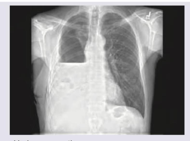
The CXR given shows presence of:

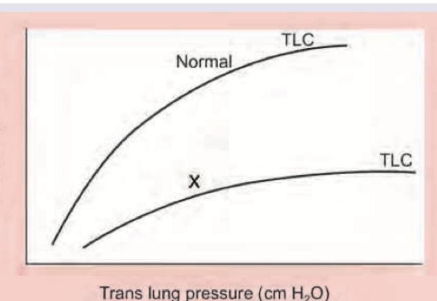
Which lung disease is shown in the image below marked as X?

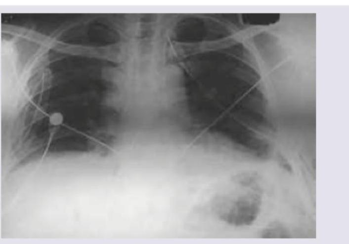
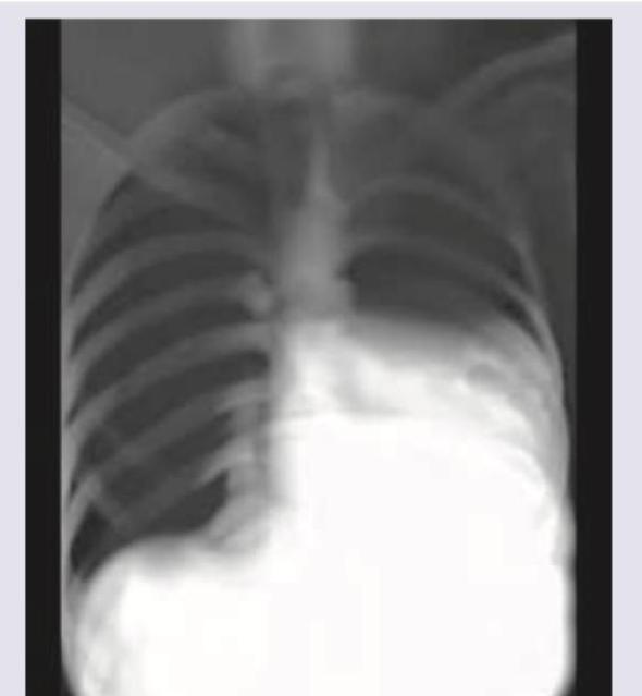
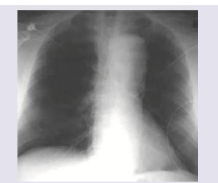
A 16-year-old boy is admitted with rapidly accumulating bilateral pleural effusion. His chest X-ray is shown below. Which of the following is incorrect about the X-ray shown?

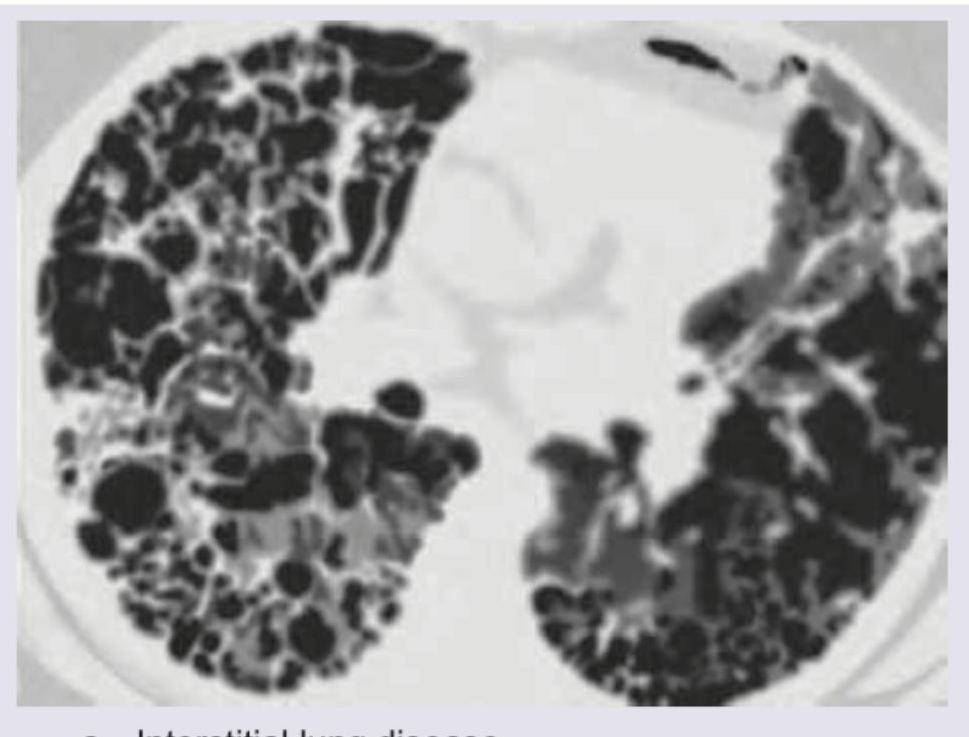
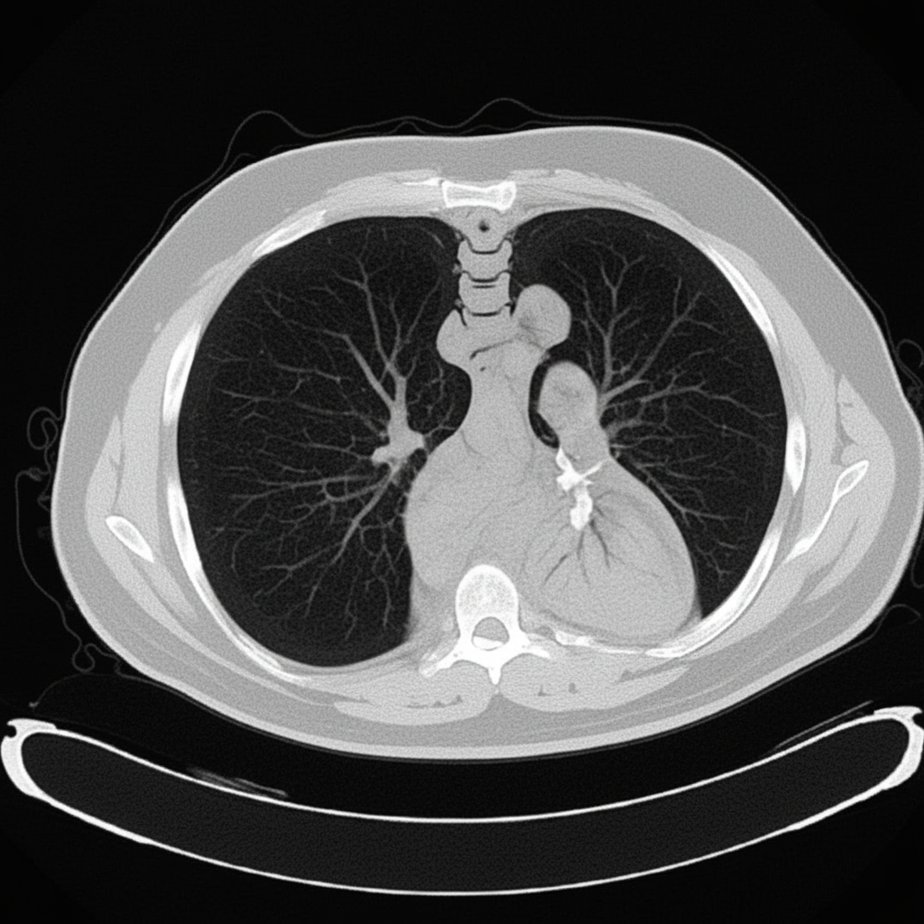
The CT chest shows presence of:

The image provided represents:

The image shows presence of:

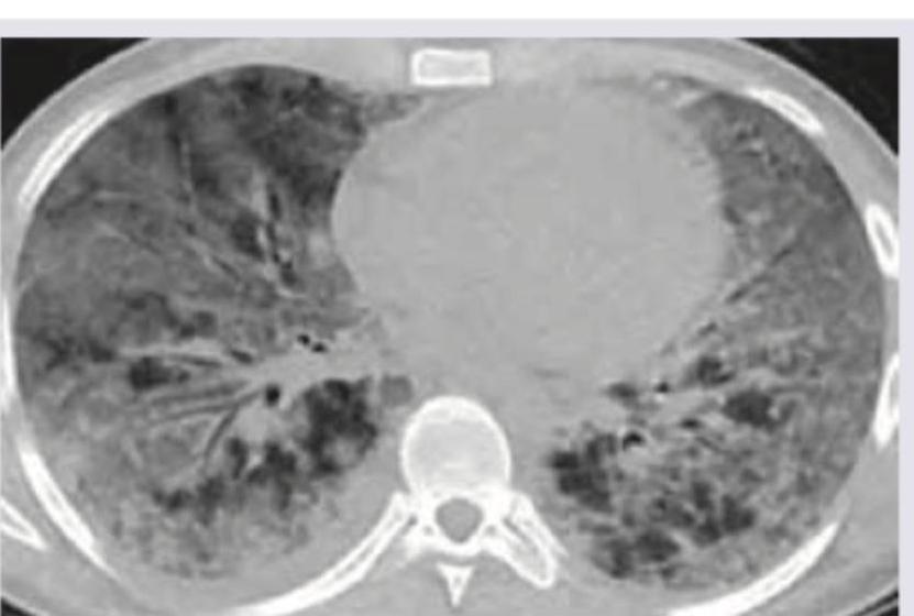
The CT chest of a patient shows:

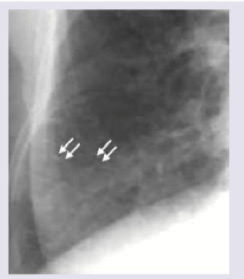
The CXR shows markings near the costophrenic angle. Which of the following is the cause of these markings? (Recent NEET Pattern 2016-17)

Chest X-ray was performed on a patient with malar flush and effort intolerance. All are true about the condition shown in the figure except:

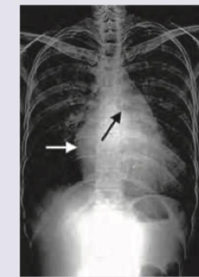
A 65-year-old hypertension patient presents with chest pain, difficulty in breathing for 1 hour. Based on the chest X-ray shown below, identify the radiological finding:

Explanation: ***Hydropneumothorax*** - The image clearly shows a **horizontal air-fluid level** within the pleural cavity on the right side. This distinct level indicates the presence of both **fluid (hydro)** and **air (pneumo)**. - The **collapsed lung** due to the presence of air and fluid is also visible, confirming the diagnosis. *Pleural effusion* - A pleural effusion would typically present with a **meniscus sign** (curvilinear upper border) rather than a straight horizontal air-fluid level. - While fluid is present, the presence of **air** is also evident and crucial for distinguishing it from a simple effusion. *Lung abscess* - A lung abscess is an **intraparenchymal collection of pus** within the lung tissue. - It would typically appear as a thick-walled cavity with an **air-fluid level inside the lung parenchyma**, not in the pleural space. *Collapse* - Lung collapse (atelectasis) involves loss of volume in part or all of a lung, often presenting as **increased opacification** and displacement of structures towards the collapsed side. - Although the lung is collapsed in this image, it is a *consequence* of the hydropneumothorax, not the primary finding on its own, and doesn't explain the distinct air-fluid level.
Explanation: ***Interstitial fibrosis*** - The curve labeled 'X' shows a **lower lung volume for a given transpulmonary pressure** compared to the 'Normal' curve, indicating **decreased lung compliance**. - A decrease in lung compliance and **reduced total lung capacity (TLC)** are hallmarks of **restrictive lung diseases** like interstitial fibrosis, where the lungs become stiff and difficult to inflate. *Emphysema* - Emphysema is an **obstructive lung disease** characterized by **increased lung compliance** and **increased total lung capacity (TLC)** due to the destruction of elastic tissue. - Its curve would be shifted to the left and up compared to normal, indicating a larger volume for a given transpulmonary pressure, which is opposite to what is shown for 'X'. *Asthma* - Asthma is also an **obstructive lung disease** primarily affecting airflow due to **bronchoconstriction** and inflammation. - While lung compliance may be slightly altered, the hallmark features of the pressure-volume curve in asthma typically involve air trapping and often a **normal or increased TLC** during an exacerbation, not a significantly reduced TLC and decreased compliance as shown. *Normal* - The curve labeled 'Normal' represents typical lung compliance, where lung volume increases appropriately with transpulmonary pressure. - Curve 'X' clearly deviates from this normal pattern, showing a **reduced capacity** to expand for the same pressure.
Explanation: ***Pneumothorax*** - A **pneumothorax** is characterized by the presence of **air in the pleural space**, which typically appears as a **lucent area** without lung markings and a displaced visceral pleural line on X-ray. - The presented chest X-ray shows **dense opacities at the lung bases**, consistent with fluid accumulation (**pleural effusion**), not air. *Left-sided pleural effusion* - The image clearly shows significant **opacification of the left hemithorax** with blunting of the **costophrenic angle** and obscuring of the left hemidiaphragm. - This finding is consistent with a large amount of fluid, indicating a **left-sided pleural effusion**. *Mediastinum shifted to right* - The **trachea** and **cardiac silhouette** are visibly displaced towards the **right side** of the chest. - This shift is a consequence of the large **left-sided pleural effusion exerting mass effect** and pushing the mediastinal structures away from the affected side. *Bilateral ICD tubes* - There are discernable radiopaque lines extending into both pleural spaces, representing **bilateral intercostal drainage (ICD) tubes**. - These tubes are placed to drain the **accumulated pleural fluid** from both the right and left sides.
Explanation: ***Pulmonary alveolar proteinosis*** - CT images of pulmonary alveolar proteinosis often show diffuse, bilateral ground-glass opacities with superimposed **interlobular and intralobular septal thickening**, creating a characteristic **crazy-paving pattern**. - This pattern, as seen in the image, is caused by the accumulation of lipoproteinaceous material within the alveoli and is a hallmark of the disease. *Interstitial lung disease* - While some forms of interstitial lung disease can cause ground-glass opacities and septal thickening, the **"crazy-paving" pattern** seen so clearly and diffusely here is not typically their most characteristic finding. - ILD often presents with **reticular opacities**, honeycombing, or traction bronchiectasis, which are less prominent or absent in this specific image. *Bronchiectasis* - Bronchiectasis is characterized by **irreversible dilation of the bronchi**, usually accompanied by wall thickening. - This condition would appear on CT as **dilated, thickened airways** (often described as "tram-track" opacities or "signet-ring" signs) and mucus plugging, which are not the predominant findings in the provided image. *Aspergillosis* - Pulmonary aspergillosis encompasses several forms, such as aspergilloma (fungus ball in a cavity), invasive aspergillosis (nodules, halo sign, or cavitation), or allergic bronchopulmonary aspergillosis (mucoid impaction, bronchiectasis). - The diffuse ground-glass opacities with septal thickening seen in the image do not typically represent the primary radiological features of aspergillosis.
Explanation: ***CT chest without contrast*** - The image displays detailed **cross-sectional anatomy** of the chest, including the lungs, mediastinum, and cardiac structures, without any evident enhancement of blood vessels or organs. - The varying **grayscale densities** accurately depict different tissue types (e.g., air in lungs as black, bone as white, soft tissues as shades of gray) as expected in an **unenhanced CT scan**. - The **cross-sectional detail** and high resolution are characteristic of a **CT scan**. *Barium swallow study* - The image provided is a **CT scan of the chest**, not a barium swallow study. A barium swallow study involves administering barium contrast and taking X-rays to visualize the esophagus. - The density and grayscale variations seen in the image are characteristic of a CT, which uses X-rays to create cross-sectional images of body structures, and not of the high-contrast white appearance of barium within an organ lumen. *CT chest with contrast* - This image does not demonstrate enhancements of vascular structures or organs typically associated with the administration of **intravenous contrast material**. - Without the presence of **hyperdense (bright) opacification of blood vessels** (like the aorta or pulmonary arteries) or soft tissues, it is less likely to be a contrast-enhanced study. *X-ray chest* - While an X-ray chest is used to visualize chest structures, this image exhibits **cross-sectional detail** and high resolution that is characteristic of a **CT scan**, rather than a plain film X-ray. - An X-ray chest provides a 2D projection, whereas this image clearly shows a 3D cut through the chest organs with distinct tissue densities.
Explanation: ***Hydropneumothorax*** - The image shows both **air and fluid levels in the pleural cavity**, indicating a hydropneumothorax. The distinct straight line separating air and fluid is characteristic. - Presence of **air (lucency)** in the superior part of the pleural space and **fluid (opacity)** forming a horizontal line below it confirms this diagnosis. *Pleural effusion* - A pleural effusion would appear as a **homogenous opacity** in the dependent portion of the pleural space, typically blunting the costophrenic angles. - It would not exhibit a distinct **air-fluid level** within the pleural cavity without an associated pneumothorax. *Pneumothorax* - A pneumothorax presents as **air in the pleural space** without fluid, resulting in increased lucency and collapse of lung tissue. - There would be **no fluid level visible**, only air separating the parietal and visceral pleura. *Pneumonia* - Pneumonia would appear as an **area of consolidation** within the lung parenchyma, often ill-defined, and without a clear air-fluid level in the pleural space. - It involves the lung tissue itself, not primarily the pleural space, and would typically present with **air bronchograms** within the consolidated area.
Explanation: ***Ground glass pattern*** - The CT scan shows diffuse hazy opacities that do not obscure the underlying bronchial and vascular markings, which is characteristic of a **ground-glass pattern**. - This pattern indicates a partial filling of airspaces, interstitial thickening, or a combination of both, often seen in conditions like **pneumonitis** or early acute respiratory distress syndrome. *Honeycomb pattern* - The image does not display the characteristic **thick-walled cystic airspaces** seen in a honeycomb pattern, which typically represents end-stage pulmonary fibrosis. - While there is some architectural distortion, it lacks the discrete, clustered cysts with clearly defined walls that define true honeycombing. *Crazy pavement pattern* - The crazy paving pattern is characterized by **ground-glass opacities superimposed with interlobular septal thickening**, which is not clearly and extensively demonstrated in this scan. - While some ground-glass is present, prominent septal thickening creating a polygonal pattern is not the predominant feature. *Normal scan* - The scan clearly shows widespread **abnormal opacities** throughout both lungs, indicating significant underlying pathology. - A normal chest CT would show uniform lung parenchyma with clear bronchial and vascular markings without any significant areas of increased attenuation.
Explanation: ***Lymphangitis carcinomatosis*** - The image shows **reticulonodular interstitial markings** with preserved lung volumes, particularly prominent near the costophrenic angle, which are classic for **lymphangitis carcinomatosis**. - This condition is caused by the infiltration of **malignant cells into the lymphatic channels** of the lung, leading to thickening of the interlobular septa and a characteristic radiographic appearance. *Pulmonary alveolar proteinosis* - This condition typically presents with diffuse ground-glass opacities and consolidation, often described as a **"crazy-paving" pattern** on CT, which is not seen here. - It involves the accumulation of lipoproteinaceous material within the **alveoli**, rather than lymphatic infiltration. *Lung abscess* - A lung abscess would appear as a **cavitated lesion** with an air-fluid level, indicating necrosis and fluid accumulation, which is distinctly different from the interstitial markings in the image. - It is typically caused by **bacterial infection** and is a focal process, not diffuse interstitial infiltration. *Pneumatocele* - A pneumatocele is a **thin-walled, air-filled cyst** commonly seen after pneumonia in children or trauma, appearing as a clear, defined space on imaging. - It does not present with the diffuse reticulonodular pattern or interstitial thickening characteristic of the image.
Explanation: ***Presystolic accentuation becomes louder with atrial fibrillation*** - In **atrial fibrillation**, the atrial kick is lost, which is responsible for presystolic accentuation. Therefore, presystolic accentuation would be **absent or diminished**, not louder. - The context of **malar flush** and **effort intolerance** points to **mitral stenosis**, a condition where presystolic accentuation is typically heard due to forceful atrial contraction against a stenotic valve. *Straightening of left heart border* - This is a common finding in **mitral stenosis** on chest X-ray, caused by the enlargement of the **left atrial appendage** and **pulmonary artery**. - It contributes to the "mitral configuration" of the heart. *Pressure half-time is used to assess severity on Doppler* - **Pressure half-time (PHT)** is the standard Doppler echocardiographic method to assess **mitral stenosis severity**. - PHT >220 ms indicates severe mitral stenosis, while PHT 130-220 ms indicates moderate stenosis. - Mitral valve area can be calculated using the formula: MVA = 220/PHT. *Pulmonary apoplexy* - **Pulmonary apoplexy** (severe pulmonary hemorrhage) is a serious complication of **mitral stenosis** due to extremely elevated pulmonary venous pressure. - The high pressure in the pulmonary circulation can cause rupture of small vessels and bleeding into the airspaces.
Explanation: ***Kerley B lines*** - The chest X-ray in a patient with acute **cardiac decompensation** and **pulmonary edema** often shows **thickened interlobular septa**, known as Kerley B lines, which are short, horizontal lines seen in the lung periphery. - These lines are indicative of **interstitial pulmonary edema**, which is highly consistent with a patient presenting with acute chest pain and difficulty breathing, especially with a history of hypertension, suggesting acute heart failure. *Widened mediastinum* - A **widened mediastinum** on chest X-ray is a classic radiological finding suggestive of **aortic dissection**, which can cause chest pain in a hypertensive patient. - However, the clinical presentation with acute heart failure symptoms and the presence of **Kerley B lines** point toward pulmonary edema rather than aortic pathology. - Widened mediastinum is not associated with Kerley B lines. *Hilar lymphadenopathy* - **Hilar lymphadenopathy** appears as enlarged lymph nodes around the hilum of the lungs, indicating conditions like sarcoidosis, tuberculosis, or malignancy. - It would not typically be an acute finding in a patient presenting with acute chest pain and difficulty breathing due to hypertension, nor would it cause Kerley B lines. *Pneumomediastinum* - **Pneumomediastinum** is characterized by the presence of air in the mediastinum, visible on a chest X-ray as a **lucent streak** outlining mediastinal structures (e.g., heart borders, aorta). - This condition does not typically present with Kerley B lines and is usually associated with symptoms like retrosternal chest pain that can radiate to the neck, dyspnea, and subcutaneous emphysema, which are different from the presented clinical picture.
Normal Chest Radiographic Anatomy
Practice Questions
Radiographic Signs in Chest Imaging
Practice Questions
Pulmonary Infections
Practice Questions
Chronic Obstructive Pulmonary Disease
Practice Questions
Interstitial Lung Diseases
Practice Questions
Pulmonary Neoplasms
Practice Questions
Pleural Diseases
Practice Questions
Mediastinal Pathology
Practice Questions
Congenital and Developmental Chest Anomalies
Practice Questions
Pulmonary Vascular Diseases
Practice Questions
Chest Trauma Imaging
Practice Questions
Post-Surgical Chest Imaging
Practice Questions
Get full access to all questions, explanations, and performance tracking.
Start For Free