
Chest Radiology — MCQs

On this page
On chest radiology, "egg-shell calcification" is seen in:
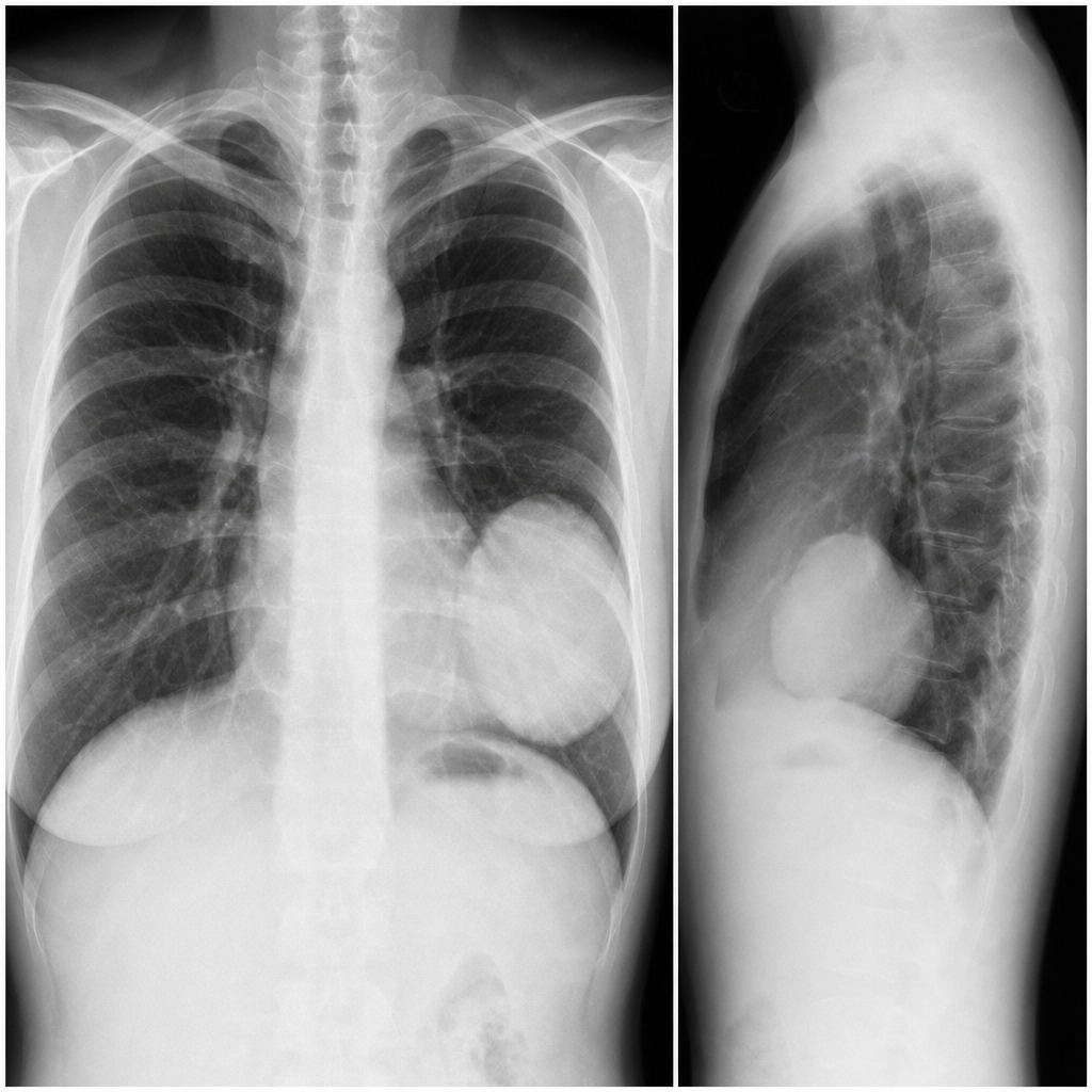
You are shown PA and lateral chest radiographs from a 53-year-old woman with mild dyspnea. Which one of the following is the most likely diagnosis based on the imaging?
Investigation of choice in bronchiectasis?
Miliary shadow in a chest X-ray is typically seen in all of the following conditions except:
The "Golden S" sign in bronchogenic carcinoma is characteristically seen in:
The air crescent sign on a chest X-ray is indicative of which condition?
Which condition is associated with pseudobronchiectasis?
Apex of the lung is best assessed by
Pleural plaques are best seen on which chest radiograph view?
Inverted mustache sign is seen in
Practice by Chapter
Normal Chest Radiographic Anatomy
Practice Questions
Radiographic Signs in Chest Imaging
Practice Questions
Pulmonary Infections
Practice Questions
Chronic Obstructive Pulmonary Disease
Practice Questions
Interstitial Lung Diseases
Practice Questions
Pulmonary Neoplasms
Practice Questions
Pleural Diseases
Practice Questions
Mediastinal Pathology
Practice Questions
Congenital and Developmental Chest Anomalies
Practice Questions
Pulmonary Vascular Diseases
Practice Questions
Chest Trauma Imaging
Practice Questions
Post-Surgical Chest Imaging
Practice Questions
Want unlimited practice?
Get full access to all questions, explanations, and performance tracking.
Scan to download app