
Chest Radiology — MCQs

On this page
To visualize vascular sling causing tracheal or external airway compression, which of the following would you best prefer?
All are radiological features of mitral stenosis except:
Which of the following conditions is most commonly associated with eggshell calcifications?
Best diagnostic aid for bronchiectasis is
The 'water lily sign' is characteristic of which condition?
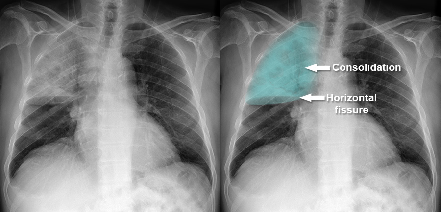
A 70-year-old male presented with complaints of breathlessness and exhibited abnormal bronchial breath sounds on examination. What is the most probable diagnosis based on the provided X-ray image?
Ring sign with dilated bronchi on CXR is a feature of
The following are direct signs of lung collapse seen on a chest X-ray, which one of the following is NOT a direct sign?
Kerley-B lines are seen when pulmonary venous pressure is what?
Which of the following conditions is most commonly associated with miliary mottling in the lungs?
Practice by Chapter
Normal Chest Radiographic Anatomy
Practice Questions
Radiographic Signs in Chest Imaging
Practice Questions
Pulmonary Infections
Practice Questions
Chronic Obstructive Pulmonary Disease
Practice Questions
Interstitial Lung Diseases
Practice Questions
Pulmonary Neoplasms
Practice Questions
Pleural Diseases
Practice Questions
Mediastinal Pathology
Practice Questions
Congenital and Developmental Chest Anomalies
Practice Questions
Pulmonary Vascular Diseases
Practice Questions
Chest Trauma Imaging
Practice Questions
Post-Surgical Chest Imaging
Practice Questions
Want unlimited practice?
Get full access to all questions, explanations, and performance tracking.
Scan to download app