
Chest Radiology — MCQs

On this page
Which of the following radiographic features is most characteristically associated with the pathophysiology of congestive heart failure?
In a patient with a suspected mediastinal mass, which imaging modality is most appropriate for initial evaluation?
Identify the most likely diagnosis based on the chest X-ray findings in a patient with low-grade fever.
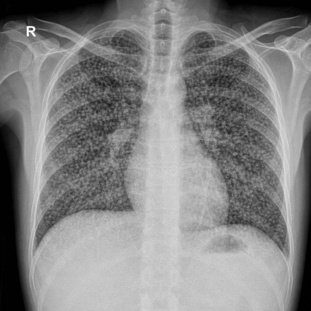
Snow storm appearance on chest X-ray is seen in -
Which radiographic sign is most characteristic of acute alveolar pulmonary edema?
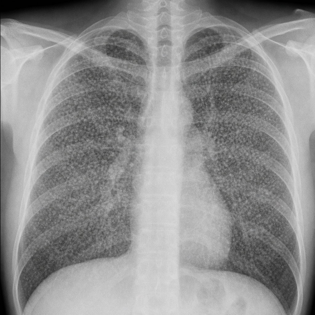
Identify the condition represented in the chest radiograph.
Kerley B lines are seen in mitral stenosis when the resting left atrial pressure exceeds which value?
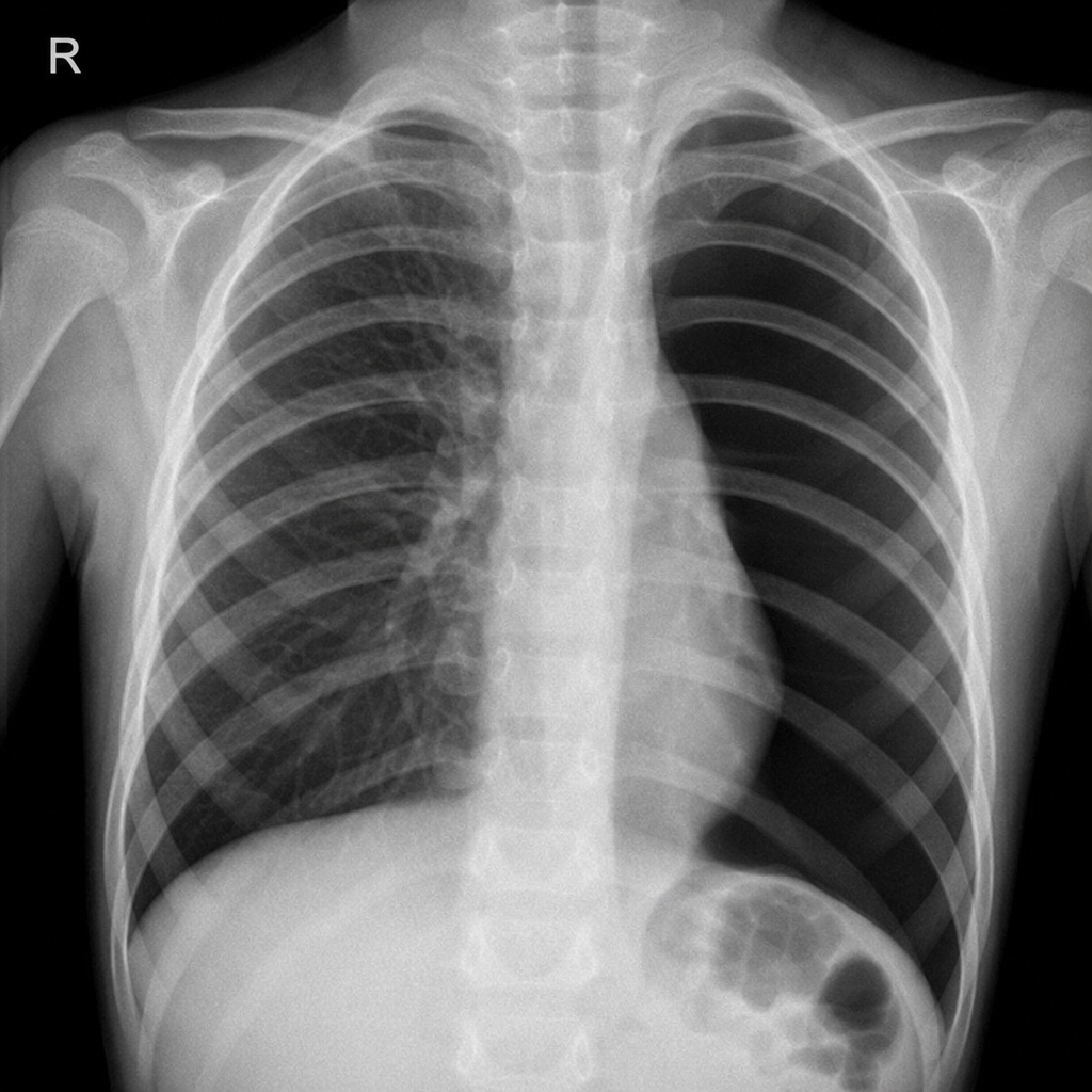
Chest x-ray of a child presenting with acute breathlessness. What is the most likely diagnosis?
What is the investigation of choice for a Pancoast tumor?
The 'Floating Water Lily' sign is associated with which of the following conditions?
Practice by Chapter
Normal Chest Radiographic Anatomy
Practice Questions
Radiographic Signs in Chest Imaging
Practice Questions
Pulmonary Infections
Practice Questions
Chronic Obstructive Pulmonary Disease
Practice Questions
Interstitial Lung Diseases
Practice Questions
Pulmonary Neoplasms
Practice Questions
Pleural Diseases
Practice Questions
Mediastinal Pathology
Practice Questions
Congenital and Developmental Chest Anomalies
Practice Questions
Pulmonary Vascular Diseases
Practice Questions
Chest Trauma Imaging
Practice Questions
Post-Surgical Chest Imaging
Practice Questions
Want unlimited practice?
Get full access to all questions, explanations, and performance tracking.
Scan to download app