
Chest Radiology — MCQs

On this page
What does the following CT chest show?
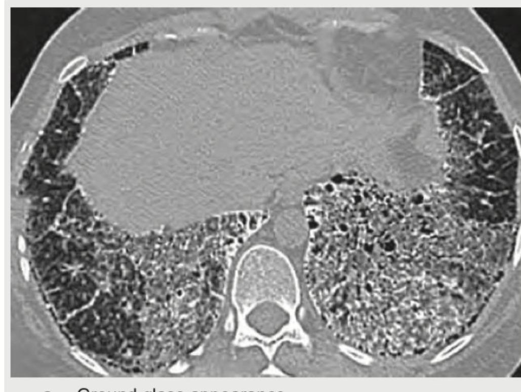
The following CT chest shows:
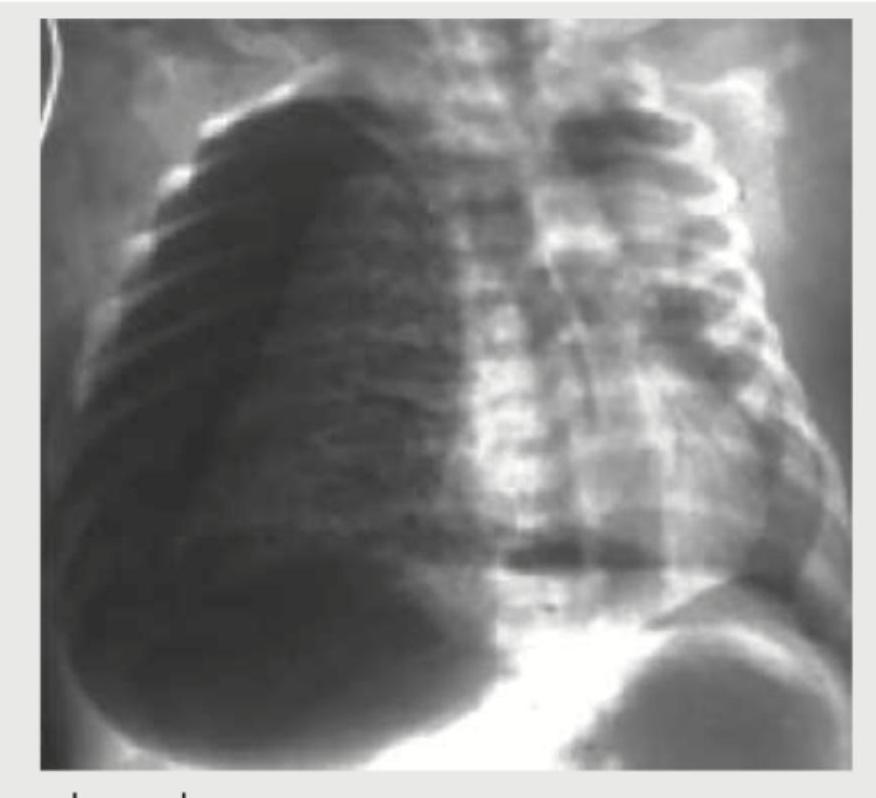
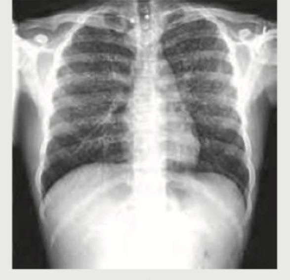
A 6-month-old child is on Vancomycin for pneumonia for 2 days. He develops worsening of respiratory distress and $\mathrm{SpO}_{2}$ falls to $80 \%$. The CXR performed is shown below. What is the diagnosis?
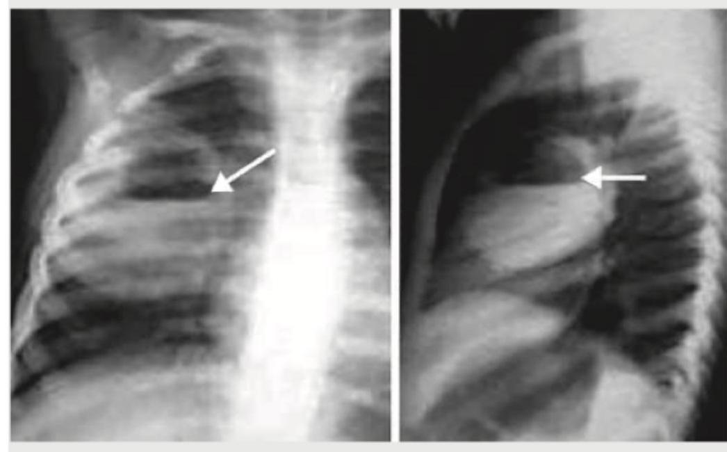
What is the most likely diagnosis based on the chest radiographs shown below?
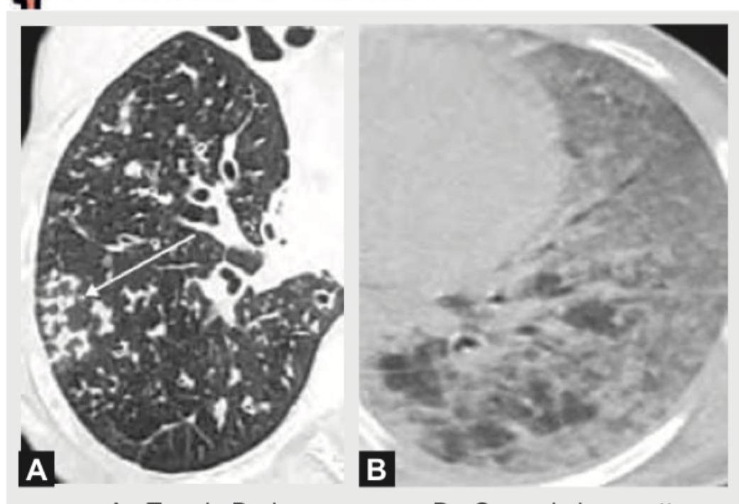
Which of the following is shown in the image below?
An AIDS patient presents with respiratory distress. CXR shows:
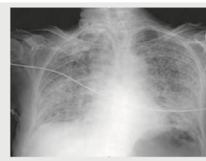
A breast cancer patient presents with difficulty in breathing. CXR shows:
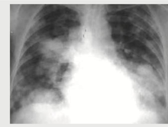
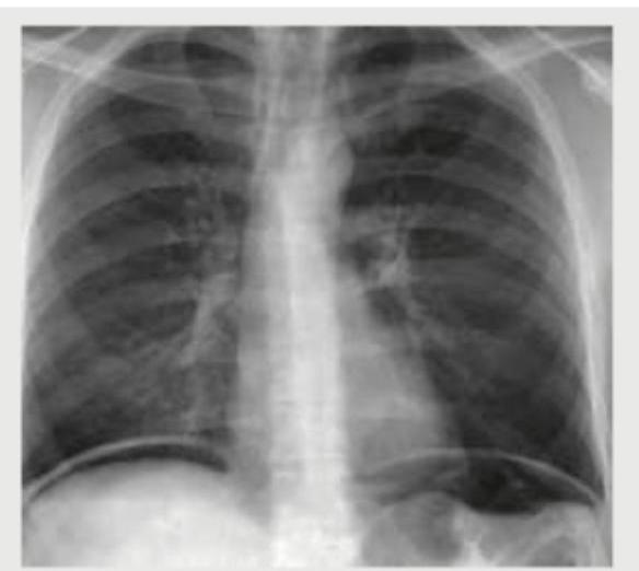
An adult undergoes multiple FFP transfusions for excessive bleeding after cardiac surgery and develops respiratory distress. CXR done is shown below. What does it indicate?
What is the diagnosis based on the CXR of a patient of cystic fibrosis shown below?

A 40-year-old male presents with sudden onset right-sided chest pain and breathlessness following a road traffic accident. On examination, breath sounds are diminished on the right side. The chest X-ray is shown below. What is the most likely diagnosis?
Practice by Chapter
Normal Chest Radiographic Anatomy
Practice Questions
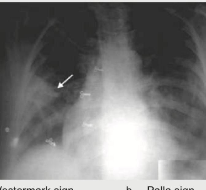
Radiographic Signs in Chest Imaging
Practice Questions
Pulmonary Infections
Practice Questions
Chronic Obstructive Pulmonary Disease
Practice Questions
Interstitial Lung Diseases
Practice Questions
Pulmonary Neoplasms
Practice Questions
Pleural Diseases
Practice Questions
Mediastinal Pathology
Practice Questions
Congenital and Developmental Chest Anomalies
Practice Questions
Pulmonary Vascular Diseases
Practice Questions
Chest Trauma Imaging
Practice Questions
Post-Surgical Chest Imaging
Practice Questions
Want unlimited practice?
Get full access to all questions, explanations, and performance tracking.
Scan to download app