
Chest Radiology — MCQs

On this page
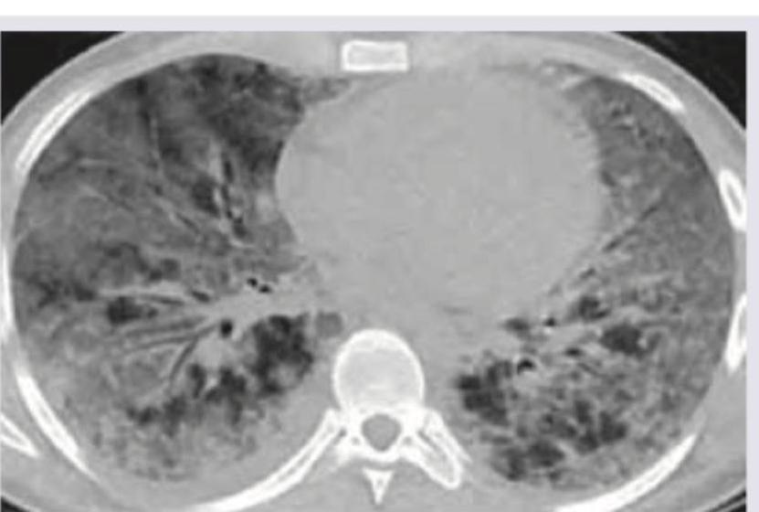
The CT chest shows presence of:
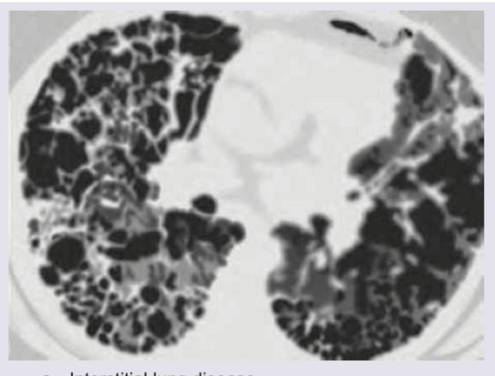
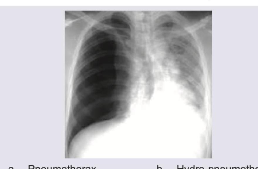
The image provided represents:
A 35-year-old coal worker presents with difficulty in breathing on exertion for last 2 years. CXR was performed. It shows:
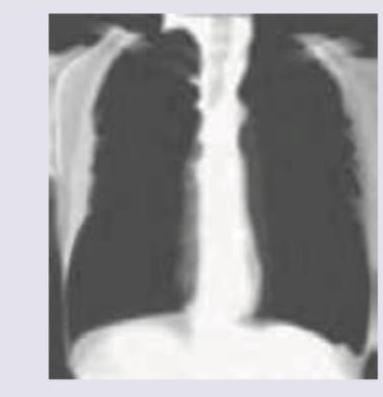
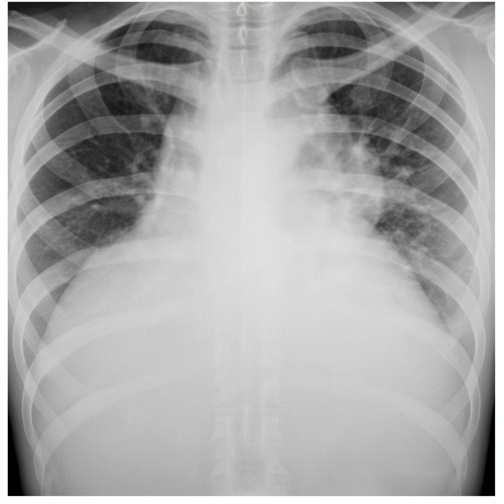
The chest X-ray of patient shows:
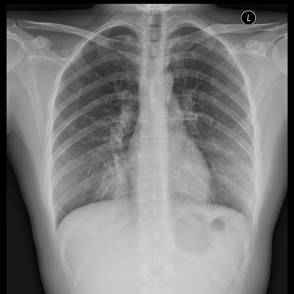
The image shows presence of:
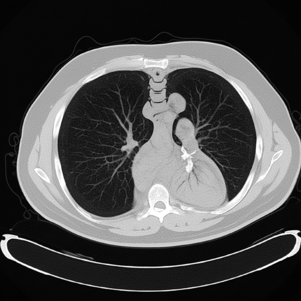
The CT chest of a patient shows:
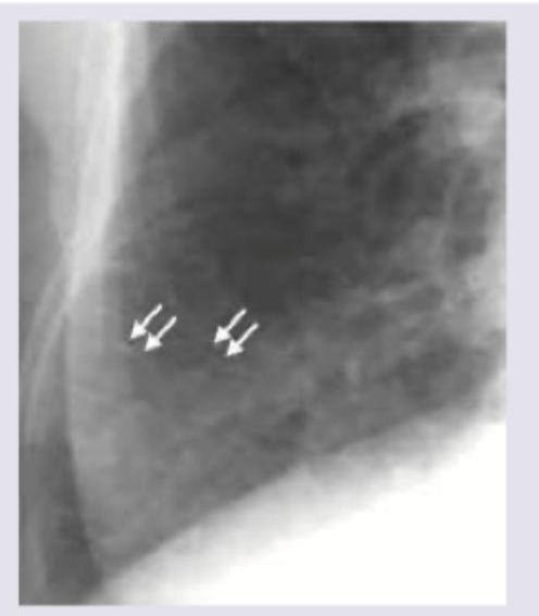
The CXR shows markings near the costophrenic angle. Which of the following is the cause of these markings? (Recent NEET Pattern 2016-17)
A 65-year-old hypertension patient presents with chest pain, difficulty in breathing for 1 hour. Based on the chest X-ray shown below, identify the radiological finding:
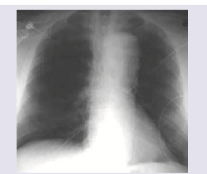
A patient presented with sudden onset difficulty in breathing with RR 28/min, normal blood pressure. X-ray was taken which is given below. What is the diagnosis?
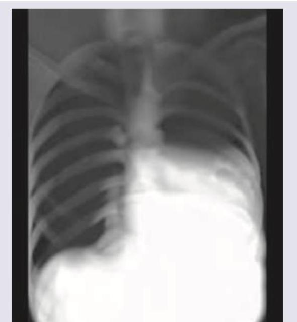
What is indicated by the 'water bottle appearance' of the heart size?
Practice by Chapter
Normal Chest Radiographic Anatomy
Practice Questions
Radiographic Signs in Chest Imaging
Practice Questions
Pulmonary Infections
Practice Questions
Chronic Obstructive Pulmonary Disease
Practice Questions
Interstitial Lung Diseases
Practice Questions
Pulmonary Neoplasms
Practice Questions
Pleural Diseases
Practice Questions
Mediastinal Pathology
Practice Questions
Congenital and Developmental Chest Anomalies
Practice Questions
Pulmonary Vascular Diseases
Practice Questions
Chest Trauma Imaging
Practice Questions
Post-Surgical Chest Imaging
Practice Questions
Want unlimited practice?
Get full access to all questions, explanations, and performance tracking.
Scan to download app