
Chest Radiology — MCQs

On this page
Cannon balls seen in the lungs are characteristic of which of the following?
Perihilar fluffy opacities on chest X-ray is seen in?
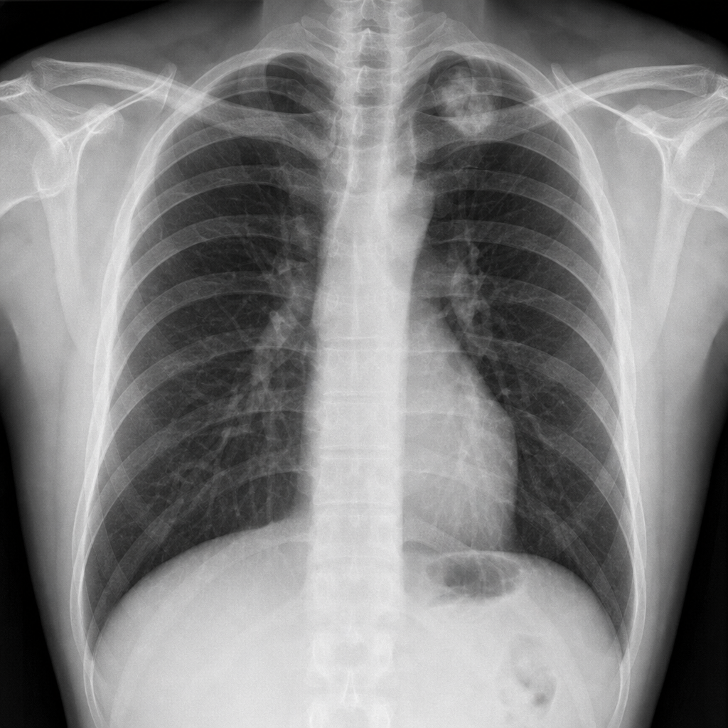
A 40-year-old man is seen for an insurance assessment. He has no past medical history and feels well. His complete physical examination is normal. His biochemistry, complete blood count (CBC), ECG, and urinalysis are also normal. His CXR is abnormal. Which of the following is the most likely diagnosis?
A patient with breathlessness shows Kerley B-lines in chest X-ray. What do these lines represent?
Inferior rib notching is seen in all except?
Which radiographic view is best for recognizing collapse of the lingula?
Unilateral hyperlucency of the lung is seen in all of the following conditions except?
Which condition presents with ipsilateral homogeneous opacification and contralateral mediastinal shift?
A middle-aged patient presents with a complaint of right hypochondrial pain. On plain chest X-ray, an elevated right hemidiaphragm is seen. Which of the following is NOT a possible diagnosis?
On barium swallow, which of the following causes a posterior impression?
Practice by Chapter
Normal Chest Radiographic Anatomy
Practice Questions
Radiographic Signs in Chest Imaging
Practice Questions
Pulmonary Infections
Practice Questions
Chronic Obstructive Pulmonary Disease
Practice Questions
Interstitial Lung Diseases
Practice Questions
Pulmonary Neoplasms
Practice Questions
Pleural Diseases
Practice Questions
Mediastinal Pathology
Practice Questions
Congenital and Developmental Chest Anomalies
Practice Questions
Pulmonary Vascular Diseases
Practice Questions
Chest Trauma Imaging
Practice Questions
Post-Surgical Chest Imaging
Practice Questions
Want unlimited practice?
Get full access to all questions, explanations, and performance tracking.
Scan to download app