Thoracotomy is indicated in all the following conditions except:
Which of the following injuries is the most serious?
The imaging modality primarily used in FAST (Focused Assessment with Sonography for Trauma) exam is:
All of the following are true regarding flail chest, EXCEPT:
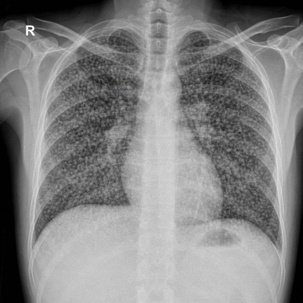
A 60-year-old woman presents with a history of smoking and cough. Chest X-ray shows a solitary pulmonary nodule. Which of the following is the most appropriate next step in management?
Identify the most likely diagnosis based on the chest X-ray findings in a patient with low-grade fever.

The following are direct signs of lung collapse seen on a chest X-ray, which one of the following is NOT a direct sign?
A patient after a heavy meal comes with epigastric pain. On examination, there is tenderness and rigidity in the upper abdomen. An X-ray shows pneumomediastinum. What could be the cause?
Most common type of shock in emergency room is
An incised-looking laceration is seen in all except:
Explanation: ***Pulmonary contusion*** - **Pulmonary contusion** is a bruise of the lung parenchyma that typically resolves with **supportive care** (oxygen, fluid management, analgesia, respiratory support) [1]. - It is generally *not* an indication for thoracotomy and is managed **conservatively** in most cases [1]. - Surgical intervention is only considered if complicated by other issues such as **uncontrolled hemorrhage**, massive hemothorax, or other injuries requiring exploration. *Penetrating chest injuries* - While approximately **85% of penetrating chest injuries** are managed conservatively with tube thoracostomy alone, **selective indications** for thoracotomy include: - **Cardiac tamponade** or suspected cardiac injury - **Great vessel injury** with hemodynamic instability - **Massive initial hemothorax** (>1500 mL) or persistent bleeding (>200 mL/hr) - **Trans-mediastinal trajectory** with suspected esophageal or major vascular injury - The key is that *specific criteria* determine need for thoracotomy, not the penetrating injury itself. *Rapidly accumulating haemothorax* - A **rapidly accumulating haemothorax** with **>1500 mL initial output** or **>200 mL/hour for 2-4 consecutive hours** indicates significant ongoing intrathoracic bleeding. - This is an **absolute indication** for thoracotomy for **source identification and hemorrhage control** [2]. - Without surgical intervention, such bleeding leads to **hemodynamic instability**, shock, and death. *Massive air leak* - A **massive persistent air leak** from chest tube, unresponsive to initial management, suggests a large **tracheobronchial injury** or major lung parenchymal disruption [3]. - This persistent leak prevents **lung re-expansion** and adequate ventilation. - Thoracotomy is indicated for **surgical repair** of the damaged bronchus, major airway, or extensive lung laceration [2].
Explanation: ***Open pneumothorax (sucking chest wound)*** - An **open pneumothorax** allows air to enter and exit the pleural space directly through a chest wall defect, leading to rapid lung collapse and severe respiratory distress. - This condition can quickly progress to a **tension pneumothorax** and compromise both ventilation and circulation, making it immediately life-threatening. *Flail chest (multiple rib fractures with paradoxical movement)* - **Flail chest** involves a segment of the thoracic cage that separates independently from the rest of the chest wall, leading to **paradoxical chest wall movement**. - While serious and often causing significant pain and respiratory compromise, it is generally less acutely life-threatening than an open pneumothorax. *Diaphragmatic injury (rupture of the diaphragm)* - A **diaphragmatic injury** can lead to herniation of abdominal contents into the chest cavity, causing respiratory distress and potential organ strangulation. - While serious and requiring surgical repair, it is often not an immediate threat to life compared to direct impairment of gas exchange seen in an open pneumothorax. *Single rib fracture (isolated rib injury)* - A **single rib fracture** is generally the least serious of the options and can cause pain, but typically does not lead to significant respiratory compromise unless associated with other complications. - Management primarily involves pain control and monitoring for potential secondary injuries like a simple pneumothorax or hemothorax.
Explanation: **USG** - **Focused Assessment with Sonography for Trauma (FAST)** exam specifically uses **ultrasound (USG)** to rapidly detect free fluid (blood) in pericardial, perihepatic, perisplenic, and pelvic spaces. - Its quick, non-invasive nature and portability make it ideal for **point-of-care assessment** in trauma settings. *X-ray* - While X-rays are useful in trauma for detecting **fractures** and some pneumothoraces, they are not the primary modality for detecting free fluid in the peritoneal or pericardial cavities during a FAST exam. - X-rays do not provide real-time, dynamic imaging of soft tissues and fluid accumulation as effectively as ultrasound. *CT* - **Computed Tomography (CT)** is a highly detailed imaging modality used in trauma for comprehensive assessment of injuries to organs, bones, and vessels. - However, it involves **radiation exposure**, takes longer to perform, and is typically reserved for hemodynamically stable patients after initial resuscitation and FAST exam. *MRI* - **Magnetic Resonance Imaging (MRI)** provides excellent soft tissue contrast, but its use in acute trauma is very limited due to its **long scan times**, high cost, and incompatibility with many metallic medical devices. - MRI is not suitable for rapid assessment of free fluid in hemodynamically unstable trauma patients.
Explanation: ***Emergency thoracotomy should be required*** - **Emergency thoracotomy** is NOT routinely required for flail chest management and represents the FALSE statement in this question. - It is reserved only for specific life-threatening complications like **massive hemothorax**, **cardiac tamponade**, or uncontrollable hemorrhage. - The primary management of flail chest involves **supportive care**, **aggressive pain control** (epidural analgesia, nerve blocks), **adequate ventilation**, and pulmonary toilet, not routine surgical intervention. *Fracture of at least three consecutive ribs in two places* - This statement is the **classic definition of flail chest**, where a segment of the thoracic cage becomes mechanically unstable and separated from the rest of the chest wall. - The free-floating segment leads to **paradoxical movement** during respiration (inward movement during inspiration, outward during expiration). *Mechanical ventilation and endotracheal intubation are not required in all cases* - While flail chest can be severe, mechanical ventilation is **selectively indicated** only in cases with significant **respiratory failure**, severe hypoxemia, or underlying pulmonary contusion. - Many patients can be managed successfully with **non-invasive positive pressure ventilation (NIPPV)**, aggressive analgesia, and pulmonary hygiene without intubation. - Modern management emphasizes avoiding unnecessary intubation when possible. *Paradoxical breathing may be less apparent in conscious patients due to chest wall splinting* - **Paradoxical motion** of the flail segment can be observed in conscious patients, but may be **less pronounced** due to pain-induced voluntary splinting and active muscle compensation. - The intercostal and accessory respiratory muscles can partially **stabilize** the chest wall, masking the full extent of paradoxical movement. - The paradoxical motion becomes more evident when the patient is sedated, fatigued, or when muscle tone decreases.
Explanation: ***CT scan of the chest*** - A **CT scan** provides a more detailed imaging of the nodule, allowing for better characterization of its size, shape, margins, and density (e.g., calcifications). - This information helps in determining the likelihood of **malignancy** and guiding further management decisions, such as surveillance or biopsy. *Sputum cytology* - **Sputum cytology** has a low diagnostic yield for solitary pulmonary nodules, especially if the nodule is not centrally located or cavitating. - It is more useful for diagnosing **central airway lesions** or widespread pulmonary infiltrates rather than discrete nodules. *Bronchoscopy* - **Bronchoscopy** is generally considered after a CT scan has provided more detailed information about the nodule's location and characteristics. - Its utility in diagnosing a **solitary pulmonary nodule** depends on the nodule's size and proximity to the bronchial tree; peripheral nodules may be difficult to reach. *PET scan* - A **PET scan** is typically used to assess the metabolic activity of a nodule and for staging once malignancy is suspected or confirmed. - It is usually performed **after a CT scan** to characterize the nodule's features, especially if the nodule is indeterminate after initial imaging.
Explanation: ***Miliary TB*** - The chest X-ray shows diffuse, small, uniformly distributed nodular opacities (2-3 mm in diameter) bilaterally, characteristic of "**millet seed**" pattern seen in **miliary tuberculosis**. - This pattern results from the hematogenous spread of *Mycobacterium tuberculosis* throughout the lungs, often presenting with **low-grade fever** and constitutional symptoms. *ILD* - **Interstitial lung disease (ILD)** typically shows reticular, nodular, or ground-glass opacities, sometimes with honeycombing, but the pattern is usually more heterogeneous and often basal or peripheral, unlike the uniform fine nodularity seen here. - While some ILDs can present with diffuse nodular patterns, the clinical context of **fever** and the classic "millet seed" appearance are more indicative of miliary TB. *Bronchopneumonia* - **Bronchopneumonia** presents as patchy, often ill-defined, multifocal areas of opacification or consolidation, usually distributed around the bronchi. - It does not typically cause the fine, diffuse, and uniform nodular pattern seen in this image, which represents widespread interstitial or alveolar involvement rather than primarily bronchial inflammation. *Consolidation* - **Consolidation** appears as a homogeneous opacification that obliterates vessels and airway walls, often with air bronchograms, typically confined to a lobe or segment. - The image shows diffuse nodular infiltrates rather than large, confluent areas of homogeneous opacification, making isolated consolidation an unlikely primary description.
Explanation: ***Mediastinal shift*** - While mediastinal shift can occur with lung collapse, it is an **indirect sign** caused by the volume loss in the affected hemithorax, pulling the mediastinum towards the collapsed lung. - Direct signs refer to changes observed *within* the collapsed lung tissue itself, such as increased density or displaced structures, whereas mediastinal shift is a secondary effect. *Crowding of the vessels* - This is a **direct sign** of lung collapse, as the pulmonary vessels become compacted due to the loss of lung volume. - The vessels appear closer together and more prominent in the area of collapse. *Loss of aeration* - This is a **direct sign** of lung collapse, as air is expelled or resorbed from the affected lung tissue, leading to increased opacity. - The collapsed lung appears denser and whiter on the X-ray compared to normally aerated lung. *Displacement of the fissure* - This is a **direct sign** of lung collapse, as the interlobar fissures are pulled towards the collapsed lobe due to volume loss. - The displacement of the fissure indicates the location and extent of the collapse.
Explanation: ***Spontaneous rupture of the esophagus*** - The presentation of **epigastric pain** after a **heavy meal**, followed by **tenderness and rigidity in the upper abdomen**, and **pneumomediastinum** on X-ray strongly suggests a **spontaneous esophageal rupture (Boerhaave syndrome)**. - This condition typically results from a sudden increase in **intra-abdominal pressure** (e.g., from vomiting after a heavy meal), leading to a full-thickness tear of the distal esophagus and leakage of gastric contents into the mediastinum. *Penetrating injury to the esophagus* - While a penetrating injury could cause esophageal rupture and pneumomediastinum, the clinical presentation does not mention any trauma or external wound. - The symptoms described are more consistent with an acute internal event rather than an external penetrating injury. *Perforated ulcer of the stomach* - A perforated stomach ulcer would cause **epigastric pain** and **abdominal rigidity**, but it typically leads to **pneumoperitoneum** (free air in the abdominal cavity), not **pneumomediastinum**. - Although there can be communication in severe cases, pneumomediastinum is not the primary radiological finding in uncomplicated perforated gastric ulcers. *Rupture of an emphysematous bulla* - Rupture of an emphysematous bulla primarily causes **pneumothorax** and/or **pneumomediastinum**, but it is generally associated with respiratory symptoms like **sudden dyspnea** and **chest pain**, which are not mentioned here. - The epigastric pain, abdominal tenderness, and association with a heavy meal point away from a primary pulmonary event, favoring an esophageal pathology.
Explanation: ***Hypovolaemic*** - **Hypovolemic shock** is the most frequent type of shock encountered in emergency rooms due to its association with a wide range of common conditions, such as **hemorrhage** (trauma, gastrointestinal bleeding) and severe dehydration. - It results from a significant **loss of circulating blood volume**, leading to inadequate tissue perfusion [2]. *Obstructive* - **Obstructive shock** occurs when there is a physical obstruction to blood flow, such as in **pulmonary embolism** [1] or **cardiac tamponade** [3]. - While serious, these conditions are less common overall in the emergency setting compared to causes of hypovolemia. *Cardiogenic* - **Cardiogenic shock** is caused by the heart's inability to pump sufficient blood, typically due to **myocardial infarction** [3] or severe heart failure. - Although life-threatening, it is less common than hypovolemic shock as a primary presenting etiology in the emergency department. *Neurogenic* - **Neurogenic shock** is a distributive shock caused by a severe injury to the **central nervous system**, leading to loss of sympathetic tone and widespread vasodilation. - While it can be seen in severe trauma, it is a specific and less common form of shock compared to hypovolemia.
Explanation: ***Chest*** - The skin and subcutaneous tissue over the chest are generally **pliable and abundant**, allowing tissues to stretch and tear irregularly rather than creating a clean, incised-looking wound. - Due to the **underlying musculature and lack of prominent bony structures** just beneath the skin, impacts tend to cause contusions, irregular lacerations, or deeper tissue damage rather than sharp, distinct cuts. *Shin* - The shin has minimal subcutaneous tissue and skin that is **tightly bound over the tibia**, a prominent bony structure. - Impacts here often cause the skin to be compressed against the bone, leading to a **clean, sharp tear that mimics an incised wound**. *Zygomatic bone* - The skin over the zygomatic bone (cheekbone) is **thin and adheres closely to the underlying bone**. - Trauma to this area can result in a **linear, incised-appearing laceration** due to the skin being split against the rigid bony surface. *Iliac crest* - Similar to the shin and zygomatic bone, the iliac crest is a **superficial bony prominence with thin skin and limited subcutaneous tissue**. - A blunt force impact can cause the skin to **split cleanly over the bone**, creating an incised-looking laceration.
Get full access to all questions, explanations, and performance tracking.
Start For Free