Time of Flight technique is employed in —
IOC for Acute Aortic Dissection in a Clinically Unstable patient is?
Which of the following is the LEAST significant risk factor for postoperative pulmonary complications?
A 68-year-old asymptomatic male is found to have an abdominal aortic aneurysm (AAA) measuring 4.5 cm on routine ultrasound screening. What is the most appropriate management?
Most sensitive investigation for abdominal trauma in a hemodynamically stable patient is-
A 58-year-old male with a history of hypertension and smoking presents with sudden severe back pain and hypotension. A CT scan reveals a 7 cm ruptured abdominal aortic aneurysm (AAA). What are the key factors in deciding whether to proceed with endovascular aneurysm repair (EVAR) or open surgical repair?
In aortic dissection, the most accurate investigation is:
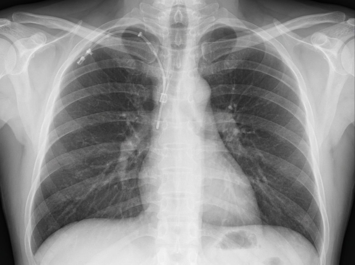
The procedure of choice for the evaluation of an aneurysm is:
A patient develops recurrent hyperparathyroidism 2 years after initial parathyroidectomy and has experienced cardiovascular complications due to persistent hypercalcemia. What is the most appropriate management?
Post parotidectomy, patient feels numb while shaving. Which nerve was involved?
Explanation: ***MR imaging*** - The **Time of Flight (TOF)** technique is a type of **magnetic resonance angiography (MRA)** that exploits the phenomenon of **flow-related enhancement** of fresh, unsaturated blood entering an imaging slice. - It is used to visualize blood flow without the need for an external contrast agent, making it particularly useful for assessing vessels in the brain and neck. *Spiral CT* - **Spiral CT** (helical CT) involves continuous data acquisition as the patient moves through the gantry, creating a spiral path of X-ray projection data. - While it has revolutionised CT angiography, it does not employ the Time of Flight principle, which is specific to MR imaging. *Digital radiography* - **Digital radiography** uses X-rays to create images, which are captured by digital sensors rather than photographic film. - This technique primarily focuses on structural imaging and does not involve the physical principles (like spin physics of protons in a magnetic field) necessary for Time of Flight applications. *CT angiography* - **CT angiography** uses **iodinated contrast material** injected intravenously to visualize blood vessels with high resolution using X-rays. - Unlike Time of Flight MRA, it relies on the contrast enhancement of flowing blood with an exogenous agent, not on the intrinsic properties of blood flow within a magnetic field.
Explanation: ***TEE (Transesophageal Echocardiography)*** - **TEE is the investigation of choice** for acute aortic dissection in **hemodynamically unstable patients** due to its **portability and rapidity**. - Can be performed at the **bedside** without transporting the critically ill patient, minimizing risk. - Provides rapid diagnosis (5-10 minutes) with **>95% sensitivity and specificity** for detecting intimal flap and false lumen. - Simultaneously assesses **complications** such as aortic regurgitation, pericardial effusion/tamponade, and ventricular function. - Particularly excellent for visualizing the **ascending aorta** and aortic root. *CT-Angio* - **CT angiography** is the **investigation of choice** for acute aortic dissection in **hemodynamically STABLE patients**. - Provides excellent anatomical detail of the entire aorta, clearly showing the intimal flap, true and false lumens, and branch vessel involvement. - Requires **patient transport** to the radiology department, which is **unsafe in unstable patients**. - Best for comprehensive surgical planning in stable patients. *MRI* - **MRI** offers the highest anatomical detail and is considered the gold standard for **chronic dissection follow-up**. - Its lengthy acquisition time (30-60 minutes) and incompatibility with monitoring equipment make it **unsuitable for acutely unstable patients**. *NCCT* - **Non-contrast CT** may show indirect signs like the **hyperdense crescent sign** in the aortic wall. - Cannot reliably differentiate true and false lumens or assess the full extent of dissection. - Insufficient for definitive diagnosis or management planning.
Explanation: ***BMI>30*** - While **obesity (BMI >30)** is associated with some surgical risks, it is generally considered a less significant independent risk factor for postoperative pulmonary complications compared to other factors like age, smoking, and surgical site. - The impact of obesity on pulmonary function is complex and varies depending on the type of surgery and presence of comorbid conditions like **sleep apnea**. *Age >70* - **Advanced age (>70)** is a significant independent risk factor due to decreased physiological reserve, reduced pulmonary function (e.g., decreased lung elasticity, impaired cough reflex), and increased prevalence of comorbidities. - Older patients are more susceptible to **atelectasis**, **pneumonia**, and **respiratory failure** postoperatively. *Patient with 7 pack years of smoking* - Even a relatively low cumulative smoking history of **7 pack-years** can impair mucociliary clearance, increase bronchial secretions, and cause airway inflammation, significantly increasing the risk of pulmonary complications. - Smoking compromises lung function and increases the risk of **bronchospasm** and infection. *Upper abdominal surgery* - **Upper abdominal surgery** is a significant risk factor because incisions close to the diaphragm interfere with diaphragmatic movement, leading to reduced lung volumes, impaired cough, and increased risk of **atelectasis** and **pneumonia**. - Pain from the incision further restricts deep breaths and coughing, contributing to pulmonary complications.
Explanation: ***Monitor regularly and consider surgery if size reaches 55mm or symptomatic*** - For **asymptomatic abdominal aortic aneurysms (AAA)** measuring less than 5.5 cm, **regular surveillance** with imaging (ultrasound or CT) is the appropriate management. - Elective surgical intervention (open repair or EVAR) is recommended when the aneurysm reaches **≥5.5 cm diameter** in men or **≥5.0 cm in women**, or if the patient becomes **symptomatic** (abdominal/back pain, tenderness). - Growth rate >1 cm/year is also an indication for repair. - The **55mm threshold** balances rupture risk against surgical mortality risk based on large randomized trials (UKSAT, ADAM). *Immediate surgical repair for all diagnosed aneurysms regardless of size* - This approach is **too aggressive** and not evidence-based. - Small AAAs (<5.5 cm) have low annual rupture rates (<1% for AAAs <5 cm), making elective surgery unjustified given operative mortality (2-5%). - Randomized trials showed **no survival benefit** from early repair of small AAAs. *Ultrasound monitoring until size exceeds 70mm* - The threshold of **70mm (7 cm) is dangerously high** and significantly increases rupture risk. - AAAs ≥5.5 cm have annual rupture rates of 3-15%, with mortality from rupture exceeding 80%. - The standard threshold for elective repair is **5.5 cm**, not 7 cm. *No treatment unless symptomatic* - This approach ignores **aneurysm size**, which is the primary predictor of rupture risk in asymptomatic patients. - Elective repair of large asymptomatic AAAs (≥5.5 cm) prevents rupture and improves survival compared to watchful waiting. - Any **symptomatic AAA** requires urgent evaluation regardless of size, as symptoms suggest impending rupture.
Explanation: ***CT Scan (Computed Tomography)*** - **CT scans** offer superior anatomical detail and can accurately detect organ damage, hemorrhage, and other injuries in **hemodynamically stable** patients with abdominal trauma. - It is considered the **most sensitive** and specific imaging modality for evaluating blunt and penetrating abdominal trauma when the patient can tolerate the study. *Ultrasonography (FAST)* - While effective for detecting **free fluid** (blood) in specific abdominal areas, **Focused Assessment with Sonography for Trauma (FAST)** has lower sensitivity for solid organ injuries or bowel perforations. - Its primary role is rapid assessment for **hemoperitoneum** to guide immediate management in unstable patients, not detailed injury characterization. *Diagnostic peritoneal lavage (DPL)* - **DPL** is an invasive procedure with high sensitivity for detecting **intraperitoneal bleeding**, but it does not identify specific organ injuries or retroperitoneal hemorrhage. - It is rarely used in hemodynamically stable patients due to its invasiveness and the availability of more detailed imaging techniques. *MRI (Magnetic Resonance Imaging)* - **MRI** provides excellent soft tissue contrast but is typically too **time-consuming** and less accessible in urgent trauma settings compared to CT. - It's generally not the first-line investigation for acute abdominal trauma due to motion artifacts and limited utility in detecting air or bone injuries.
Explanation: ***Patient's hemodynamic stability, anatomy of the aneurysm, and access to EVAR equipment*** - **Hemodynamic stability** is crucial; unstable patients may benefit from more rapid intervention, potentially open repair, or require stabilization before EVAR. - The **anatomy of the aneurysm** (e.g., neck length, angulation, iliac artery access) dictates suitability for EVAR, as specific morphological criteria must be met for stent-graft placement. - **Access to EVAR equipment and trained personnel** is also a practical consideration for emergency intervention. *Patient's hemodynamic stability and anatomy of the aneurysm* - While **hemodynamic stability** and **aneurysm anatomy** are critical factors, access to specialized EVAR equipment and facilities is also a practical determinant of whether EVAR can even be attempted, especially in an emergent setting. - This option overlooks the logistical requirements necessary for performing an **EVAR procedure**. *Access to EVAR equipment and patient's age* - **Access to EVAR equipment** is important, but **patient's age** is generally less critical than factors like physiological status, comorbidities, and aneurysm morphology when deciding between EVAR and open repair for ruptured AAAs. - Younger patients may tolerate open surgery better, but age alone does not preclude EVAR if anatomy is suitable. *Surgeon's experience with EVAR procedures* - While **surgeon experience** is important for procedural success and outcomes, it is considered secondary to the immediate patient-centered and anatomical factors. - In emergency settings, the decision primarily hinges on the **patient's hemodynamic status**, **aneurysm anatomical suitability**, and **immediate availability of EVAR resources**, rather than being driven by surgeon preference based on experience alone. - Institutional protocols typically guide whether EVAR or open repair should be attempted based on the factors in the correct answer.
Explanation: ***CT scan*** - **CT angiography** of the chest is the **gold standard** and most accurate readily available imaging modality for diagnosing acute aortic dissection, with sensitivity and specificity both >95%. - It offers **rapid acquisition** (3-5 minutes), high spatial resolution, and is widely available in emergency settings. - It clearly visualizes the **true and false lumens**, intimal flap, entry/re-entry tears, extent of the dissection (Stanford/DeBakey classification), involvement of branch vessels, and any associated complications like pericardial effusion or mediastinal hematoma. *MRI scan* - **MRI/MRA** offers comparable diagnostic accuracy (sensitivity ~98%, specificity ~95%) without radiation exposure and is excellent for chronic dissections or surveillance. - However, its use in acute settings is limited by **longer acquisition times** (20-30 minutes), limited availability in emergency departments, and contraindications (pacemakers, metallic implants, claustrophobia). - It is **not feasible** in hemodynamically unstable patients requiring rapid diagnosis and intervention. *ECG* - An **ECG** is routinely performed to evaluate chest pain and rule out acute coronary syndrome, but it does **not visualize** the aorta or diagnose dissection. - It may show non-specific ST-T changes or signs of **myocardial ischemia** if coronary ostia are involved in the dissection, but these findings are neither sensitive nor specific for aortic dissection. *Aortography* - **Conventional aortography** (invasive catheter-based angiography) was historically the gold standard but has been **replaced by CT and MRI** as first-line imaging. - It has lower sensitivity (~85-90%) than modern cross-sectional imaging and carries procedural risks including **arterial access complications**, contrast-induced nephropathy, and stroke. - Currently reserved for cases where intervention is planned or when non-invasive imaging is inconclusive.
Explanation: ***Computed tomography*** **Computed tomography (CT)**, particularly **CT angiography (CTA)**, is widely considered the procedure of choice for evaluating aneurysms due to its **rapid acquisition**, **high spatial resolution**, and ability to visualize the vessel lumen and surrounding structures. **Key advantages:** - Particularly useful for assessing aneurysm size, morphology, thrombus formation, and rupture - Excellent for both emergent and elective settings - Widely available and fast imaging acquisition - Provides comprehensive anatomical detail *Ultrasonography* **Ultrasonography** is an excellent and cost-effective **screening tool for abdominal aortic aneurysms (AAA)** because it is non-invasive and does not involve radiation. However, its utility is limited for: - Complex aneurysms requiring detailed anatomical information - Less accessible locations (e.g., thoracic, cerebral aneurysms) - **Operator dependence** and **limited field of view** restrict its use as a definitive diagnostic tool *Magnetic resonance imaging* **Magnetic resonance imaging (MRI)** and **magnetic resonance angiography (MRA)** provide excellent soft tissue contrast without ionizing radiation and can accurately evaluate aneurysm morphology and flow characteristics. However, MRI is: - More time-consuming and expensive - May be contraindicated in patients with metallic implants or claustrophobia - Less suitable for initial acute evaluation compared to CT *Angiography* **Angiography**, traditionally a catheter-based invasive procedure, provides detailed images of the vessel lumen and is excellent for evaluating precise anatomy and planning endovascular repair. While it offers highly detailed images, its: - Invasiveness - Exposure to radiation and contrast agents - Potential for complications These factors typically reserve it for **interventional planning** or when non-invasive methods are inconclusive, rather than as the primary diagnostic tool.
Explanation: ***Repeat parathyroidectomy after medical optimization*** - Recurrent **hyperparathyroidism** often requires repeat surgery, particularly in patients who have experienced cardiovascular events, as persistent hypercalcemia can exacerbate cardiac risk. - **Medical optimization** of cardiovascular conditions and metabolic status before reoperation is crucial to minimize surgical risks and improve outcomes. *Repeat neck surgery* - While repeat neck surgery is often necessary, this option is incomplete as it does not sufficiently emphasize the importance of **medical optimization** in patients with a history of cardiovascular events. - Performing surgery without adequate pre-operative evaluation and optimization can lead to increased **perioperative complications** in this high-risk group. *Observation and repeat serum Ca2+ in two months* - **Observation** is generally not appropriate for recurrent hyperparathyroidism, especially when it has already led to cardiovascular events, as continued hypercalcemia poses significant long-term health risks. - Delaying definitive treatment allows for ongoing end-organ damage, including worsening **cardiovascular disease** and bone complications. *Medical management with calcimimetics (cinacalcet)* - **Calcimimetics** like **cinacalcet** can reduce parathyroid hormone (PTH) and calcium levels, but they are typically used as an adjunct or for patients who are not surgical candidates. - In cases of recurrent hyperparathyroidism, especially with clinical sequelae like cardiovascular events, **surgical removal of the adenoma** remains the definitive treatment to achieve a cure.
Explanation: ***Greater auricular*** - The **greater auricular nerve** provides sensory innervation to the skin over the angle of the mandible, parotid gland, and mastoid process, as well as the lower half of the auricle. - Due to its superficial course over the **sternocleidomastoid muscle** and proximity to the parotid gland, it is frequently damaged during parotidectomy, leading to **numbness** in its distribution. *Mandibular* - The **mandibular nerve** (V3) is a branch of the trigeminal nerve that provides motor innervation to the muscles of mastication and sensory innervation to the lower face and chin. - While it has sensory branches to the lower lip and chin, it is not directly involved in the sensory innervation of the skin over the parotid gland. *Facial* - The **facial nerve (CN VII)** is primarily a motor nerve, responsible for facial expression, and also carries taste sensations from the anterior two-thirds of the tongue. - Damage to the facial nerve during parotidectomy would result in **facial paralysis** (e.g., drooping of the mouth, inability to close the eye), not numbness. *Auriculotemporal* - The **auriculotemporal nerve**, a branch of the mandibular nerve (V3), supplies sensory innervation to the skin anterior to the ear, the temporomandibular joint, and the parotid gland capsule. - While it does innervate the parotid region, damage to this nerve is more typically associated with **Frey's syndrome** (gustatory sweating) rather than simple numbness after parotidectomy.
Get full access to all questions, explanations, and performance tracking.
Start For Free