
Cardiovascular Radiology — MCQs

On this page
For which of the following conditions is CT thorax considered the gold standard imaging modality for diagnosis?
Which of the following is a radiological feature of coarctation of the aorta?
Which view is taken for aortic window?
Which imaging modality is most effective for assessing plaque morphology in coronary arteries?
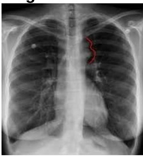
The chest radiograph shown below is from a 25-year-old male patient presenting with hypertension. The image demonstrates bilateral inferior rib notching. What is the most likely diagnosis?
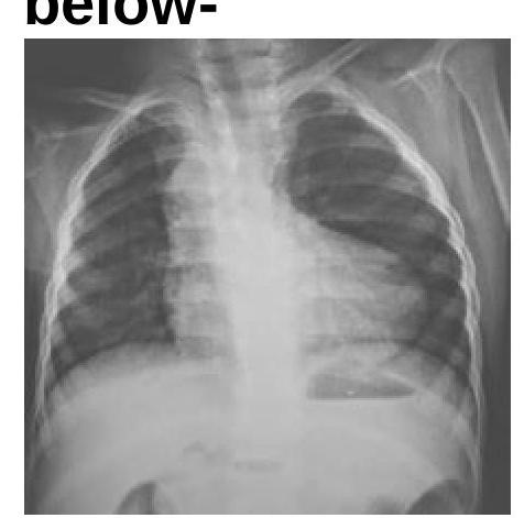
Identify the condition in the X-ray given below:
Pulmonary plethora is typically seen in all of the following conditions except:
Boot shape of heart in TOF is due to:
Which of the following conditions is NOT associated with a flask-shaped heart?
For pericardial calcifications, which is the best investigation?
Practice by Chapter
Cardiovascular Anatomy
Practice Questions
Cardiac CT Techniques
Practice Questions
Cardiac MRI Techniques
Practice Questions
Ischemic Heart Disease Imaging
Practice Questions
Valvular Heart Disease
Practice Questions
Cardiomyopathies
Practice Questions
Pericardial Diseases
Practice Questions
Congenital Heart Disease
Practice Questions
Aortic and Great Vessel Imaging
Practice Questions
Peripheral Vascular Imaging
Practice Questions
Cardiovascular Interventional Procedures
Practice Questions
Post-Surgical Cardiovascular Imaging
Practice Questions
Want unlimited practice?
Get full access to all questions, explanations, and performance tracking.
Scan to download app