
Cardiovascular Radiology — MCQs

On this page
IOC for Acute Aortic Dissection in a Clinically Unstable patient is?
Earliest sign of left atrial enlargement is -
Investigation of choice for vascular ring around airway:
In aortic dissection, the most accurate investigation is:
Double atrial shadow in mitral stenosis due to -
Which one of the following investigations is considered to be "Gold standard" technique for diagnosis of arterial occlusive disease –
In which of the following a 'Coeur en Sabot' shape of the heart is seen:
In coarctation of aorta the rib changes are seen from:
Gas shadow in the heart and great vessels on chest imaging most commonly appears in association with-
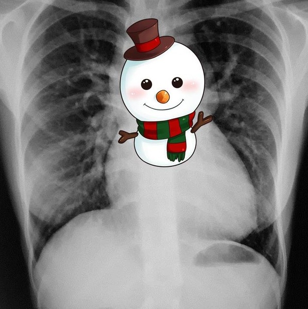
Snowman appearance on x-ray is seen in which cardiac pathology -
Practice by Chapter
Cardiovascular Anatomy
Practice Questions
Cardiac CT Techniques
Practice Questions
Cardiac MRI Techniques
Practice Questions
Ischemic Heart Disease Imaging
Practice Questions
Valvular Heart Disease
Practice Questions
Cardiomyopathies
Practice Questions
Pericardial Diseases
Practice Questions
Congenital Heart Disease
Practice Questions
Aortic and Great Vessel Imaging
Practice Questions
Peripheral Vascular Imaging
Practice Questions
Cardiovascular Interventional Procedures
Practice Questions
Post-Surgical Cardiovascular Imaging
Practice Questions
Want unlimited practice?
Get full access to all questions, explanations, and performance tracking.
Scan to download app