A 2-month-old infant is brought to the clinic because of poor feeding, sweating, and difficulty breathing. The parents state that she was doing very well, and has actually been a "very easy going baby", until about a week ago. They assumed that she was developing a "cold", but it has not passed, and the symptoms have been worsening. Cardiac examination reveals a loud, harsh systolic murmur with a thrill that is heard best at the left sternal border. The most likely underlying abnormality is
A 2-week-old girl is found to have a harsh murmur along the left sternal border. The parents report that the baby gets "bluish" when she cries or drinks from her bottle. Echocardiogram reveals a congenital heart defect associated with pulmonary stenosis, ventricular septal defect, dextroposition of the aorta, and right ventricular hypertrophy. What is the appropriate diagnosis?
In a child with coarctation of aorta, all the following are seen in plain chest radiograph except:
A child presented at 10 weeks with recurrent episode of pneumonia and failure to thrive. X-ray shows cardiomegaly & pulmonary plethora. What is the diagnosis?
Which of the following is the most common congenital cardiac malformation?
A 45-year-old woman presents with clubbing and cyanosis. Chest X-ray shows a prominent pulmonary artery. Which congenital heart disease should be considered?
Flask shaped heart is seen in –
Boot shape of heart in TOF is due to:
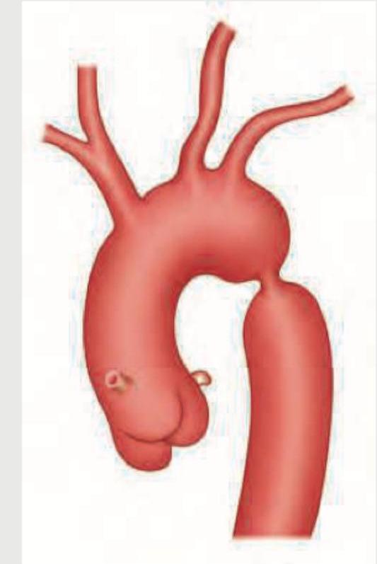
In the condition shown below, rib notching is present in which of the following ribs? (AIIMS Nov 2015)

Radiological features of left ventricular heart failure are all, except -
Explanation: ***ventricular septal defect*** - A **ventricular septal defect (VSD)** causes a **harsh, holosystolic (pansystolic) murmur** best heard at the **left sternal border**, often with a palpable thrill, due to turbulent blood flow through the defect from the high-pressure left ventricle to the lower-pressure right ventricle. - Symptoms like **poor feeding, sweating, and difficulty breathing** in an infant, especially with recent onset and worsening, are consistent with **congestive heart failure** secondary to a large VSD causing significant left-to-right shunting. - The timing of symptom onset (around 2 months) is typical, as pulmonary vascular resistance drops after the first 4-6 weeks of life, increasing the left-to-right shunt and precipitating heart failure. *aortic stenosis* - **Aortic stenosis** typically presents with a **systolic ejection murmur** loudest at the **right upper sternal border** with radiation to the neck, not a harsh holosystolic murmur at the left sternal border. - While severe aortic stenosis can lead to heart failure symptoms, the murmur's location and quality are inconsistent with this diagnosis. *atrial septal defect* - An **atrial septal defect (ASD)** typically causes a **systolic ejection murmur** at the **pulmonary area** (left upper sternal border) due to increased flow across the pulmonic valve, often accompanied by a **fixed, wide splitting of S2**. - Significant heart failure in infancy is rare with an isolated ASD, as the pressure gradient across the atria is usually low, leading to asymptomatic presentation until later childhood or adulthood. *patent ductus arteriosus* - A **patent ductus arteriosus (PDA)** is characterized by a **continuous, machine-like murmur** loudest below the left clavicle (infraclavicular area), not a harsh systolic murmur with a thrill at the left sternal border. - While a large PDA can cause heart failure symptoms in infancy, the distinctive continuous murmur differentiates it from a VSD.
Explanation: ***Tetralogy of Fallot*** - The combination of **pulmonary stenosis**, **ventricular septal defect**, **dextroposition of the aorta** (overriding aorta), and **right ventricular hypertrophy** is the classic definition of Tetralogy of Fallot. - The "bluish" episodes (cyanosis) when crying or feeding are characteristic of **tet spells**, indicating right-to-left shunting and reduced pulmonary blood flow, exacerbated by activity. *Atrial septal defect* - An ASD primarily involves a **left-to-right shunt** and typically presents with a **fixed, split S2** and a **pulmonic flow murmur**, usually without cyanosis in infancy. - It does not involve the characteristic four defects seen in this patient, particularly the significant pulmonary stenosis and cyanosis. *Coarctation of aorta, postductal* - **Postductal coarctation** typically presents in older children or adults with **hypertension in the upper extremities** and **diminished or absent femoral pulses**, often without cyanosis. - This condition is a narrowing of the aorta **distal to the ductus arteriosus** and does not involve the four specific intracardiac defects described. *Coarctation of aorta, preductal* - **Preductal coarctation** can present in neonates with **heart failure** and **differential cyanosis** (upper body pink, lower body blue), or signs of shock if the ductus arteriosus closes. - This condition involves a narrowing of the aorta **proximal to the ductus arteriosus** and is not characterized by the four specific tetralogy defects.
Explanation: ***'E' Sign or 'Reverse Three sign' (Reverse ε sign)*** - The **'E' sign** or **'reverse three sign'** is seen on **barium esophagram** (lateral view), NOT on a plain chest X-ray - On barium swallow, the esophagus shows indentation creating a reverse '3' or 'ε' shape due to impression from the dilated pre-stenotic aorta, the coarctation site, and the dilated post-stenotic aorta - **This is the correct answer** as the question asks specifically about plain chest radiograph findings - This sign requires contrast study and cannot be visualized on plain radiography *Three sign ('3' sign)* - The **'three sign'** is a **classic finding** in coarctation on plain chest X-ray (PA view) - Seen on the **left heart border** representing: (1) dilated left subclavian artery, (2) indentation at coarctation site, (3) post-stenotic dilation of descending aorta - Creates the shape of the numeral '3' along the aortic knuckle region - This is directly visible on plain radiograph *Prominent ascending aorta* - **Commonly seen** in coarctation due to increased afterload on the left ventricle - Results in **left ventricular hypertrophy** and dilation of the ascending aorta - Part of the cardiovascular remodeling in response to chronic pressure overload - Visible as widening of the superior mediastinum on plain chest X-ray *Rib notching* - **Classic finding** in long-standing coarctation of the aorta (usually after 5-6 years of age) - Due to **collateral circulation** through dilated intercostal arteries that erode the inferior rib margins - Typically affects **ribs 3-9** bilaterally - Represents chronic compensatory mechanism to bypass the obstruction
Explanation: ***VSD*** - **Ventricular septal defect (VSD)** is the most common cause of this presentation in early infancy (symptoms typically appear at **6-10 weeks** of age). - Large VSDs cause significant **left-to-right shunt** leading to pulmonary overcirculation, resulting in **recurrent pneumonia** and **failure to thrive**. - **Cardiomegaly** (due to volume overload of left atrium and ventricle) and **pulmonary plethora** (increased pulmonary vascular markings) on X-ray are classic findings. - The infant may also present with tachypnea, feeding difficulties, and poor weight gain. *TOF* - **Tetralogy of Fallot (TOF)** is a **cyanotic heart defect** with right-to-left shunt, presenting with cyanosis and hypoxic spells, not recurrent pneumonia. - X-ray shows **boot-shaped heart** and **pulmonary oligemia** (decreased pulmonary vascular markings), not pulmonary plethora. - Does not typically cause failure to thrive in the same manner as acyanotic left-to-right shunt lesions. *Patent foramen ovale* - A **patent foramen ovale (PFO)** is a normal variant in infants and typically remains **asymptomatic**. - Does not cause significant hemodynamic shunting in the absence of elevated right atrial pressure. - Does not cause **cardiomegaly**, **pulmonary plethora**, recurrent pneumonia, or failure to thrive. *ASD* - An **atrial septal defect (ASD)** also causes left-to-right shunt with pulmonary plethora, but the shunt develops **gradually** over time. - ASD typically presents **later in childhood or adulthood** with milder symptoms (fatigue, exercise intolerance) due to lower pressure gradient across atria. - **Recurrent pneumonia and failure to thrive at 10 weeks** are uncommon with isolated ASD, as the hemodynamic changes are less pronounced in early infancy compared to VSD. - When symptomatic in infancy, large ASDs present later (around 6 months to 1 year) rather than at 10 weeks.
Explanation: ***Ventricular septal defect (VSD)*** - VSDs are the **most common congenital heart defect**, accounting for approximately 25-30% of all congenital cardiac malformations. - They involve a **hole in the septum** separating the left and right ventricles, leading to a left-to-right shunt. *Persistent truncus arteriosus (PTA)* - PTA is a rare congenital heart defect where a **single arterial trunk** arises from the heart, supplying both systemic and pulmonary circulation. - Its incidence is much **lower than VSD**, representing less than 1% of congenital heart defects. *Common ventricle (CV)* - A common ventricle, also known as **single ventricle**, is a complex and rare congenital defect where only one functional ventricle is present. - It is a **severe malformation** and much less common than VSD. *Atrial septal defect (ASD)* - ASDs are congenital heart defects involving a **hole in the wall between the atria** of the heart. - While relatively common, ASDs are **less frequent than VSDs**, accounting for about 5-10% of congenital heart defects.
Explanation: Atrial septal defect (ASD) with Eisenmenger syndrome. - Clubbing, cyanosis, and a prominent pulmonary artery in a 45-year-old suggest long-standing pulmonary hypertension with shunt reversal, characteristic of Eisenmenger syndrome [1]. - An ASD is a common congenital heart defect that can lead to significant left-to-right shunting, eventually causing pulmonary hypertension [1]. and reversed shunt flow (Eisenmenger syndrome) over decades [2]. *Patent ductus arteriosus (PDA) with Eisenmenger syndrome.* - While PDA can also lead to Eisenmenger syndrome, it typically presents with a continuous murmur and often causes symptoms earlier in life if the shunt is large. - The patient's age and the specific features presented are more classic for an ASD progressing to Eisenmenger syndrome. *Tetralogy of Fallot* - This is a cyanotic congenital heart disease from birth or early childhood, commonly presenting with cyanosis and "tet spells" [3]. - While it causes cyanosis, a prominent pulmonary artery is not a typical feature; instead, there is often reduced pulmonary blood flow and a small pulmonary artery due to pulmonary outflow obstruction [3]. *Pulmonary stenosis (PS)* - Pulmonary stenosis primarily causes obstruction to blood flow out of the right ventricle, leading to right ventricular hypertrophy and potentially a systolic murmur. - While severe PS can cause cyanosis due to a right-to-left shunt through an ASD (if present), isolated PS is typically acyanotic and does not usually present with a prominent pulmonary artery or clubbing unless it's very severe and associated with other defects.
Explanation: ***Pericardial effusion*** - A **"flask-shaped"** or **"water bottle-shaped" heart** on chest X-ray is a classic finding in significant pericardial effusion. - This appearance results from the accumulation of a large amount of fluid in the **pericardial sac**, which causes the cardiac silhouette to enlarge symmetrically and assume a globular shape. *TOF (Tetralogy of Fallot)* - TOF typically presents with a **"boot-shaped" heart** (coeur en sabot) on chest X-ray due to right ventricular hypertrophy and a concave pulmonary artery segment. - This morphology is distinctly different from the flask-shaped appearance of pericardial effusion. *Ebstein anomaly* - Ebstein anomaly is characterized by apical displacement of the tricuspid valve, leading to **massive right atrial enlargement**. - On chest X-ray, this often results in a **markedly enlarged oval-shaped heart**, which can be quite massive but does not typically have the distinct flask/water bottle shape. *TAPVC (Total Anomalous Pulmonary Venous Connection)* - TAPVC can present with different X-ray findings depending on the type, but a classic finding for the supracardiac type is a **"snowman" or "figure-of-8" heart** in children. - This appearance is due to the dilated superior vena cava and anomalous veins draining to it, not a flask shape.
Explanation: ***Right ventricular hypertrophy*** - The characteristic **boot-shaped heart (coeur en sabot)** seen in Tetralogy of Fallot (TOF) on a chest X-ray is primarily due to **right ventricular hypertrophy** and the small pulmonary artery. - The hypertrophied right ventricle lifts the cardiac apex, while the concavity in the area of the pulmonary artery (due to **pulmonary stenosis**) gives the heart its distinctive shape. *Enlargement of the left atrium* - Left atrial enlargement is not a feature of **Tetralogy of Fallot**; in fact, chronic pulmonary outflow obstruction often leads to a relatively normal or small left atrium. - This condition involves right-sided heart abnormalities, and left atrial enlargement would suggest increased left-sided pressures, which are not typical for TOF. *Enlargement of the right atrium* - While right atrial enlargement can occur in severe cases of TOF due to increased resistance to blood flow, it is **right ventricular hypertrophy** that is the primary determinant of the classic boot-shaped cardiac silhouette. - Right atrial enlargement alone does not create the specific "boot" appearance which is largely due to the ventricular contour. *Hypertrophy of both ventricles* - In Tetralogy of Fallot, the primary ventricular abnormality is **right ventricular hypertrophy**, driven by the need to pump blood through a stenosed pulmonary artery. - The left ventricle typically maintains a normal size and function, as it primarily pumps into the systemic circulation and is not directly affected by the primary defects in the same way as the right ventricle.
Explanation: **3rd to 9th ribs** - The image provided depicts **coarctation of the aorta**, characterized by a narrowing of the aorta, typically distal to the origin of the left subclavian artery. - In coarctation of the aorta, collateral circulation develops through the **intercostal arteries** to bypass the constriction, leading to their enlargement and subsequent erosion of the inferior margins of the **3rd to 9th ribs**, a finding known as "rib notching." *1st to 9th ribs* - While rib notching affects upper ribs, it typically **spares the 1st and 2nd ribs** because the superior intercostal arteries (which supply these ribs) originate directly from the subclavian artery, often proximal to the coarctation, so they do not participate in collateral circulation as significantly. - The pattern of notching is usually more concentrated in the mid-thoracic region. *11th and 12th ribs* - Rib notching from coarctation of the aorta is rarely observed in the **floating ribs** (11th and 12th ribs). - These ribs have a different anatomical relationship with the pleura and typically do not bear the brunt of increased collateral flow from the intercostal arteries in the same way as the higher ribs. *All ribs* - Rib notching is a localized phenomenon reflecting increased blood flow through specific intercostal arteries involved in collateral circulation due to aortic coarctation. - Therefore, it does **not affect all ribs**, and its absence in certain ribs (like the 1st, 2nd, 11th, and 12th) helps differentiate this condition radiologically.
Explanation: ***Oligemic lung fields*** - **Oligemic lung fields** are characteristic of conditions like severe **pulmonary hypertension** or **pulmonary embolism** post-embolus, leading to reduced blood flow to the lungs, not left ventricular heart failure. - In left ventricular heart failure, the primary issue is **pulmonary venous congestion** and **edema**, leading to increased, not decreased, pulmonary vascular markings. *Kerley B lines* - **Kerley B lines** are often seen in left ventricular heart failure, indicating **interstitial pulmonary edema**. - They represent thickened, edematous interlobular septa due to increased hydrostatic pressure in the pulmonary capillaries. *Cardiomegaly* - **Cardiomegaly** (enlarged heart) on chest X-ray is a common finding in left ventricular heart failure, reflecting ventricular dilation and/or hypertrophy due to chronic increased workload. - This enlargement is often due to the heart's compensatory mechanisms attempting to maintain cardiac output. *Increased flow in upper lobe veins* - **Increased flow in upper lobe veins** (cephalization of pulmonary vessels) is an early sign of pulmonary venous hypertension in left ventricular heart failure. - Due to elevated left atrial pressure, blood is preferentially shunted to the less gravitationally dependent upper lobes.
Get full access to all questions, explanations, and performance tracking.
Start For Free