Posterior cardinal veins develop into:
All the following openings in the right atrium are guarded by a valve except
Left anterior descending artery is a direct branch of
What is the cardiothoracic ratio in children?
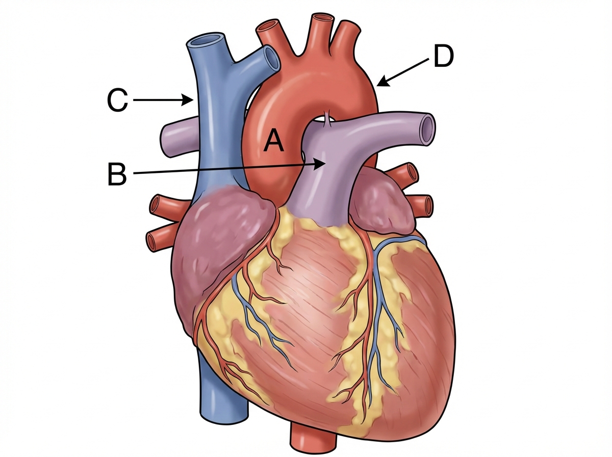
For pericardial calcifications, which is the best investigation?
Which X-ray finding is more characteristic of ASD compared to VSD?
All of the following arteries are common sites of occlusion by a thrombus except:
Which heart chamber has the thickest wall?
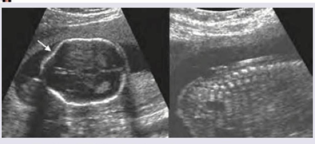
Keyhole sign on fetal ultrasound is seen in:

Which one of the following is used in Cardiovascular imaging?
Explanation: ***Common iliac vein*** - The **posterior cardinal veins** are paired primitive veins in the embryo that drain the caudal body. - The **caudal portions** of the posterior cardinal veins persist and directly form the **common iliac veins** and contribute to the internal iliac veins [1]. - This is the **primary and most direct derivative** of the posterior cardinal veins, making it the best answer. *Azygos vein* - The **azygos vein** develops from the **right supracardinal vein** + **cranial portion of the right posterior cardinal vein**. - While posterior cardinal veins do contribute to its formation, this is not the primary derivative. - The middle portions of posterior cardinal veins regress, and the supracardinal contribution is more significant. *Hemiazygos vein* - The **hemiazygos vein** is derived from the **left supracardinal vein** + **cranial portion of the left posterior cardinal vein**. - Similar to the azygos, posterior cardinal veins contribute but are not the primary source. - The supracardinal vein provides the major contribution. *Parts of inferior vena cava* - The **IVC** forms from multiple embryonic veins: right vitelline vein (hepatic segment), right subcardinal vein (renal segment), right supracardinal vein (infrarenal segment), and hepatic veins. - While the common iliac veins (derived from posterior cardinal veins) drain into the IVC, the posterior cardinal veins themselves do **not directly form the IVC proper**. - The posterior cardinal veins largely regress in their middle portions.
Explanation: ***Superior vena cava*** - The opening of the **superior vena cava** into the right atrium is generally not guarded by a valve. - Its blood flow into the heart is maintained by relatively low pressure and directly continuous with the right atrium. *Inferior vena cava* - The opening of the **inferior vena cava** into the right atrium is guarded by a rudimentary valve called the **Eustachian valve** [1]. - This valve is more prominent in fetal life, helping to direct oxygenated blood from the umbilical vein to the foramen ovale [1]. *Coronary sinus* - The opening of the **coronary sinus** into the right atrium is guarded by the **Thebesian valve**. - This valve's function is to prevent regurgitation of blood from the right atrium into the coronary sinus during atrial systole. *Atrioventricular opening* - The **right atrioventricular opening** is guarded by the **tricuspid valve** [2]. - This valve prevents backflow of blood from the right ventricle into the right atrium during ventricular systole [2].
Explanation: ***Left coronary artery*** - The left coronary artery (LCA) is a major coronary artery that arises from the **aorta** and quickly branches into two main arteries: the **left anterior descending (LAD) artery** [1] and the circumflex artery. - The LAD artery, also known as the **"widowmaker"**, supplies oxygenated blood to the **anterior wall of the left ventricle** and the interventricular septum, making it crucial for heart function. *Right coronary artery* - The **right coronary artery (RCA)** typically supplies the **right atrium**, most of the **right ventricle**, and the inferior wall of the left ventricle, which are distinct areas from the LAD's supply. - The RCA originates from the **right sinus of Valsalva** and travels in the atrioventricular groove, while the LAD originates from the left main coronary artery [1]. *Circumflex artery* - The circumflex artery is another main branch of the **left coronary artery**, typically supplying the **lateral and posterior walls of the left ventricle** and the left atrium. - While it branches from the same parent vessel as the LAD, it is a direct branch itself, not the origin of the LAD [1]. *Ascending aorta* - The ascending aorta is the initial part of the aorta that originates from the **left ventricle** and gives rise to the **coronary arteries** (both left and right coronary arteries). - It is the source from which the **left coronary artery** (and thus the LAD indirectly) originates, but it is not a direct branch itself.
Explanation: ***50-55%*** - The normal **cardiothoracic ratio** in children is generally considered to be **50-55%** on a **posterior-anterior (PA) chest X-ray**. - In **infants and young children (under 2 years)**, the ratio can be **up to 55-60%** due to relatively larger cardiac size and more horizontal positioning of the heart. - In **older children (over 2 years)**, the normal ratio approaches adult values of **less than 50%**. - A ratio consistently greater than 55% could indicate **cardiomegaly**, which warrants further investigation. *30-35%* - This range is typically too low and would suggest an **unusually small heart** for the chest cavity, which is not a normal finding in children. - A very low ratio like this is not characteristic of the pediatric population and may indicate technical issues with the radiograph. *40-45%* - While this range approaches normal values for **older children and adults**, it is generally on the lower side for the average **pediatric cardiothoracic ratio**. - This percentage alone is not the best answer when considering the entire pediatric age spectrum, including infants and younger children. *60-65%* - A cardiothoracic ratio in this range would typically be considered **abnormal** and indicative of **cardiomegaly** in children beyond infancy. - Such an elevated ratio would suggest an enlarged heart, prompting further cardiac evaluation including **echocardiography** to assess for structural heart disease.
Explanation: ***Correct: CT scan*** - **CT scans** are highly sensitive and specific for detecting **pericardial calcifications** due to their excellent spatial resolution and ability to measure calcium density (Hounsfield units). - They provide detailed anatomical information about the **pericardium** and can accurately map the extent, location, and thickness of calcified areas. - **CT is the gold standard** for detecting and quantifying pericardial calcification, particularly in constrictive pericarditis. *Incorrect: Ultrasound* - While ultrasound (echocardiography) can visualize the pericardium and may detect calcifications, its ability to definitively identify and characterize **calcifications** is limited compared to CT. - **Acoustic shadowing** from calcifications can obscure underlying structures, making a precise assessment challenging. - Useful for detecting pericardial effusion and thickening, but not optimal for calcification assessment. *Incorrect: MRI* - **MRI excels** in visualizing soft tissues, pericardial inflammation, and fluid collections, but it is **poor at detecting calcium**. - Calcifications typically appear as signal voids (black) on MRI, making it difficult to differentiate them from other structures, air, or motion artifacts. - MRI is valuable for assessing pericardial inflammation and constriction but not the preferred method for calcification. *Incorrect: Transesophageal echocardiography* - TEE offers high-resolution images of cardiac structures and is primarily used for assessing valve function, intracardiac masses, endocarditis, and aortic pathology. - Its utility in detecting and characterizing **pericardial calcifications** is limited compared to CT, especially for diffuse or subtle calcifications. - The pericardium is not optimally visualized with TEE compared to transthoracic echocardiography.
Explanation: ***Normal LA*** - In an uncomplicated **atrial septal defect (ASD)**, blood shunts from the left atrium to the right atrium, decompressing the left atrium. - This decompression results in a **normal-sized left atrium** on chest X-ray, distinguishing it from conditions with **left ventricular overload**. *Enlarged LA* - An **enlarged left atrium (LA)** is more characteristic of conditions causing **left-sided volume or pressure overload**, such as **ventricular septal defect (VSD)** with significant left-to-right shunt. - In VSD, blood shunts from the left ventricle to the right ventricle, increasing **pulmonary blood flow** and pressure, ultimately leading to LA enlargement. *Aortic shadow* - The **aortic shadow** on X-ray reflects the size and position of the aorta, and while some cardiac conditions can affect it, changes in its size are not a primary distinguishing feature between ASD and VSD. - A subtle **aortic knuckle** may be seen, but it does not differentiate the two defects. *Pulmonary Congestion* - While both ASD and VSD can cause increased **pulmonary blood flow**, **pulmonary congestion** (interstitial or alveolar edema) is more likely to be prominent in a **large VSD** due to the higher pressure shunt. - ASD typically leads to **pulmonary arterial hypertension** over time, but less frank congestion unless there's associated left-sided heart failure.
Explanation: ***Marginal*** - The **marginal arteries** are typically small and supply a smaller portion of the right ventricle, making them less likely sites for **major clinical occlusion** compared to larger, more critical coronary vessels. - While occlusion can occur, it usually causes less extensive damage and is therefore **less common** as a primary site of acute thrombus-related myocardial infarction. *Posterior interventricular* - The **posterior interventricular artery (PDA)** is a major coronary artery, responsible for supplying the posterior walls of the ventricles and the posterior one-third of the interventricular septum. - Occlusion of the PDA, often a branch of the right coronary artery (RCA) or circumflex artery, can lead to **significant infarction** in these critical areas, making it a common site of thrombus formation. *Circumflex* - The **circumflex artery (Cx)** is a major branch of the left main coronary artery that supplies the left atrium and the posterior and lateral walls of the left ventricle. - Occlusion of the circumflex artery can result in **lateral or posterior myocardial infarction**, making it a frequent site for thrombus formation. *Anterior interventricular* - The **anterior interventricular artery (LAD)**, also known as the left anterior descending artery, is the most common site of coronary artery occlusion. - It supplies the anterior wall of the left ventricle and the anterior two-thirds of the interventricular septum, and its occlusion is often referred to as the **"widowmaker"** due to the extensive damage and high mortality associated with it.
Explanation: ***Left ventricle*** - The left ventricle is responsible for pumping **oxygenated blood** to the entire systemic circulation, requiring significant force. - Its muscular wall is the **thickest** to generate the high pressures needed to overcome systemic vascular resistance [1]. *Right atrium* - The right atrium receives deoxygenated blood from the body and pumps it to the right ventricle, which is a **low-pressure circuit** [2]. - Its walls are relatively thin compared to the ventricles, as it only needs to provide a small "kick" to fill the right ventricle. *Left atrium* - The left atrium receives oxygenated blood from the lungs and pumps it to the left ventricle, operating under **low pressure**. - Its walls are thin, similar to the right atrium, as it does not need to generate high pressures. *Right ventricle* - The right ventricle pumps deoxygenated blood to the **pulmonary circulation**, which is a **low-pressure system** [1]. - While thicker than the atria, its wall is thinner than the left ventricle because it faces less resistance and pumps against lower pressures to the lungs.
Explanation: ***Dandy-Walker syndrome*** - The ultrasound image shows **enlargement of the posterior fossa** with a **large cyst occupying the space normally taken by the cerebellum**, and a **keyhole sign** (arrow pointing to a defect where the cerebellar vermis should be). This is characteristic of Dandy-Walker syndrome. - Absence or **hypoplasia of the cerebellar vermis** is a hallmark feature, leading to communication of the fourth ventricle with a posterior fossa cyst. *Arnold-Chiari malformation* - Characterized by **herniation of cerebellar tonsils** through the foramen magnum and often associated with myelomeningocele. - Key ultrasound findings include a **lemon sign** (flattened frontal bones) and **banana sign** (anteriorly curved cerebellum), which are not depicted here. *Spina bifida* - This is a **neural tube defect** involving incomplete closure of the spinal column. - While it can be associated with Arnold-Chiari malformation, the primary features of spina bifida (e.g., a **sacral defect** with a mass) are not shown in these images. *Aqueductal stenosis* - Results in **dilation of the lateral and third ventricles** due to obstruction of cerebrospinal fluid flow in the aqueduct of Sylvius. - It primarily affects the supratentorial ventricular system and does not typically involve the **posterior fossa cyst** and **vermic hypoplasia** seen in the image.
Explanation: **Explanation:** **Multidetector CT (MDCT)** is the gold standard for cardiovascular imaging because it overcomes the two biggest challenges in cardiac radiology: **cardiac motion** and **respiratory motion**. 1. **Why MDCT is correct:** MDCT utilizes multiple rows of detectors, allowing for sub-millimeter isotropic resolution and high temporal resolution. When combined with **ECG-gating** (synchronizing data acquisition with the diastolic phase of the cardiac cycle), it allows for motion-free imaging of the coronary arteries. Modern MDCT (64-slice and above) can image the entire heart in a single breath-hold, making it essential for Coronary CT Angiography (CCTA). 2. **Why other options are incorrect:** * **Second and Third Generation CT:** These are historical iterations. Second-generation used a "translate-rotate" motion with a fan beam, and Third-generation used a "rotate-rotate" motion. While Third-generation is the basis for modern scanners, the basic configuration lacked the speed and detector density required to freeze cardiac motion. * **Spiral (Helical) CT:** While a prerequisite for MDCT, early single-slice spiral CTs were too slow to capture the heart without significant motion artifacts and could not provide the necessary spatial resolution for small vessels like the coronary arteries. **High-Yield Clinical Pearls for NEET-PG:** * **Temporal Resolution:** The time required to acquire data for one image. High temporal resolution is vital to "freeze" the heart. * **Electron Beam CT (EBCT):** Historically known as the "Ultrafast CT," it was the previous gold standard for Calcium Scoring but has been largely replaced by MDCT. * **Calcium Scoring (Agatston Score):** Performed on MDCT to predict the risk of future adverse cardiac events. * **Beta-blockers:** Often administered before a Cardiac CT to lower the heart rate (ideally <60-65 bpm) to improve image quality.
Get full access to all questions, explanations, and performance tracking.
Start For Free