A patient presents with engorged neck veins, a blood pressure of 80/50 mmHg, and a pulse rate of 100 beats per minute following blunt trauma to the chest. The diagnosis is:
The procedure of choice for the evaluation of aortic aneurysms is -
A 68-year-old asymptomatic male is found to have an abdominal aortic aneurysm (AAA) measuring 4.5 cm on routine ultrasound screening. What is the most appropriate management?
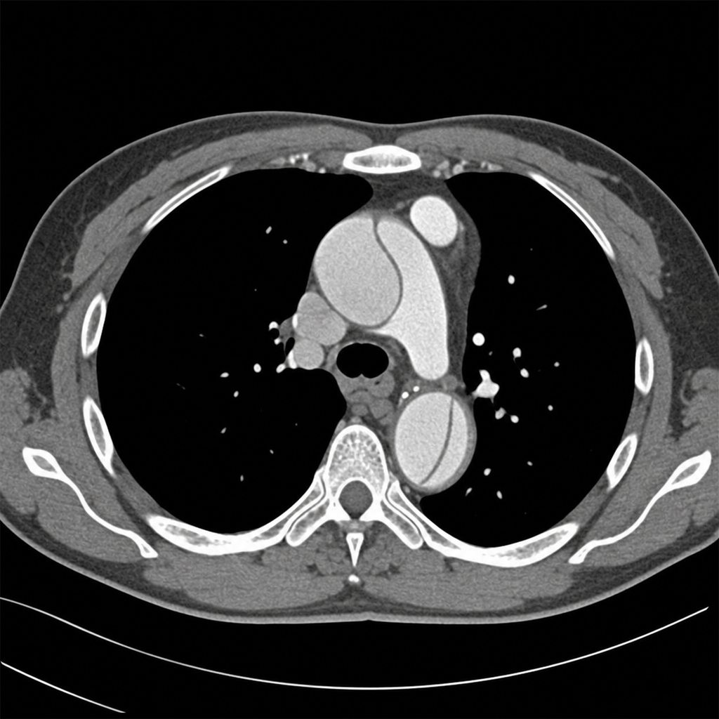
What is the cutoff for surgery in an abdominal aortic aneurysm in asymptomatic patients?
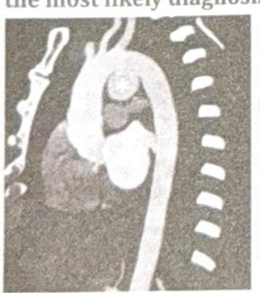
A 38-year-old patient presents with chest pain and hoarseness of voice for the past month. Based on the radiographic image below, what is the most likely diagnosis?

In Marfan's syndrome, Aortic aneurysm occurs most commonly in:
Single heart sound (S2) is heard in:
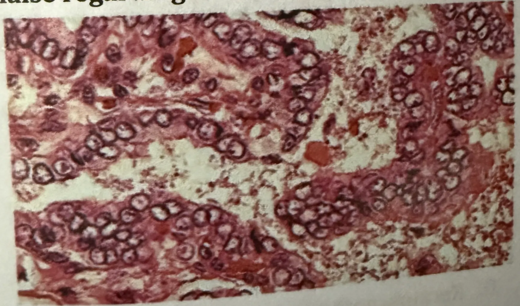
A middle aged male patient presents with painless slow growing neck swelling. On examination, lymph nodes are positive. Surgery is done and biopsy is shown in the image below. Which of the following is false regarding the HPE findings?

The thymus is located in which part of the body?
An asymptomatic old patient presents with bruit in the carotid artery. Which of the following is the investigation of choice?
Explanation: ***Cardiac tamponade*** - The clinical presentation shows **two components of Beck's triad**: **engorged neck veins (elevated JVP)** and **hypotension** (80/50 mmHg). While muffled heart sounds (the third component) are not mentioned, this is not required for diagnosis. - The combination of **blunt chest trauma** and these symptoms strongly suggests fluid accumulation in the pericardial sac, compressing the heart and impairing its filling. - **Tachycardia** (100 bpm) represents a compensatory response to reduced cardiac output. *Pneumothorax* - While pneumothorax can cause respiratory distress and hypotension, it typically presents with **absent breath sounds** on the affected side and **hyperresonance to percussion**, which are not described. - Engorged neck veins are not characteristic of simple pneumothorax. **Tension pneumothorax** can cause distended neck veins and severe hypotension, but would also present with severe respiratory distress and tracheal deviation away from the affected side. *Right ventricular failure* - Right ventricular failure can cause **engorged neck veins** but usually presents with signs of systemic congestion like **peripheral edema** and hepatomegaly, developing over time. - This is not typically an acute, immediate consequence of blunt chest trauma. The **acute hypotension** and **tachycardia** are more indicative of obstructive shock (cardiac tamponade) rather than pump failure. *Hemothorax* - Hemothorax involves blood accumulation in the pleural space, leading to **absent breath sounds** and **dullness to percussion** on the affected side. - While it can cause hypotension and tachycardia due to **hypovolemic shock** from blood loss, **engorged neck veins** are not a feature. In fact, significant blood loss typically causes **flat or collapsed neck veins** due to reduced venous return.
Explanation: ***Computed tomography*** - **Computed tomography (CT)** offers excellent spatial resolution and is the gold standard for diagnosing, staging, and pre-operative planning for aortic aneurysms. - It precisely measures aneurysm size, detects mural thrombus, assesses rupture risk, and evaluates the extent of involvement with surrounding structures. *Ultrasonography* - While useful for initial screening and serial monitoring of known abdominal aortic aneurysms due to its non-invasiveness and cost-effectiveness, its accuracy can be limited by **patient body habitus** and **bowel gas**. - It may not reliably visualize the entire aorta or accurately assess complex anatomy and rupture. *Magnetic resonance imaging* - **Magnetic resonance imaging (MRI)** provides detailed anatomical information and avoids radiation exposure, but it is typically more expensive and time-consuming than CT. - It is often reserved for patients with **renal insufficiency** where iodinated contrast is a concern or when evaluating specific tissue characteristics not well seen on CT. *Arteriography* - **Arteriography** (angiography) is an invasive procedure involving direct contrast injection, carrying risks such as arterial injury and nephrotoxicity. - While it can visualize the aortic lumen, it primarily shows the patent lumen and may **underestimate the true aneurysm size** due to mural thrombus. It is typically used for intervention planning or specific contexts rather than initial diagnosis.
Explanation: ***Monitor regularly and consider surgery if size reaches 55mm or symptomatic*** - For **asymptomatic abdominal aortic aneurysms (AAA)** measuring less than 5.5 cm, **regular surveillance** with imaging (ultrasound or CT) is the appropriate management. - Elective surgical intervention (open repair or EVAR) is recommended when the aneurysm reaches **≥5.5 cm diameter** in men or **≥5.0 cm in women**, or if the patient becomes **symptomatic** (abdominal/back pain, tenderness). - Growth rate >1 cm/year is also an indication for repair. - The **55mm threshold** balances rupture risk against surgical mortality risk based on large randomized trials (UKSAT, ADAM). *Immediate surgical repair for all diagnosed aneurysms regardless of size* - This approach is **too aggressive** and not evidence-based. - Small AAAs (<5.5 cm) have low annual rupture rates (<1% for AAAs <5 cm), making elective surgery unjustified given operative mortality (2-5%). - Randomized trials showed **no survival benefit** from early repair of small AAAs. *Ultrasound monitoring until size exceeds 70mm* - The threshold of **70mm (7 cm) is dangerously high** and significantly increases rupture risk. - AAAs ≥5.5 cm have annual rupture rates of 3-15%, with mortality from rupture exceeding 80%. - The standard threshold for elective repair is **5.5 cm**, not 7 cm. *No treatment unless symptomatic* - This approach ignores **aneurysm size**, which is the primary predictor of rupture risk in asymptomatic patients. - Elective repair of large asymptomatic AAAs (≥5.5 cm) prevents rupture and improves survival compared to watchful waiting. - Any **symptomatic AAA** requires urgent evaluation regardless of size, as symptoms suggest impending rupture.
Explanation: ***5.5cm*** - For **asymptomatic patients**, an abdominal aortic aneurysm (AAA) measuring **5.5 cm or larger** is generally considered the threshold for surgical repair. - This cutoff is based on studies showing that the risk of rupture significantly increases beyond this size, outweighing the risks of elective repair. *6.5cm* - While a 6.5 cm AAA would certainly warrant repair, the **standard cutoff for elective repair is 5.5 cm** to prevent rupture. - Delaying repair until this size would expose the patient to an unnecessarily higher risk of complications. *7.5cm* - An aneurysm of 7.5 cm carries a **very high risk of rupture**, making emergency repair almost inevitable if it is not addressed proactively. - This size is well beyond the recommended threshold for elective intervention. *8.5cm* - An 8.5 cm AAA has an **extremely high and imminent risk of rupture**, which would be a life-threatening event. - Surgical intervention would be considered urgent in this scenario, as it is far past the ideal window for elective repair.
Explanation: ***Aortic dissection of the arch*** - The image suggests a dissection flap within the **aortic arch**, creating a true and false lumen, which is characteristic of an aortic dissection. - Chest pain and **hoarseness of voice** (due to recurrent laryngeal nerve compression by the expanding aorta) are classic symptoms of aortic dissection affecting the aortic arch. - Aortic dissection involves an **intimal tear** with blood entering the media, creating separate lumens, which differentiates it from a simple aneurysm. *Saccular aneurysm of distal arch* - A **saccular aneurysm** would appear as a focal, out-pouching dilatation of the aorta, without evidence of an intimal flap or separate lumens seen in the image. - While an aneurysm can cause symptoms like chest pain or hoarseness, the imaging features specifically point to dissection rather than a simple saccular aneurysm. *Coarctation of the aorta* - **Coarctation of the aorta** is a congenital narrowing of the aorta, typically distal to the left subclavian artery, which would appear as a localized constriction, not a dissection. - While it can manifest with chest pain, hoarseness is not a typical symptom, and classic imaging would show a "shelf-like" indentation or rib notching on X-ray. *Stenosis of the aorta* - **Aortic stenosis** usually refers to narrowing of the aortic valve or a focal narrowing of the aorta. The image displays a complex abnormality of the aortic wall and lumen, not simple stenosis. - While severe aortic stenosis can cause chest pain (angina), hoarseness is not a common associated symptom.
Explanation: ***Ascending aorta*** - The **ascending aorta** is the most common site for aortic aneurysm and dissection in Marfan syndrome due to cystic medial degeneration weakening the vessel wall [1]. - This predisposition is linked to defects in the **fibrillin-1 gene (FBN1)**, severely impacting the structural integrity of the arterial media primarily in the ascending aorta [1]. *Descending aorta* - While possible, **descending aortic** involvement is less common than ascending aortic involvement in Marfan syndrome [2]. - Aneurysms here are more frequently associated with atherosclerosis or other connective tissue disorders. *Abdominal aorta* - **Abdominal aortic aneurysms** are relatively rare in Marfan syndrome and are more typically seen in older patients with atherosclerosis [3]. - The disease primarily affects the elastic tissue content, which is most abundant in the proximal aorta. *Arch of aorta* - Aortic arch aneurysms can occur, but they are still less frequent than those in the **ascending aorta** as the primary initial site of dilation and dissection in Marfan syndrome. - Arch involvement often represents an extension of a more proximal ascending aortic pathology.
Explanation: ***Tetralogy of Fallot*** - A **single S2 heart sound** is characteristic of Tetralogy of Fallot due to the **pulmonary stenosis** (or atresia) which prevents the closure sound of the pulmonary valve from being heard [1]. - The single S2 heard is typically the **aortic component** (A2), as the pulmonary component (P2) is diminished or absent [1]. *Transposition of great vessels* - This condition is often associated with a **loud, single S2** because the aorta arises from the right ventricle, but a split S2 can occur if there is a large patent ductus arteriosus or ventricular septal defect. - The S2 is usually composed mainly of the **aortic component**, which is anteriorly placed. *Ebstein's anomaly* - Characterized by the downward displacement of the **tricuspid valve leaflets** into the right ventricle. - This typically results in a **wide, fixed splitting of S2** and can be associated with a gallop rhythm due to S3 and S4 sounds [2]. *TAPVC (Total Anomalous Pulmonary Venous Connection)* - TAPVC typically presents with a **widely split and fixed S2** due to increased blood flow through the pulmonary circulation. - When there is an obstruction, the P2 component can be louder, and a **gallop rhythm** might be present, but a single S2 is not a primary feature.
Explanation: ***Fine needle aspiration cytology (FNAC) is not diagnostic*** - FNAC can often provide significant insights, but in cases of **specific malignancies** or certain lesions, it may not yield definitive diagnoses [1]. - Diagnostic challenges arise as **cellular architecture** or certain **nuclear features** may not be appreciated in FNAC samples [1]. *It spreads quickly via lymphatics* - This condition can indeed spread via lymphatics, making it **aggressive** in nature [1]. - **Lymphatic spread** is a common pathway for many head and neck conditions, particularly malignancies [1]. *Excellent prognosis is associated with this condition* - While some conditions may have favorable prognoses, many midline neck lesions can have **serious implications** depending on their nature [1]. - Prognosis often varies widely and may not always be classified as **excellent** based solely on initial presentation [1]. *Nuclear characteristics are used for the identification* - Nuclear morphology is critical for identifying various **neoplastic conditions**, aiding in differentiation from benign lesions [1][2]. - Many pathologies, especially those involving **malignancy**, rely heavily on **nuclear features** for accurate diagnosis [1][2]. **References:** [1] Kumar V, Abbas AK, et al.. Robbins and Cotran Pathologic Basis of Disease. 9th ed. The Endocrine System, pp. 1101-1102. [2] Kumar V, Abbas AK, et al.. Robbins and Cotran Pathologic Basis of Disease. 9th ed. The Endocrine System, pp. 1100-1101.
Explanation: ***Correct: Anterior mediastinum*** - The **thymus** is primarily located in the **anterior mediastinum** (also called the prevascular compartment) [1] - It lies behind the **sternum** and in front of the **pericardium** and great vessels [1] - In children, the thymus is large and may extend upward into the **superior mediastinum** and inferiorly to the level of the 4th costal cartilage [2] - In adults, the thymus undergoes **involution** but remains primarily an anterior mediastinal structure - This is the standard classification in modern anatomy texts including **Gray's Anatomy** *Incorrect: Superior mediastinum* - The **superior mediastinum** extends from the thoracic inlet to the **sternal angle** (level of T4/T5) - While the thymus may extend into the superior mediastinum, especially in children, it is **not primarily classified** as a superior mediastinal structure [2] - Superior mediastinum contains: thymus (upper portion), great vessels (aortic arch, brachiocephalic vessels, SVC), trachea, esophagus, thoracic duct, vagus and phrenic nerves [2] *Incorrect: Middle mediastinum* - The **middle mediastinum** contains the **heart within the pericardium** and the **phrenic nerves** [2] - It extends from the **sternal angle** superiorly to the **diaphragm** inferiorly - The thymus lies **anterior** to the pericardium, not within the middle mediastinum *Incorrect: Posterior mediastinum* - The **posterior mediastinum** lies behind the pericardium and contains the **descending thoracic aorta**, **esophagus**, **thoracic duct**, **azygos venous system**, and **sympathetic chains** - The thymus is located in the **most anterior** part of the mediastinum, far from the posterior compartment
Explanation: ***Doppler ultrasonography*** - **Doppler ultrasonography** is a non-invasive, quick, and accurate method for assessing the degree of stenosis in the carotid arteries. - It provides information on **blood flow velocity** and plaque morphology, which is crucial for determining the need for intervention in **asymptomatic carotid bruits**. *Internal carotid angiography* - **Internal carotid angiography** is an invasive procedure with a risk of complications such as stroke. - It is typically reserved for cases where non-invasive imaging is inconclusive or as a preliminary step to endovascular intervention, not as a primary diagnostic tool for an **asymptomatic patient**. *Aortic arch angiography* - **Aortic arch angiography** is also an invasive procedure primarily used to visualize the great vessels originating from the aortic arch. - While it can show the proximal carotid arteries, it is not the initial investigation of choice for focusing specifically on **carotid stenosis**. *Spiral CT angiography* - **Spiral CT angiography** involves radiation exposure and intravenous contrast material, making it less suitable as a first-line screening tool. - While it provides detailed anatomical information, **Doppler ultrasonography** is preferred as the initial investigation due to its safety, lack of radiation, and effectiveness for **asymptomatic carotid disease**.
Get full access to all questions, explanations, and performance tracking.
Start For Free