Which one of the following is used in Cardiovascular imaging?
Which of the following is NOT a characteristic feature of mitral stenosis on X-ray?
Transesophageal echocardiogram (TEE) is preferred to transthoracic echocardiogram (TTE) in which of the following evaluations?
Which of the following statements is true regarding cardiac MRI?
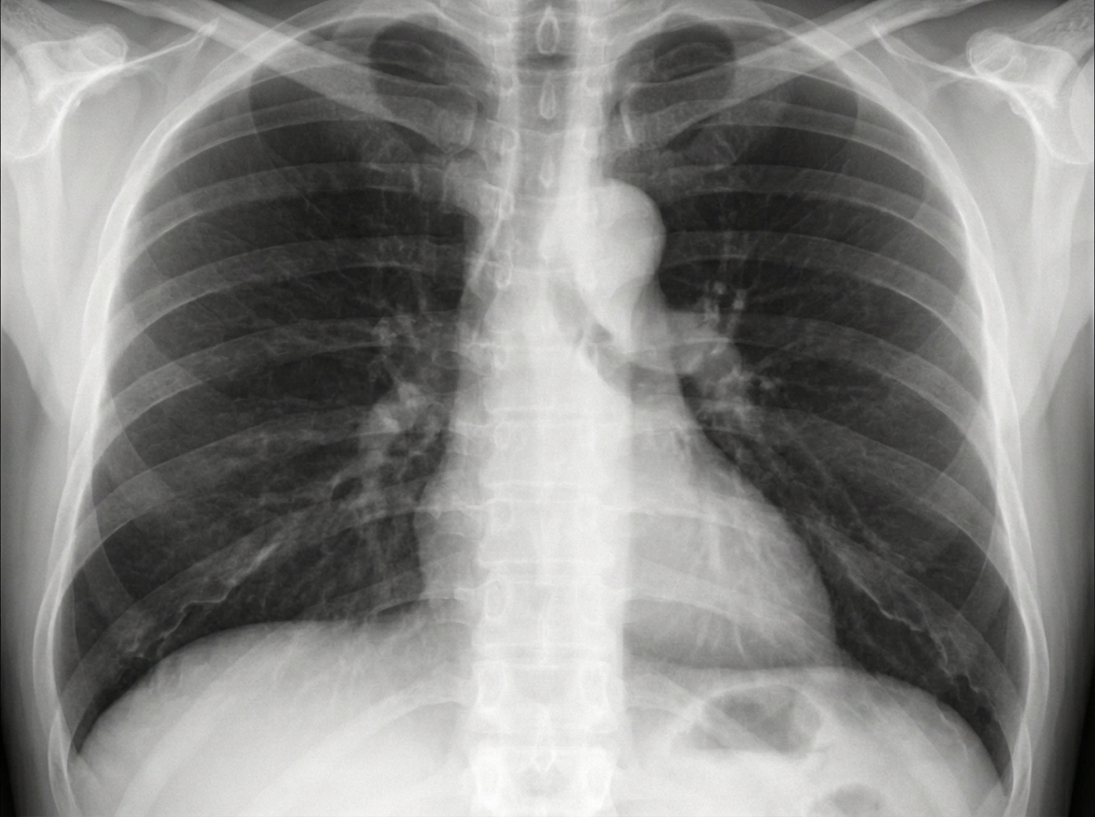
A patient presents with chest pain. What is the diagnosis?

Classification of aortic dissection depends upon?
Which drug is used to perform stress echocardiography?
Which of the following is NOT a sign of right atrial enlargement?
A 40-year-old patient presents with a swelling on the left lateral aspect of the neck. Examination reveals a painless swelling that moves side-to-side but not up-and-down. There is no history of tuberculosis. Laboratory investigations, including thyroid profile and CBC, are normal. Ultrasound shows a highly vascular hypoechoic mass lesion anterior to the sternocleidomastoid muscle. A CT angiogram was subsequently performed. What is the most likely diagnosis?
A triangular shaped heart is a radiological finding of which of the following conditions?
Explanation: **Explanation:** **Multidetector CT (MDCT)** is the gold standard for cardiovascular imaging because it overcomes the two biggest challenges in cardiac radiology: **cardiac motion** and **respiratory motion**. 1. **Why MDCT is correct:** MDCT utilizes multiple rows of detectors, allowing for sub-millimeter isotropic resolution and high temporal resolution. When combined with **ECG-gating** (synchronizing data acquisition with the diastolic phase of the cardiac cycle), it allows for motion-free imaging of the coronary arteries. Modern MDCT (64-slice and above) can image the entire heart in a single breath-hold, making it essential for Coronary CT Angiography (CCTA). 2. **Why other options are incorrect:** * **Second and Third Generation CT:** These are historical iterations. Second-generation used a "translate-rotate" motion with a fan beam, and Third-generation used a "rotate-rotate" motion. While Third-generation is the basis for modern scanners, the basic configuration lacked the speed and detector density required to freeze cardiac motion. * **Spiral (Helical) CT:** While a prerequisite for MDCT, early single-slice spiral CTs were too slow to capture the heart without significant motion artifacts and could not provide the necessary spatial resolution for small vessels like the coronary arteries. **High-Yield Clinical Pearls for NEET-PG:** * **Temporal Resolution:** The time required to acquire data for one image. High temporal resolution is vital to "freeze" the heart. * **Electron Beam CT (EBCT):** Historically known as the "Ultrafast CT," it was the previous gold standard for Calcium Scoring but has been largely replaced by MDCT. * **Calcium Scoring (Agatston Score):** Performed on MDCT to predict the risk of future adverse cardiac events. * **Beta-blockers:** Often administered before a Cardiac CT to lower the heart rate (ideally <60-65 bpm) to improve image quality.
Explanation: In Mitral Stenosis (MS), the narrowing of the mitral valve leads to increased pressure in the left atrium (LA), causing **Left Atrial Enlargement (LAE)** and subsequent pulmonary venous hypertension. ### Why Option D is the Correct Answer In MS, pulmonary venous hypertension leads to a phenomenon called **Cephalization** (Antler sign). Due to perivascular edema in the lower lobes, there is reflex vasoconstriction of the lower lobe veins and compensatory dilatation of the **upper lobe veins**. Therefore, prominence of veins in the *lower* lobes is incorrect; they are actually constricted, while upper lobe veins are prominent. ### Explanation of Incorrect Options (Features of LAE) * **Double contour of the right border:** As the LA enlarges, its right border pushes toward the right and overlaps the right atrium, creating a "double shadow" or "double density" sign on a PA view. * **Elevation of the left main bronchus:** An enlarging LA pushes the left main bronchus upward, making it more horizontal. * **Widening of the carinal angle:** The subcarinal angle (normally 60–75°) increases to >90° as the LA expands superiorly between the two main bronchi. ### High-Yield Clinical Pearls for NEET-PG * **Straightening of the left cardiac border:** This occurs due to a prominent pulmonary artery segment and an enlarged left atrial appendage (filling the "mitral surf"). * **Kerley B Lines:** Horizontal lines at the lung bases indicating chronic pulmonary venous hypertension and interstitial edema. * **Most sensitive view for LAE:** Lateral view with barium swallow (shows posterior indentation of the esophagus). * **Walking Man Sign:** On a lateral X-ray, the posterior displacement of the left main bronchus resembles a person walking.
Explanation: **Explanation:** The preference for Transesophageal Echocardiography (TEE) over Transthoracic Echocardiography (TTE) is primarily determined by the anatomical proximity of the esophagus to the posterior structures of the heart. **Why Option D is Correct:** The **Left Atrial Appendage (LAA)** is a posterior structure that is difficult to visualize clearly on TTE due to its depth and interference from ribs and lung tissue. TEE uses a high-frequency transducer positioned in the esophagus, directly behind the left atrium. This provides superior spatial resolution, making TEE the **gold standard** for detecting LAA thrombi, especially in patients with atrial fibrillation prior to cardioversion. **Analysis of Incorrect Options:** * **A. Pericardial diseases:** TTE is the initial investigation of choice for pericardial effusion and tamponade as it provides an excellent global view of the pericardial space and hemodynamics. * **B. Tricuspid wall vegetations:** The tricuspid valve is an anterior structure. While TEE is more sensitive for small vegetations, TTE is generally sufficient and preferred as the first-line evaluation for right-sided valves. * **C. Left ventricular (LV) apical thrombi:** The LV apex is the structure furthest from the TEE probe but closest to the TTE transducer (apical four-chamber view). TTE (often with contrast) is superior for visualizing the LV apex. **High-Yield Clinical Pearls for NEET-PG:** * **TEE is superior for:** Left atrial appendage thrombi, prosthetic valve dysfunction, infective endocarditis (detecting small vegetations/abscesses), and aortic dissection. * **TTE is superior for:** Routine screening, apical thrombi, and initial assessment of pericardial effusions. * **Rule of Thumb:** If the structure is **posterior** (LA, LAA, Mitral Valve, Aorta), think **TEE**. If the structure is **anterior** (RV, LV apex, Pericardium), think **TTE**.
Explanation: **Explanation:** **Cardiac MRI (CMR)** is the gold standard for assessing myocardial viability and tissue characterization. **1. Why Option B is correct:** CMR uses **Late Gadolinium Enhancement (LGE)** to differentiate viable from infarcted myocardium. Gadolinium is an extracellular contrast agent that cannot enter intact cell membranes. In **infarcted/scarred tissue**, the cell membranes are ruptured or replaced by collagen, allowing gadolinium to accumulate. This results in "bright" (hyperintense) areas. Viable myocardium excludes the dye and appears dark. This is crucial for deciding whether a patient will benefit from revascularization. **2. Why the other options are incorrect:** * **Option A:** Infarcted myocardium (especially in the acute phase) is typically **hyperintense** on T2-weighted images due to **myocardial edema**. * **Option C:** Most modern prosthetic heart valves are made of non-ferromagnetic materials (like titanium or St. Jude valves) and are **MRI-conditional**, not an absolute contraindication. Absolute contraindications usually include older pacemakers, metallic intraocular foreign bodies, or cochlear implants. * **Option D:** **CT Coronary Angiography (CTCA)** is currently superior to MRI for assessing coronary artery anatomy and stenosis due to its higher spatial resolution and faster acquisition time. **High-Yield Clinical Pearls for NEET-PG:** * **LGE Patterns:** * *Subendocardial/Transmural:* Ischemic (Infarction). * *Mid-myocardial/Epicardial:* Non-ischemic (e.g., Myocarditis, Hypertrophic Cardiomyopathy). * **T1 Mapping:** Used to detect diffuse myocardial fibrosis. * **Adenosine Stress MRI:** Used to detect inducible ischemia (functional assessment).
Explanation: ***Ascending aortic aneurysm*** - Chest X-ray shows **widened mediastinum** and **prominent right paracardiac border** due to dilatation of the ascending aorta. - Often presents with **chest pain** and may cause compression of surrounding structures, leading to characteristic radiological findings. *Acute pulmonary embolism* - Chest X-ray is typically **normal** or may show subtle signs like **Westermark sign** (oligemia) or **Hampton's hump** (wedge-shaped opacity). - Diagnosis primarily relies on **CT pulmonary angiogram (CTPA)** or **V/Q scan**, not chest X-ray findings. *Hypertrophic cardiomyopathy* - Chest X-ray may show **left ventricular enlargement** with a **boot-shaped heart** configuration. - Lacks the characteristic **mediastinal widening** seen with aortic pathology, instead showing cardiac silhouette changes. *Coarctation of the aorta* - Chest X-ray demonstrates **rib notching** due to collateral circulation and **figure-of-3 sign** from pre- and post-stenotic dilatation. - Shows **left heart enlargement** but lacks the prominent right paracardiac border characteristic of ascending aortic aneurysm.
Explanation: Aortic dissection is classified based on the **anatomical location** of the intimal tear and the extent of the involvement of the aorta. This is because the anatomical level determines the clinical management (surgical vs. medical). ### Why the Correct Answer is Right: The two primary classification systems used in clinical practice are: 1. **Stanford Classification:** The most widely used system. It divides dissections into **Type A** (involves the ascending aorta, regardless of the site of origin) and **Type B** (involves only the aorta distal to the left subclavian artery). 2. **DeBakey Classification:** Categorizes based on the origin and extent. **Type I** involves the ascending aorta, arch, and descending aorta; **Type II** is confined to the ascending aorta; **Type III** is confined to the descending aorta. ### Why Other Options are Wrong: * **A. Cause of dissection:** While hypertension, Marfan syndrome, and trauma are common causes, they do not dictate the classification. * **C. Percentage of aorta affected:** Classification depends on *which* specific segments are involved (ascending vs. descending), not the total percentage of the vessel's length. ### High-Yield Facts for NEET-PG: * **Management Rule:** Stanford Type A is a **surgical emergency** (due to risk of aortic regurgitation, tamponade, or coronary involvement). Stanford Type B is generally managed **medically** with aggressive blood pressure control (Beta-blockers). * **Gold Standard Investigation:** **CT Angiography (CTA)** is the investigation of choice. * **Classic Sign:** On X-ray, look for **mediastinal widening**. On CT, the pathognomonic finding is an **intimal flap** separating the true and false lumens. * **Double Barrel Aorta:** A term used when there are two distinct flow channels.
Explanation: **Explanation:** **Stress Echocardiography** is a diagnostic tool used to assess myocardial wall motion abnormalities under physiological stress. When a patient is unable to exercise, pharmacological agents are used to simulate the heart's response to exertion. **Why Dobutamine is correct:** **Dobutamine** is a synthetic catecholamine that acts primarily as a **$\beta_1$-receptor agonist**. It increases myocardial oxygen demand by increasing heart rate (chronotropy) and contractility (inotropy). In a stress echo, dobutamine is infused in escalating doses to provoke ischemia in areas supplied by stenosed coronary arteries, which is then visualized as new or worsening regional wall motion abnormalities (RWMA) on ultrasound. **Analysis of Incorrect Options:** * **Thallium (A):** This is a radioactive isotope used in **Nuclear Medicine (SPECT)** for myocardial perfusion imaging, not as a stress-inducing agent. * **Adrenaline (C):** While it has $\beta_1$ effects, it also has significant $\alpha$-adrenergic effects (vasoconstriction), making it hemodynamically unpredictable and unsuitable for routine stress testing. * **Adenosine (D):** This is a potent vasodilator used in **Cardiac MRI** or **Nuclear Stress tests** (MPI). It works by causing "coronary steal" rather than increasing myocardial oxygen demand. It is rarely used in echocardiography because it does not reliably induce wall motion abnormalities. **High-Yield Pearls for NEET-PG:** * **Antidote:** If a patient develops severe arrhythmia or ischemia during a Dobutamine stress test, **Esmolol** (a short-acting $\beta$-blocker) is the drug of choice to reverse the effects. * **Contraindication:** Dobutamine stress echo should be avoided in patients with severe tachyarrhythmias or significant outflow tract obstruction (e.g., HOCM). * **Viability:** Low-dose dobutamine is specifically used to identify "hibernating myocardium" (contractile reserve).
Explanation: **Explanation:** Right atrial (RA) enlargement is primarily assessed on the Chest X-ray (CXR) by observing changes in the right heart border and the retrosternal space. **Why Option D is the correct answer:** The **Hoffman-Rigler sign** is a classic radiological sign used to identify **Left Ventricular (LV) enlargement**, not right atrial enlargement. It is measured on a lateral CXR: if the distance between the posterior border of the LV and the posterior border of the Inferior Vena Cava (IVC) is greater than 1.8 cm (at a point 2 cm above the diaphragm), it indicates LV hypertrophy/dilatation. **Analysis of Incorrect Options:** * **Option A:** In the PA view, the right heart border is formed entirely by the RA. Enlargement causes the border to shift further to the right (more than 5.5 cm from the midline) with increased convexity. * **Option B & C:** On a lateral view, the RA forms the upper portion of the anterior cardiac contour. When enlarged, it expands anteriorly and superiorly, encroaching upon and eventually **obliterating the retrosternal clear space** (the lucent area between the heart and the sternum). **High-Yield Clinical Pearls for NEET-PG:** * **Right Ventricular (RV) Enlargement:** Also obliterates the retrosternal space but typically causes an "upturned apex" (Coeur en Sabot) on PA view. * **Left Atrial (LA) Enlargement:** Look for the "Double atrial contour" sign, "Splaying of the carina," and "Walking cane appearance" of the left main bronchus. * **Most sensitive view for RA enlargement:** PA view (displacement of the right heart border). * **Most sensitive view for LA enlargement:** Lateral view (posterior displacement of the esophagus if barium swallow is used).
Explanation: ### Explanation The clinical presentation and imaging findings are classic for a **Carotid Body Paraganglioma** (also known as a Chemodectoma). **Why the Correct Answer is Right:** * **Clinical Sign:** The hallmark of this tumor is the **Fontaine Sign**—the mass is mobile horizontally (side-to-side) but fixed vertically because it is tethered to the carotid bifurcation. * **Location:** It typically presents at the level of the hyoid bone, anterior to the sternocleidomastoid muscle, within the carotid triangle. * **Imaging:** Ultrasound shows a **highly vascular**, hypoechoic mass. The gold standard for diagnosis is CT or MR Angiography, which reveals the pathognomonic **"Lyre Sign"**—the splaying of the internal and external carotid arteries at the bifurcation. **Why Incorrect Options are Wrong:** * **Fibromatosis colli:** This is a benign proliferation of the sternocleidomastoid muscle seen in **neonates/infants** (congenital muscular torticollis), not in 40-year-olds. * **Tubercular lymph node:** Usually presents with constitutional symptoms (fever, weight loss), may show central necrosis on imaging, and is not typically hypervascular on angiography. * **Soft tissue sarcoma:** While these can be vascular, they do not specifically splay the carotid bifurcation or demonstrate the specific horizontal-only mobility characteristic of carotid body tumors. **NEET-PG High-Yield Pearls:** 1. **Lyre Sign:** Widening of the carotid bifurcation on angiography. 2. **Salt-and-Pepper Appearance:** Seen on T1-weighted MRI (Salt = hemorrhage/slow flow; Pepper = flow voids of high-velocity vessels). 3. **Shamblin Classification:** Used to grade these tumors based on their involvement/encasement of the carotid arteries. 4. **Origin:** Arises from the **neural crest cells** (chemoreceptors) located at the carotid body.
Explanation: **Explanation:** The correct answer is **Constrictive Pericarditis**. In this condition, the pericardium becomes thickened, fibrotic, and often calcified, which restricts the diastolic filling of the heart chambers. On a chest X-ray, this rigid "shell" prevents the normal rounded contour of the heart, resulting in a flattened, **triangular, or "tent-shaped" cardiac silhouette**. **Analysis of Options:** * **Constrictive Pericarditis:** The hallmark radiological feature is **pericardial calcification** (best seen on a lateral X-ray). The heart size is usually normal or small, but the rigid pericardium gives it a characteristic straight-edged, triangular appearance. * **Mitral Incompetence (Regurgitation):** This leads to left atrial and left ventricular enlargement, resulting in a **globular or enlarged heart** with a "double atrial shadow." * **TAPVC (Supracardiac type):** This presents with the classic **"Snowman sign" or "Figure-of-8" appearance** due to the dilation of the vertical vein, innominate vein, and superior vena cava. * **Ebstein’s Anomaly:** Characterized by massive right atrial enlargement, leading to a massive, **"box-shaped" heart**. **High-Yield Clinical Pearls for NEET-PG:** * **Pericardial Effusion:** Shows a **"Water-bottle" or "Money-bag"** appearance. * **Tetralogy of Fallot:** Shows a **"Boot-shaped" heart (Coeur en sabot)** due to RVH and upturned apex. * **Transposition of Great Arteries (TGA):** Shows an **"Egg-on-side"** appearance. * **Coarctation of Aorta:** Shows the **"Figure of 3" sign** on X-ray and **Rib notching** (inferior aspect of 3rd to 8th ribs).
Cardiovascular Anatomy
Practice Questions
Cardiac CT Techniques
Practice Questions
Cardiac MRI Techniques
Practice Questions
Ischemic Heart Disease Imaging
Practice Questions
Valvular Heart Disease
Practice Questions
Cardiomyopathies
Practice Questions
Pericardial Diseases
Practice Questions
Congenital Heart Disease
Practice Questions
Aortic and Great Vessel Imaging
Practice Questions
Peripheral Vascular Imaging
Practice Questions
Cardiovascular Interventional Procedures
Practice Questions
Post-Surgical Cardiovascular Imaging
Practice Questions
Get full access to all questions, explanations, and performance tracking.
Start For Free