On mammogram, all of the following are the features of a malignant tumor except:
Mammography can be best used in?
Risk of malignancy in BIRADS score 2 is
On mammogram all of the following are the features of a malignant tumor except:
The most appropriate technique for mammography is:
ACR score 4 in breast imaging indicates
Which finding excludes BIRADS 3 categorization?
A 40-year-old woman with breast implants presents with breast pain after a recent fall. MRI shows the 'linguine sign.' What does this indicate?
A 60-year-old woman with a family history of breast cancer presents with a newly detected breast mass. What is the first-line imaging modality for its evaluation?
A 50-year-old female presents with a new-onset palpable lump in her breast. What is the most appropriate initial imaging study?
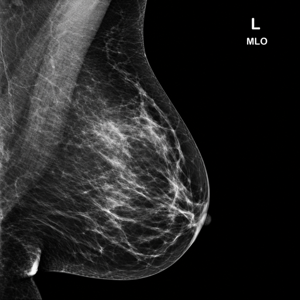
Explanation: ***Macrocalcification*** - **Macrocalcifications** are typically **benign** and are often associated with involutional changes in the breast, such as aging or fibroadenomas. - These are usually larger, coarser calcifications that are easily seen and rarely indicate malignancy. *Microcalcification* - **Microcalcifications**, especially when **pleomorphic**, **linear**, or grouped, are a significant indicator of potential malignancy, such as **ductal carcinoma in situ (DCIS)**. - They represent calcium deposits within the ducts or stromal tissue, which can be associated with rapidly proliferating cells. *Irregular mass* - An **irregularly shaped mass** with ill-defined margins is highly suspicious for malignancy because it suggests invasive growth into surrounding tissues. - Unlike benign lesions which tend to be round or oval with smooth borders, malignant tumors often grow in an uncontrolled, infiltrative manner. *Spiculation* - **Spiculation** refers to **radiating lines or projections** extending from the borders of a mass, indicating an infiltrative process highly suggestive of malignancy. - These spicules represent fibrous tissue reaction to an invading tumor and are a strong predictor of breast cancer.
Explanation: ***Early breast carcinoma*** - **Mammography** is the gold standard for **early detection of breast carcinoma**, particularly for identifying **microcalcifications** and small masses before they are palpable. - It plays a crucial role in **screening asymptomatic women** to reduce breast cancer mortality. *Mastitis* - **Mastitis** is an **inflammatory condition** of the breast, often associated with infection, which is usually diagnosed clinically. - While mammography might show diffuse **increased density**, it is not the primary diagnostic tool and often has limited value due to inflammatory changes masking pathology. *Fibroadenoma* - **Fibroadenomas** are **benign breast tumors** common in younger women, typically appearing as well-circumscribed masses on mammography. - While mammography can detect them, their characterization often requires **ultrasound** and **biopsy** for definitive diagnosis, as differentiation from malignant lesions can be challenging. *Phylloides tumor* - A **Phylloides tumor** is a rare tumor that can be benign, borderline, or malignant, and it typically presents as a rapidly growing, palpable mass. - Mammography may show a well-defined mass, but **ultrasound** and **core needle biopsy** are essential for accurate diagnosis and distinction from fibroadenomas or malignancy.
Explanation: ***0%*** - A **BIRADS score of 2** indicates a **definitively benign finding**, meaning there is **essentially 0% risk of malignancy**. - BIRADS 2 is assigned to findings that are clearly benign such as **simple cysts, intramammary lymph nodes, calcified fibroadenomas**, and other characteristically benign lesions. - **No follow-up or intervention is required** for BIRADS 2 findings. *2-4%* - This risk range is typically associated with **BIRADS 4A** lesions (low suspicion for malignancy), which require **tissue diagnosis/biopsy**. - BIRADS 2 findings are definitively benign and have no measurable risk of malignancy. *10%* - A 10% risk of malignancy is aligned with **BIRADS 4A-4B** lesions (low to moderate suspicion), which require **biopsy**. - This percentage indicates a suspicious finding, which is completely different from a definitively benign BIRADS 2 finding. *50%* - A 50% risk of malignancy corresponds to a highly suspicious finding, typically **BIRADS 4C**, demanding **immediate biopsy**. - This level of risk is far too high for a benign finding like BIRADS 2, which carries no risk of malignancy.
Explanation: ***Smooth margin*** - A **smooth margin** on a mammogram is typically a feature of a **benign mass**, indicating that the lesion is well-defined and not invading surrounding tissues. - Malignant tumors usually exhibit **irregular, ill-defined, or spiculated margins** due to their invasive growth patterns. *Spiculation* - **Spiculation** is a **highly suspicious feature** of malignancy, characterized by sharp, radiating lines extending from the mass into the surrounding breast tissue. - It represents tumor invasion and desmoplastic reaction, indicating an aggressive growth pattern. *High density* - **High density** of a mass on mammography, appearing brighter than the surrounding breast tissue, is a **common feature of malignancy**. - This increased density is often due to higher cellularity or desmoplastic reaction within the tumor. *Ill-defined margin* - An **ill-defined margin** is a strong indicator of **malignancy** as it suggests infiltrative growth into adjacent tissues. - The borders of the mass are difficult to distinguish clearly from the surrounding breast parenchyma.
Explanation: ***Mediolateral oblique view*** - This view captures the **most breast tissue**, from the axilla down to the inframammary fold, as it visualizes both the upper outer quadrant and the posterior breast. - It allows for comprehensive imaging of the breast, which is crucial for **screening and diagnostic mammography**, enabling detection of abnormalities that might be missed on other views. *Spot Compression view* - This technique is used for **further evaluation of suspicious areas**, not as a primary screening or diagnostic view. - It applies localized compression to a smaller area of breast tissue to improve lesion visibility and definition. *Lateral view* - While it provides an orthogonal view to the craniocaudal projection, the standard lateral view (specifically, the medial lateral projection) **does not include as much axillary tissue** as the mediolateral oblique view. - It is often used as a supplementary view to help **localize lesions** in the mediolateral dimension. *Craniocaudal view* - This view is a standard part of mammography, providing a superior-inferior projection, but it **misses a significant portion of the axillary tail and posterior breast tissue**, especially in the upper outer quadrant. - It is usually performed in conjunction with the mediolateral oblique view to provide a **two-dimensional assessment** of the breast.
Explanation: ***Suspicious abnormality*** - An **ACR BI-RADS category 4** indicates a **suspicious abnormality** that necessitates a biopsy to rule out malignancy. - The risk of malignancy in this category ranges from **2% to 94%**, representing findings that do not have the classic appearance of malignancy but have a definite probability of being cancer. *Probably benign* - This description corresponds to an **ACR BI-RADS category 3**, which suggests a less than 2% chance of malignancy. - Category 3 findings are usually followed up with **short-term interval imaging** (e.g., 6 months) to assess stability. *Highly suggestive of malignancy* - This corresponds to an **ACR BI-RADS category 5**, where the findings almost certainly represent **malignancy** (at least 95% probability). - Category 5 lesions require **appropriate action**, such as biopsy or definitive treatment, based on the highest level of suspicion. *Negative* - This description is for an **ACR BI-RADS category 1**, meaning there are **no significant findings** and the breast is normal. - Category 1 indicates that the study is completed and no further action is needed beyond routine screening.
Explanation: ***Architectural distortion*** - **Architectural distortion** refers to a disruption of the normal breast parenchymal architecture in the absence of a discrete mass, often indicating an underlying malignancy. - This finding is suspicious enough to warrant a recommendation of **biopsy (BI-RADS 4 or 5)**, thus **excluding BI-RADS 3**, which implies a probably benign finding with a low likelihood of malignancy (<2%). - Architectural distortion has a high association with malignancy and cannot be categorized as BI-RADS 3. *Normal lymph node* - A **normal lymph node** within the breast or axilla is a common and benign finding, characterized by an oval shape, fatty hilum, and thin cortex. - Its presence does not increase the suspicion of malignancy and is classified as **BI-RADS 1 or 2** (definitely benign). - This does NOT exclude BI-RADS 3; it is simply a more benign finding. *Simple cyst* - A **simple cyst** is a very common and benign fluid-filled sac, readily identifiable by clear sonographic criteria (anechoic, thin smooth walls, posterior acoustic enhancement). - It is classified as **BI-RADS 2** (benign finding), meaning it is definitely not malignant. - This does NOT exclude BI-RADS 3; it is simply a more benign finding that does not require follow-up. *Focal asymmetry* - **Focal asymmetry** refers to an area of fibroglandular tissue that is visible on only one mammographic projection or is less conspicuous than a mass. - If it has **no associated suspicious features**, it can be appropriately categorized as **BI-RADS 3**, requiring short interval follow-up. - This does NOT exclude BI-RADS 3 categorization.
Explanation: ***Implant rupture*** - The **"linguine sign"** on MRI is a classic finding for an **intracapsular rupture** of a silicone breast implant, where the collapsed implant shell is seen floating within the silicone gel. - The recent fall and localized breast pain are consistent with a traumatic event leading to implant compromise. *Fibroadenoma* - A fibroadenoma is a **benign solid tumor** of the breast that does not typically cause the "linguine sign." - Its appearance on imaging is usually a well-defined, smooth or lobulated mass, not indicative of implant rupture. *Breast carcinoma* - Breast carcinoma typically presents as an **irregular mass** with spiculations or microcalcifications, and does not cause the "linguine sign." - While breast pain can be a symptom, the specific MRI finding points away from malignancy. *Breast abscess* - A breast abscess is a localized collection of **pus**, usually presenting with signs of infection such as redness, warmth, and fever, along with a focal fluctuating mass. - It would appear as a fluid collection on MRI, but not with the characteristic "linguine sign" of implant rupture.
Explanation: ***Mammography*** - **Mammography** is the gold standard and first-line imaging modality for evaluating a newly detected breast mass, especially in women over 40. - It is effective in detecting **calcifications** and architectural distortions, which are key indicators of breast cancer. *Ultrasound* - **Ultrasound** is often used as a complementary modality to further characterize findings from a mammogram, differentiate between solid and cystic masses, and guide biopsies. - It is particularly useful in women with **dense breast tissue** where mammography may be less sensitive but is not typically the first-line alone. *MRI* - **MRI** is highly sensitive for breast cancer detection but is usually reserved for specific situations like screening high-risk individuals, evaluating extent of disease in newly diagnosed cancer, or assessing implant integrity. - It is not the initial imaging choice for a newly palpable mass due to its **cost**, potential for **false positives**, and common need for contrast injection. *CT scan* - A **CT scan** of the breast is generally not used for primary evaluation of a breast mass. - It is primarily utilized for staging cancer, assessing distant metastasis, or evaluating the chest wall.
Explanation: ***Mammography*** - **Mammography** is the **most appropriate initial imaging study** for a new breast lump in a 50-year-old female, as it serves as the primary modality for both **screening and diagnostic evaluation** of breast abnormalities. - It has high sensitivity for detecting **masses and calcifications** in this age group, where decreased breast density improves visualization of lesions. - In practice, for a palpable lump, mammography is often performed along with targeted ultrasound as part of comprehensive diagnostic evaluation. *Ultrasound* - While ultrasound is an essential complementary tool, especially for evaluating **palpable masses** and differentiating **solid from cystic lesions**, it is typically considered an **adjunct to mammography** rather than the initial or sole imaging study. - Ultrasound is operator-dependent and has lower sensitivity for detecting **microcalcifications** compared to mammography. - However, it is highly valuable for further characterization of masses identified on clinical examination or mammography. *MRI* - **MRI** is highly sensitive for breast cancer detection but is generally reserved for **high-risk screening**, **staging of known cancers**, evaluating extent of disease, or problem-solving in inconclusive cases. - Its high cost, longer examination time, and potential for **false positives** make it unsuitable as a primary imaging modality for initial evaluation of a breast lump. *CT scan* - A **CT scan** is not routinely used for the initial evaluation of breast lesions. - It involves **ionizing radiation** without providing adequate soft tissue resolution for breast tissue characterization. - CT is primarily used for **staging known breast cancers** to assess for locoregional or distant metastasis, rather than for primary diagnosis.
Breast Anatomy and Physiology
Practice Questions
Male Breast Imaging
Practice Questions
Mammography Techniques
Practice Questions
BI-RADS Classification
Practice Questions
Breast Ultrasonography
Practice Questions
Breast MRI
Practice Questions
Digital Breast Tomosynthesis
Practice Questions
Benign Breast Diseases
Practice Questions
Breast Cancer Detection and Diagnosis
Practice Questions
Interventional Breast Procedures
Practice Questions
Breast Cancer Screening
Practice Questions
Male Breast Imaging
Practice Questions
Post-treatment Breast Imaging
Practice Questions
Get full access to all questions, explanations, and performance tracking.
Start For Free