Linguine sign is seen in which of the following conditions?
Which of the following investigations is NOT indicated in a lactating woman presenting with painful breasts?
What does BI-RADS 4 classification indicate for a breast lesion?
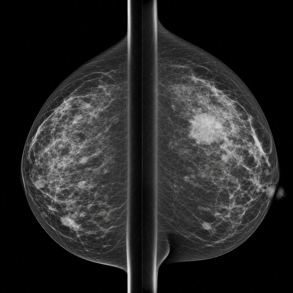
A 55-year-old post-menopausal woman, on hormone replacement therapy, presents with heaviness in both breasts. A screening mammogram reveals a high density speculated mass with a cluster of pleomorphic microcalcification and ipsilateral large axillary lymph nodes. The mass described here most likely represents?
A 55-year-old post-menopausal woman, on hormone replacement therapy (HRT), presents with heaviness in both breasts. A screening mammogram reveals a high density speculated mass with a cluster of pleomorphic microcalcification and ipsilateral large axillary lymph nodes. The mass described here most likely represents?
What are the standard views for mammography?
In a mammogram, all of the following are features of carcinoma breast EXCEPT?
What is true about screening mammography?
Diffuse increase in the parenchymal density of the breast is seen in which of the following conditions?
Which of the following conditions does not contain fat on mammography?
Explanation: **Explanation:** The **Linguine sign** is a classic radiological finding on **MRI** (the gold standard for implant evaluation) that indicates an **intracapsular rupture** of a silicone breast implant. When an intracapsular rupture occurs, the silicone gel leaks out of the elastomer shell but remains contained within the fibrous capsule formed by the body. The elastomer shell collapses and becomes wavy or folded within the silicone gel. On T2-weighted or silicone-selective MRI sequences, these collapsed, curvilinear membranes resemble strands of linguine pasta. **Analysis of Incorrect Options:** * **A. Duct ectasia:** Characterized by the dilation of subareolar ducts, often showing "cigar-shaped" or "rod-like" calcifications (Secretory calcifications) on mammography. * **B. Duct papilloma:** Typically presents with pathological nipple discharge. On galactography, it appears as a small filling defect within a duct. * **C. Breast carcinoma:** Radiologically presents as an irregular, spiculated mass with pleomorphic microcalcifications or architectural distortion. **High-Yield Clinical Pearls for NEET-PG:** * **Intracapsular Rupture:** Linguine sign (MRI), Stepladder sign (Ultrasound). * **Extracapsular Rupture:** Silicone leaks beyond the fibrous capsule into the breast parenchyma. It is characterized by the **"Snowstorm appearance"** on Ultrasound due to intense echogenic noise. * **Teardrop sign:** Seen on MRI when a small amount of silicone is trapped between the folds of the collapsed shell (early sign of rupture). * **Water-lily sign:** Though similar in description (collapsed membranes), this is associated with **Hydatid cysts** in the lung or liver, not breast implants.
Explanation: **Explanation:** The correct answer is **A. Ultrasound (USG)**. In a lactating woman presenting with painful breasts, the most common clinical concerns are **mastitis** or a **breast abscess**. For these conditions, **Ultrasound (USG)** is the **investigation of choice** (first-line imaging). It is highly sensitive for detecting fluid collections (abscesses), does not involve ionizing radiation (safe for the mother and breastfeeding infant), and is not hindered by the increased glandular density of the lactating breast. **Why the other options are NOT the primary indication:** * **B. Mammography:** In lactating women, the breasts are physiologically dense due to glandular proliferation and milk production. This significantly decreases the sensitivity of mammography, making it difficult to distinguish between normal tissue and pathology. It is generally reserved for cases where malignancy is strongly suspected. * **C. Computed Tomography (CT):** CT is not a standard tool for breast imaging due to high radiation doses and poor soft-tissue resolution compared to USG or MRI. It is typically used only for staging systemic disease (metastasis). * **D. Magnetic Resonance Imaging (MRI):** While highly sensitive, MRI is not indicated for acute painful conditions like mastitis. Furthermore, increased vascularity during lactation leads to significant background parenchymal enhancement, which can result in false positives. **High-Yield Clinical Pearls for NEET-PG:** * **First-line imaging for any woman <30 years or pregnant/lactating:** Ultrasound. * **Breast Abscess:** USG shows a hypoechoic collection with posterior acoustic enhancement. * **Management:** If an abscess is confirmed, **USG-guided needle aspiration** is preferred over incision and drainage to allow continued breastfeeding. * **Galactocele:** The most common benign mass in a lactating woman; appears on USG as a well-defined mass with internal echoes (due to fat-fluid levels).
Explanation: **Explanation:** The **BI-RADS (Breast Imaging-Reporting and Data System)** is a standardized reporting tool developed by the American College of Radiology to communicate the risk of malignancy and guide clinical management. **1. Why "Suspicious of Malignancy" is Correct:** BI-RADS 4 is defined as a lesion that does not have the classic appearance of malignancy but has a sufficiently high level of suspicion to warrant a **tissue biopsy**. The risk of malignancy in this category ranges from **>2% to <95%**. It is further subdivided into: * **4A:** Low suspicion (2–10% risk) * **4B:** Moderate suspicion (10–50% risk) * **4C:** High suspicion (50–95% risk) **2. Analysis of Incorrect Options:** * **A. Benign (BI-RADS 2):** Indicates findings like a simple cyst or stable fibroadenoma. Risk of malignancy is 0%. * **B. Probably Benign (BI-RADS 3):** Indicates a <2% risk of malignancy. Management involves short-interval follow-up (usually 6 months) rather than immediate biopsy. * **D. Malignant (BI-RADS 5):** Indicates findings "highly suggestive of malignancy" with a risk of **≥95%**. BI-RADS 6 is used for biopsy-proven malignancy. **Clinical Pearls for NEET-PG:** * **BI-RADS 0:** Incomplete assessment; requires further imaging (e.g., additional views or ultrasound). * **Management Rule:** BI-RADS 1 & 2 require routine screening; BI-RADS 3 requires follow-up; BI-RADS 4 & 5 require tissue diagnosis (biopsy). * **Gold Standard:** While BI-RADS categorizes risk, the definitive diagnosis for any suspicious lesion remains histopathology.
Explanation: ### Explanation The clinical and radiological presentation strongly points toward **Breast Carcinoma**. **1. Why Carcinoma is correct:** The mammographic findings described are classic "red flags" for malignancy (BI-RADS 5): * **High-density spiculated mass:** Spiculation (radiating lines from the margin) is the most specific mammographic sign of breast cancer, representing desmoplastic reaction or infiltration into surrounding tissue. * **Pleomorphic microcalcifications:** Fine, irregular, and varying shapes/sizes of calcifications are highly suspicious for ductal carcinoma in situ (DCIS) or invasive cancer. * **Axillary Lymphadenopathy:** The presence of large ipsilateral nodes suggests lymphatic spread. * **Risk Factors:** Post-menopausal status and **Hormone Replacement Therapy (HRT)** are known risk factors for developing breast cancer. **2. Why other options are incorrect:** * **Fibroadenoma:** Typically presents as a well-circumscribed, oval, low-density mass with "popcorn" calcifications in older women. It is benign and does not cause spiculation or pleomorphic microcalcifications. * **Lymphoma:** Primary breast lymphoma is rare. It usually presents as a large, non-spiculated, circumscribed mass without microcalcifications. * **Cystosarcoma Phylloides:** These are large, rapidly growing, well-circumscribed fibroepithelial tumors. They rarely show microcalcifications or spiculation and typically spread hematogenously rather than to axillary nodes. **Clinical Pearls for NEET-PG:** * **Most common mammographic sign of malignancy:** A mass with spiculated margins. * **Most common type of breast cancer:** Invasive Ductal Carcinoma (NOS). * **BI-RADS Classification:** Remember that **BI-RADS 5** indicates a >95% probability of malignancy. * **HRT:** Long-term combined HRT (Estrogen + Progesterone) increases the risk of breast cancer and can increase breast density, making mammography harder to interpret.
Explanation: ### Explanation **1. Why Carcinoma is the Correct Answer:** The clinical and radiological features described are classic "red flags" for **Invasive Breast Carcinoma**. * **Demographics:** Post-menopausal status and the use of **Hormone Replacement Therapy (HRT)** are significant risk factors for breast cancer. * **Mammographic Findings:** A **high-density spiculated mass** is the most specific sign of malignancy (representing desmoplastic reaction). **Pleomorphic microcalcifications** (varying in size and shape) are highly suspicious (BI-RADS 4/5) for Ductal Carcinoma in Situ (DCIS) or invasive cancer. * **Lymphadenopathy:** The presence of large ipsilateral axillary lymph nodes strongly suggests metastatic spread. **2. Why Other Options are Incorrect:** * **Cystosarcoma Phylloides:** Typically presents as a large, rapidly growing, well-circumscribed, lobulated mass. It rarely shows microcalcifications or spiculated margins and usually spreads hematogenously rather than to axillary nodes. * **Lymphoma:** Primary breast lymphoma is rare. It usually appears as a large, non-calcified, circumscribed mass. While it involves lymph nodes, it lacks the spiculated margins and pleomorphic calcifications characteristic of epithelial carcinoma. * **Fibroadenoma:** These are benign tumors common in younger women. On mammography, they appear as well-defined "popcorn" calcifications (in involuting stages) or oval masses, not spiculated masses with pleomorphic calcifications. **3. Clinical Pearls for NEET-PG:** * **Most common mammographic sign of malignancy:** Spiculated mass. * **Most common type of breast cancer:** Invasive Ductal Carcinoma (NOS). * **BI-RADS Classification:** Pleomorphic and linear branching calcifications are high-probability markers for malignancy. * **HRT Risk:** Long-term combined HRT increases the risk of breast cancer and can increase breast density, making mammographic detection more challenging.
Explanation: **Explanation:** In screening mammography, the goal is to visualize the maximum amount of breast tissue with optimal compression. The standard "two-view" screening protocol consists of: 1. **Cranio-caudal (CC) View:** The X-ray beam passes from superior to inferior. This view is best for visualizing the **medial aspect** of the breast. 2. **Medio-lateral Oblique (MLO) View:** The X-ray beam passes from the superomedial to the inferolateral aspect (usually at a 30-60° angle). This is the most important view as it visualizes the **maximum amount of breast tissue**, including the **Upper Outer Quadrant (UOQ)** and the **Axillary Tail of Spence**, where most breast cancers occur. **Analysis of Incorrect Options:** * **AP and Lateral (A & B):** Anteroposterior (AP) views are not used in mammography because the breast must be compressed to reduce thickness and radiation dose; the CC view serves this purpose. * **Cranio-caudal and Medio-lateral (C):** While the Medio-lateral (ML) view exists, it is a **supplemental/diagnostic view** used to localize lesions (the "muffins rise, lead falls" rule) or perform biopsies. It does not capture the axillary tail as effectively as the MLO view. **High-Yield Clinical Pearls for NEET-PG:** * **BI-RADS:** The standard reporting system for mammography (0: Incomplete, 1: Normal, 2: Benign, 3: Probably Benign, 4: Suspicious, 5: Highly Suggestive of Malignancy, 6: Known Biopsy-proven Malignancy). * **Calcifications:** "Pleomorphic" or "Fine Linear Branching" calcifications are highly suspicious for DCIS/Malignancy. * **Screening Age:** Usually starts at 40 years (as per ACR guidelines). * **Younger Patients:** Ultrasound is the primary modality for women <30 years due to dense breast tissue and radiation sensitivity.
Explanation: **Explanation:** In breast imaging, distinguishing between benign and malignant features is crucial for accurate diagnosis. **Why "Areas of macrocalcification" is the correct answer:** Macrocalcifications (typically >0.5 mm) are usually coarse, large, and "popcorn-like." These are generally associated with **benign conditions** such as involuting fibroadenomas, fat necrosis, or secretory calcifications. They are rarely associated with malignancy. **Analysis of other options (Malignant Features):** * **A. Solid lesion with ill-defined/stellate configuration:** A spiculated or stellate mass is the most specific mammographic sign of breast cancer. It represents the infiltration of tumor cells into the surrounding stroma (desmoplastic reaction). * **B. True microcalcification:** These are tiny, calcium deposits (<0.5 mm). Malignant microcalcifications are typically pleomorphic (varying shapes/sizes) or fine-linear branching, often representing Ductal Carcinoma In Situ (DCIS). * **D. Increased skin thickness:** Skin thickening (>2 mm) and retraction are secondary signs of malignancy, often seen in inflammatory breast cancer or due to lymphatic obstruction by the tumor. **Clinical Pearls for NEET-PG:** * **BI-RADS Classification:** Used for standardized reporting. BI-RADS 1 is normal; BI-RADS 5 is highly suggestive of malignancy; BI-RADS 6 is biopsy-proven malignancy. * **Most common mammographic finding in malignancy:** A mass with spiculated margins. * **Popcorn calcification:** Pathognomonic for an involuting (old) fibroadenoma. * **Egg-shell/Rim calcification:** Characteristic of fat necrosis or oil cysts. * **Screening:** Mammography is the gold standard for screening women over 40 years. For younger women with dense breasts, Ultrasound is the preferred initial modality.
Explanation: ### **Explanation** **1. Why Option A is Correct:** Screening mammography is the gold standard for the early detection of breast cancer in asymptomatic women. While guidelines vary slightly (e.g., ACR, ACS, and WHO), the age group of **50–60 years** represents the peak window where screening has the highest sensitivity and significantly reduces mortality. In this age group, breast tissue becomes less dense (fatty replacement), making mammography highly effective at detecting microcalcifications and small masses. **2. Why Other Options are Incorrect:** * **Option B:** While mammography uses ionizing radiation, the dose is extremely low (approx. 0.4 mSv). The benefit of early cancer detection far outweighs the negligible theoretical risk of radiation-induced malignancy. * **Option C:** MRI is highly sensitive but has lower specificity (higher false positives) and is significantly more expensive. It is reserved for high-risk screening (e.g., BRCA mutations) or as an adjunct, not as a primary screening tool for the general population. * **Option D:** Ultrasound (USG) is the investigation of choice for women **<35 years** (dense breasts) and for differentiating cystic from solid lesions. However, it cannot reliably detect **microcalcifications**, which are the earliest sign of ductal carcinoma in situ (DCIS) seen on mammography. **High-Yield Clinical Pearls for NEET-PG:** * **Standard Views:** Craniocaudal (CC) and Mediolateral Oblique (MLO). * **Earliest Sign of Malignancy:** Fine, pleomorphic, or linear branching **microcalcifications**. * **BI-RADS Classification:** A standardized reporting system (0: Incomplete; 1: Normal; 2: Benign; 3: Probably Benign; 4: Suspicious; 5: Highly Suggestive of Malignancy; 6: Known Biopsy-proven Malignancy). * **Tomosynthesis:** 3D mammography that reduces the effect of overlapping tissue.
Explanation: **Explanation:** The mammographic density of the breast is determined by the ratio of radiopaque fibroglandular tissue to radiolucent adipose (fatty) tissue. A **diffuse increase in parenchymal density** occurs when there is either an increase in glandular elements or a significant loss of subcutaneous and retromammary fat. 1. **Hormone Replacement Therapy (HRT):** Estrogen and progesterone stimulate the proliferation of acinar and ductal structures. This leads to an increase in fibroglandular volume, making the breast appear more "dense" or "white" on a mammogram. This is a common pharmacological cause of increased density in postmenopausal women. 2. **Lipodystrophy:** This condition involves the abnormal distribution or loss of adipose tissue. Since fat provides the primary radiolucency in a mammogram, its loss results in a relative concentration of the remaining stromal and glandular elements, leading to increased parenchymal density. 3. **Cachexia:** In states of extreme malnutrition or systemic illness, the body mobilizes fat stores from the breast. The depletion of the fatty background causes the remaining parenchymal structures to appear more compact and dense. **High-Yield Clinical Pearls for NEET-PG:** * **BI-RADS Density Categories:** Breast density is classified from A (entirely fatty) to D (extremely dense). High density is a known independent risk factor for breast cancer and can mask small lesions (the "masking effect"). * **Other causes of diffuse density:** Pregnancy, lactation, and inflammatory breast cancer (due to edema/lymphatic obstruction). * **Weight Gain vs. Loss:** Weight gain typically increases fatty deposition (decreasing density), while weight loss increases apparent parenchymal density.
Explanation: **Explanation:** The correct answer is **D. Seborrheic keratosis.** On mammography, fat appears **radiolucent (black)**. Identifying fat within a lesion is a crucial diagnostic sign because it almost always indicates a benign process. **Why Seborrheic Keratosis is the correct answer:** Seborrheic keratosis is a common, benign **skin lesion** (epithelial origin). While it can appear on a mammogram as a well-defined mass, it is composed of keratinocytes and keratin-filled "horn cysts." It does not contain adipose tissue. Its characteristic mammographic appearance is a "mulberry" or "air-etched" look due to air trapped within the crevices of the lesion’s surface, not internal fat. **Why the other options are incorrect:** * **Hamartoma (Fibroadenolipoma):** Classically described as a **"breast within a breast."** It is a benign proliferation of fibrous, glandular, and fatty tissue surrounded by a thin capsule. * **Galactocele:** This is a milk-containing cyst. Because milk has a high fat content, a galactocele often shows a **fat-fluid level** on a lateral decubitus film or appears radiolucent. * **Post-traumatic cyst (Oil Cyst):** These form due to fat necrosis. As the necrotic fat liquefies, it forms a smooth-walled, **radiolucent** (fat-containing) cyst, often with peripheral "eggshell" calcification. **NEET-PG High-Yield Pearls:** 1. **Fat-containing benign lesions:** Remember the mnemonic **"G-H-O-L-F"** (Galactocele, Hamartoma, Oil cyst, Lipoma, Fat necrosis). 2. **Malignancy Rule:** A lesion that is purely fat-density (radiolucent) is virtually never malignant. 3. **Skin Lesions:** Always correlate mammographic findings with physical exams; skin lesions like moles or seborrheic keratosis can mimic intraparenchymal masses. Use skin markers if necessary.
Breast Anatomy and Physiology
Practice Questions
Male Breast Imaging
Practice Questions
Mammography Techniques
Practice Questions
BI-RADS Classification
Practice Questions
Breast Ultrasonography
Practice Questions
Breast MRI
Practice Questions
Digital Breast Tomosynthesis
Practice Questions
Benign Breast Diseases
Practice Questions
Breast Cancer Detection and Diagnosis
Practice Questions
Interventional Breast Procedures
Practice Questions
Breast Cancer Screening
Practice Questions
Male Breast Imaging
Practice Questions
Post-treatment Breast Imaging
Practice Questions
Get full access to all questions, explanations, and performance tracking.
Start For Free