Identify the imaging modality given below.

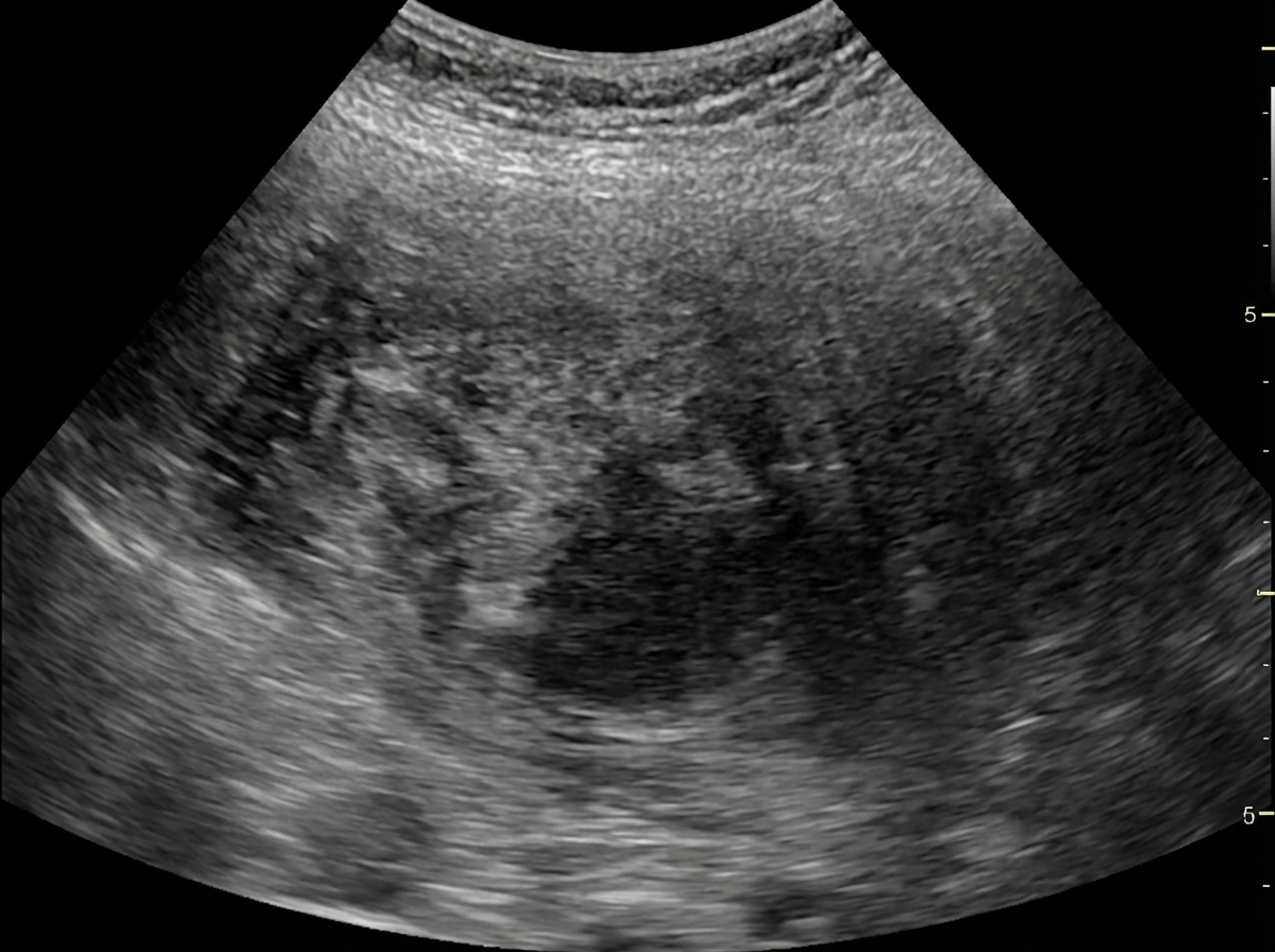
20 years old female came with complaint of a palpable painless mass in right breast. On examination, mass was mobile and hard in consistency. Ultrasound of right breast was performed . Most likely diagnosis is?
What is not an advantage of USG over mammography?
Which of the following features on mammogram would suggest malignancy?
Fat-containing breast lesions are seen in:
Gold standard investigation for breast carcinoma screening in a patient with silicone breast implants
Mammography can be best used in?
On mammogram, all of the following are the features of a malignant tumor except:
In a child, non-functioning kidney is best diagnosed by
Which is not echogenic while doing ultrasonography:
Explanation: ***USG*** - The image displays characteristic **gray-scale imaging** with an **echogenic appearance** of tissues, typical of an ultrasound. - Presence of annotations like "10 MHz G 64%" for **frequency and gain**, and "PRC" suggest ultrasound parameters. *Fluoroscopy* - Fluoroscopy provides **real-time X-ray images** and often involves the use of contrast agents, appearing as a dynamic, darker image with high contrast. - The image lacks the distinct bone and air contrast and dynamic motion typical of fluoroscopy. *X-Ray* - X-ray images depict a **static shadowgram** of dense structures like bones as white, and air as black, with sharp delineation. - The image shows a **granular texture** and fluid-filled structures that are characteristic of soft tissue imaging through ultrasound, not X-ray. *MRI* - MRI produces **cross-sectional images** with high soft tissue contrast in multiple planes (axial, sagittal, coronal). - The image shows real-time B-mode ultrasound characteristics with **probe frequency notation**, not the slice-based imaging of MRI.
Explanation: ***Fibroadenoma*** - A **painless**, **mobile**, and **hard mass** in a 20-year-old female is highly characteristic of a fibroadenoma, which is a common **benign breast tumor**. - Its **mobility** (often described as "breast mouse") is a key distinguishing feature from malignant masses. *Breast cyst* - While breast cysts can be **palpable** and **painless**, they are typically **soft or fluctuant** on palpation, not hard. - Cysts are fluid-filled sacs, and their mobile nature is less pronounced than the discrete, solid feel of a fibroadenoma. *Phyllodes tumor* - Phyllodes tumors can present as a **palpable mass**, but they usually grow **rapidly** and tend to be larger, often feeling lobulated. - While they can be benign, they have a potential for **malignant transformation** and are less common than fibroadenomas, especially in a 20-year-old. *Ductal papilloma* - Ductal papillomas are typically very small, located within milk ducts, and often present with **nipple discharge**, particularly bloody discharge, rather than a palpable mass. - They are rarely felt as a discrete, mobile, hard mass on routine physical examination.
Explanation: ***Superior detection of microcalcifications*** - **Mammography** is the gold standard for detecting **microcalcifications**, which can be a key indicator of **ductal carcinoma in situ (DCIS)** or early invasive breast cancer. - **Ultrasound (USG)** has limited sensitivity for detecting and characterizing microcalcifications. *Can be used for guided biopsy* - **USG-guided biopsy** is a common and advantageous technique for obtaining tissue samples from suspicious lesions in the breast or other organs. - This allows for **real-time visualization** of the needle, improving accuracy and reducing complications. *Can be used to differentiate solid VS cystic* - **USG** excels at distinguishing between **solid masses and fluid-filled cysts** due to differences in sound wave reflection. - This capability is crucial in characterizing breast lesions and often eliminates the need for further invasive procedures for benign cysts. *In young females with dense breasts* - **Dense breast tissue** in young females can obscure lesions on mammography, making interpretation difficult. - **USG** is particularly valuable in this population because it is not hindered by breast density and can provide a clearer view of underlying pathology.
Explanation: ***Areas of spiculated microcalcifications*** - **Spiculated microcalcifications** are highly suspicious for malignancy due to their irregular shape, distribution, and association with rapid, uncontrolled cell growth. - These calcifications often represent **necrotic cells** within rapidly growing tumors, which can deposit calcium. *Smooth borders* - **Smooth borders** typically indicate a benign lesion, such as a cyst or fibroadenoma, as they suggest gradual, uniform growth rather than invasive spread. - Malignant lesions tend to have **irregular** or ill-defined borders due to their infiltrative nature. *Well defined lesion* - A **well-defined lesion** usually suggests a benign process, as it indicates a mass that is clearly demarcated from surrounding tissue and is likely encapsulated. - Malignancies, conversely, often exhibit **indistinct or irregular margins** as they invade adjacent structures. *A mass of decreased density* - A mass of **decreased density** is generally considered a benign finding, often representing a **cyst** or an area of normal fatty tissue. - Malignant tumors typically present as a **mass of increased density** due to their cellular proliferation and desmoplastic reaction.
Explanation: ***All of the options*** - **Galactocele**, **fat necrosis**, and **hamartoma** are all types of breast lesions that can contain fat, making this the correct comprehensive answer. - Understanding that various benign breast conditions can present with a fat component is important for differential diagnosis. *Galactocele* - A **galactocele** is a benign **milk-filled cyst** that can develop in lactating or recently lactating women. - While primarily fluid-filled, it can sometimes contain areas of fat due to the milk's content (fat globules). *Fat necrosis* - **Fat necrosis** is a benign condition that occurs due to **trauma** or **ischemia** to breast tissue, leading to the breakdown of fat cells. - Imaging often reveals **oil cysts** or **calcifications** within areas of necrotic fat. *Hamartoma* - A **hamartoma** (also known as a fibroadenolipoma) is a benign mixed tumor composed of varying amounts of **glandular tissue**, **fibrous stroma**, and **fat**. - The fat component is typically well-defined and can give it a characteristic appearance on imaging.
Explanation: ***MRI*** - **MRI** is considered the **gold standard** for breast cancer screening in patients with silicone breast implants due to its superior ability to visualize breast tissue through the implant and detect subtle lesions. - It offers **high sensitivity** in detecting both implant rupture and early malignancies, often providing better clarity than mammography in augmented breasts where implants can obscure tissue. *Mammography* - While a standard screening tool, **mammography** can be limited in patients with silicone implants because the implants can **obscure adjacent breast tissue**, making detection of small masses challenging. - Special views (e.g., **Eklund views**) can be used, but sensitivity is still reduced compared to MRI in augmented breasts. *CT scan* - **CT scans** are not routinely used for primary breast cancer screening due to their use of **ionizing radiation** and lower sensitivity for detecting early breast lesions compared to MRI. - CT is more commonly used for **staging** advanced cancers or evaluating complex masses detected by other modalities. *USG* - **Ultrasound (USG)** is a valuable complementary tool, especially for evaluating palpable lumps or clarifying findings from mammography, but it is **operator-dependent** and has a lower overall sensitivity for general screening compared to MRI. - It is particularly useful for differentiating between **cystic and solid masses** and detecting implant ruptures but is not the gold standard for comprehensive screening in augmented breasts.
Explanation: ***Early breast carcinoma*** - **Mammography** is the gold standard for **early detection of breast carcinoma**, particularly for identifying **microcalcifications** and small masses before they are palpable. - It plays a crucial role in **screening asymptomatic women** to reduce breast cancer mortality. *Mastitis* - **Mastitis** is an **inflammatory condition** of the breast, often associated with infection, which is usually diagnosed clinically. - While mammography might show diffuse **increased density**, it is not the primary diagnostic tool and often has limited value due to inflammatory changes masking pathology. *Fibroadenoma* - **Fibroadenomas** are **benign breast tumors** common in younger women, typically appearing as well-circumscribed masses on mammography. - While mammography can detect them, their characterization often requires **ultrasound** and **biopsy** for definitive diagnosis, as differentiation from malignant lesions can be challenging. *Phylloides tumor* - A **Phylloides tumor** is a rare tumor that can be benign, borderline, or malignant, and it typically presents as a rapidly growing, palpable mass. - Mammography may show a well-defined mass, but **ultrasound** and **core needle biopsy** are essential for accurate diagnosis and distinction from fibroadenomas or malignancy.
Explanation: ***Macrocalcification*** - **Macrocalcifications** are typically **benign** and are often associated with involutional changes in the breast, such as aging or fibroadenomas. - These are usually larger, coarser calcifications that are easily seen and rarely indicate malignancy. *Microcalcification* - **Microcalcifications**, especially when **pleomorphic**, **linear**, or grouped, are a significant indicator of potential malignancy, such as **ductal carcinoma in situ (DCIS)**. - They represent calcium deposits within the ducts or stromal tissue, which can be associated with rapidly proliferating cells. *Irregular mass* - An **irregularly shaped mass** with ill-defined margins is highly suspicious for malignancy because it suggests invasive growth into surrounding tissues. - Unlike benign lesions which tend to be round or oval with smooth borders, malignant tumors often grow in an uncontrolled, infiltrative manner. *Spiculation* - **Spiculation** refers to **radiating lines or projections** extending from the borders of a mass, indicating an infiltrative process highly suggestive of malignancy. - These spicules represent fibrous tissue reaction to an invading tumor and are a strong predictor of breast cancer.
Explanation: ***DTPA renogram*** - A **DTPA renogram** (diethylene triamine pentaacetic acid scan) is a nuclear medicine study that assesses **renal blood flow** and **glomerular filtration rate (GFR)**. - It is highly effective in determining if a kidney is non-functioning because it directly measures the **uptake and excretion of a radiotracer** by the kidney, providing quantitative data on its functional capacity. *Creatinine clearance* - **Creatinine clearance** is a measure of overall kidney function, reflecting the GFR of **both kidneys combined**. - It cannot specifically identify a non-functioning individual kidney, as the other kidney might compensate for the non-functioning one, leading to a near-normal overall creatinine clearance. *Ultrasonography* - **Ultrasonography** is excellent for evaluating **renal anatomy**, such as size, shape, and presence of cysts, hydronephrosis, or stones. - While it can show structural abnormalities, it provides limited direct information about the **functional status** of the kidney, and a structurally normal kidney can still be non-functional. *IVU (Intravenous Urography)* - **Intravenous Urography (IVU)** uses contrast dye injected intravenously to visualize the kidneys, ureters, and bladder, assessing both anatomy and some aspects of function. - If a kidney is non-functioning, it would show **no uptake or excretion of the contrast dye**, but IVU involves radiation exposure and nephrotoxic contrast, making DTPA renogram often preferred in children for functional assessment.
Explanation: ***Bile*** - Bile is largely composed of **water**, which allows ultrasound waves to pass through it with minimal reflection, appearing **anechoic** (black) on ultrasound. - This property makes the gallbladder lumen, when filled with bile, appear anechoic, which is crucial for identifying structures like gallstones. *Bone* - **Bone** is highly dense and reflects a significant portion of ultrasound waves, making it appear very **echogenic** (bright) on ultrasonography. - Due to its high reflectivity, bone often produces a strong **acoustic shadow** behind it, obscuring deeper structures. *Gas* - **Gas** (air) is a strong reflector of ultrasound waves and appears brightly echogenic, often with a characteristic **dirty shadowing** or **reverberation artifact**. - The presence of gas can significantly hinder visualization of underlying tissues due to its strong reflection and scatter of the ultrasound beam. *Gall stones* - **Gallstones** are solid concretions that are highly reflective of ultrasound waves, appearing as bright, **echogenic foci** within the gallbladder lumen. - A classic ultrasound sign of gallstones is an echogenic structure with strong **posterior acoustic shadowing**.
Get full access to all questions, explanations, and performance tracking.
Start For Free