Causes of thickened gallbladder wall on ultrasound examination are all except:
The X-ray appearance of a CBD stone on cholangiography is:
What is the best way to diagnose gallbladder stones?
Which of the following is not a risk factor for cholangiocarcinoma?
In a patient presenting with jaundice, the HIDA scan would be most useful for which of the following:
Which of the following statements about CT imaging is the MOST accurate?
What is the investigation of choice in a patient with blunt abdominal trauma with hematuria?
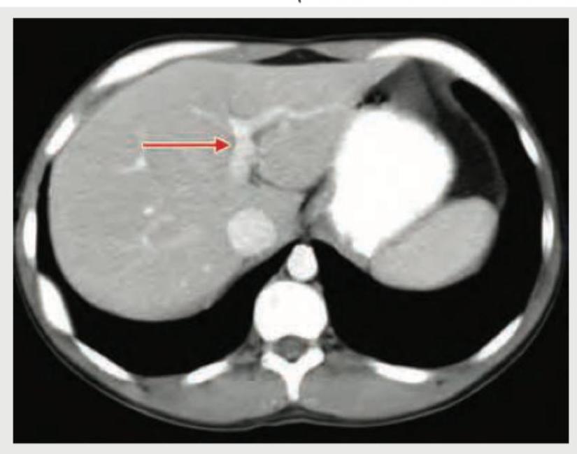
Identify the structure shown in CT abdomen section. (Recent NEET Pattern 2018-19)

Causes of thickened gallbladder wall on ultrasound examination are all except:
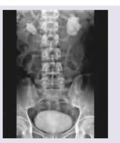
The following IVU shows:

Explanation: ***Kawasaki disease*** (Correct - NOT a typical cause of gallbladder wall thickening) * Although **Kawasaki disease** can cause systemic inflammation and **gallbladder hydrops** (distension) in children, it typically does NOT present with primary gallbladder wall thickening as a diagnostic finding. * The gallbladder manifestation in Kawasaki disease is **hydrops** (distension without stones), which is distinct from wall thickening. * Other hallmark features include **coronary artery aneurysms**, mucocutaneous manifestations, rash, and lymphadenopathy. * Gallbladder wall thickening is **not a recognized criterion** or common ultrasound finding in Kawasaki disease. --- *Congestive cardiac failure* (Incorrect - DOES cause thickening) * **Right-sided heart failure** leads to systemic venous congestion, causing **edema** in the gallbladder wall and subsequent thickening on ultrasound. * The thickening results from increased **hydrostatic pressure** and fluid accumulation in the wall. * This is a well-recognized cause of gallbladder wall thickening in the setting of hepatic congestion and ascites. *Postprandial state* (Incorrect - DOES cause thickening) * After a meal, especially one rich in fats, the gallbladder **contracts physiologically** to expel bile into the duodenum. * The contracted gallbladder wall appears **thickened** due to muscular contraction and reduced luminal volume. * This is a **normal physiological response**, not pathology; the thickening is *transient*, *symmetric*, and resolves on follow-up imaging after fasting. *Cholecystitis* (Incorrect - DOES cause thickening) * **Acute cholecystitis** involves inflammation of the gallbladder, directly causing **edema** and cellular infiltration of the gallbladder wall. * Wall thickening (>3 mm) is a **primary ultrasound diagnostic criterion** for acute cholecystitis. * Often accompanied by *pericholecystic fluid*, gallstones, and positive **sonographic Murphy's sign**.
Explanation: ***Meniscus sign*** - A **meniscus sign** on cholangiography is characteristic of a **filling defect** caused by a gallstone within the **common bile duct (CBD)**. - The contrast agent outlines the stone, creating a crescent-shaped or **meniscus-like appearance** around it. *Non-central occlusion* - This term usually refers to an incomplete or **partial blockage** that doesn't necessarily have the distinct outline of a stone. - While a stone can cause occlusion, "meniscus sign" specifically describes the **radiographic appearance** of the stone itself. *Abrupt cut-off* - An **abrupt cut-off** suggests a sudden and complete obstruction, often seen with **malignant strictures** or **impacted stones** without clear outlining. - It does not specifically describe the characteristic rimming of contrast around a stone that defines a meniscus sign. *Benign tapering* - **Benign tapering** typically indicates a gradual, smooth narrowing of the duct, often associated with **inflammatory strictures** or **fibrotic changes**. - This appearance is different from the distinct filling defect caused by a stone.
Explanation: ***USG*** - **Ultrasound (USG)** is the preferred and most **sensitive imaging modality** for detecting gallbladder stones due to its non-invasive nature and high diagnostic accuracy. - It can effectively visualize stones as **echogenic foci** with **posterior acoustic shadowing** within the gallbladder lumen. *Oral cholecystography* - This method involves ingesting a contrast agent, which is then absorbed and excreted by the liver into the bile, outlining the gallbladder. - It has largely been replaced by ultrasound due to its **lower sensitivity** for small stones, **patient inconvenience**, and potential for **adverse reactions** to the contrast. *Percutaneous transhepatic cholangiography* - **PTC** is an **invasive procedure** where a needle is inserted through the skin and liver into the bile ducts to inject contrast. - It is typically reserved for cases where **other imaging modalities are inconclusive** or for **therapeutic interventions** in patients with biliary obstruction, not routine stone detection. *Intravenous cholangiogram* - This involves intravenous administration of a contrast agent that is concentrated in the bile and allows visualization of the bile ducts and gallbladder. - It has also been **largely replaced by USG and ERCP** due to the risk of **allergic reactions** to contrast, **poor visualization** in cases of liver dysfunction, and lower sensitivity than ultrasound.
Explanation: ***Aflatoxin*** - **Aflatoxin** is a potent **hepatocarcinogen** produced by *Aspergillus* species that is specifically and strongly linked to **hepatocellular carcinoma (HCC)** [1], NOT cholangiocarcinoma. - This is the **most clearly unrelated** risk factor to cholangiocarcinoma among the options, as its carcinogenic mechanism targets hepatocytes specifically [1], [2]. - It contaminates crops in warm, humid regions and is a well-established cause of liver cancer in endemic areas [1]. *Thorotrast* - **Thorotrast** (thorium dioxide) was a radioactive contrast agent used until the 1950s that **IS a known risk factor** for cholangiocarcinoma. - Due to prolonged retention in the liver and biliary system, it significantly increases the risk of both **cholangiocarcinoma** and **hepatic angiosarcoma** [3]. - Its use was discontinued precisely because of its strong carcinogenic potential. *Radon* - **Radon** is a naturally occurring radioactive gas that is primarily and overwhelmingly associated with **lung cancer** from inhalation exposure. - While a potent carcinogen, it has **no established epidemiological link** to cholangiocarcinoma due to its route of exposure and target organ. *Dioxin* - **Dioxins** are environmental pollutants with documented carcinogenic effects. - While some studies have explored potential links to various cancers, dioxin is **not recognized as an established risk factor** for cholangiocarcinoma in major medical references. **References:** [1] Kumar V, Abbas AK, et al.. Robbins and Cotran Pathologic Basis of Disease. 9th ed. Liver and Gallbladder, pp. 876-877. [2] Kumar V, Abbas AK, et al.. Robbins and Cotran Pathologic Basis of Disease. 9th ed. Neoplasia, pp. 331-332. [3] Cross SS. Underwood's Pathology: A Clinical Approach. 6th ed. (Basic Pathology) introduces the student to key general principles of pathology, both as a medical science and as a clinical activity with a vital role in patient care. Part 2 (Disease Mechanisms) provides fundamental knowledge about the cellular and molecular processes involved in diseases, providing the rationale for their treatment. Part 3 (Systematic Pathology) deals in detail with specific diseases, with emphasis on the clinically important aspects., pp. 216-217.
Explanation: ***Biliary atresia*** - A **HIDA scan** (hepatobiliary iminodiacetic acid scan) is instrumental in diagnosing biliary atresia by demonstrating the **absence of bile flow** into the duodenum. - In infants with persistent jaundice, the failure of the tracer to appear in the small bowel after a prolonged period strongly suggests this condition, indicating **obstructed or absent bile ducts**. *Cholelithiasis* - While HIDA scans can detect **cystic duct obstruction** in acute cholecystitis, they are less definitive for uncomplicated cholelithiasis (gallstones without acute inflammation). - **Ultrasound** is typically the primary imaging modality for diagnosing gallstones due to its non-invasiveness and ability to visualize stones directly. *Benign biliary disease* - This is a broad category, and while a HIDA scan can assess bile flow, it's not the **primary diagnostic tool** for all benign biliary conditions. - For most benign biliary diseases (e.g., choledocholithiasis without acute cholecystitis), **ultrasound, ERCP, or MRCP** often provide more detailed anatomical information. *Bile duct carcinoma* - A HIDA scan might show **obstructed bile flow** in bile duct carcinoma (cholangiocarcinoma), but it does not provide the detailed anatomical information or staging necessary for diagnosis and treatment planning. - **CT, MRI, MRCP, or ERCP** with biopsy are far more effective for identifying, characterizing, and staging bile duct malignancies.
Explanation: ***Water has a Hounsfield unit (HU) of zero.*** - The **Hounsfield unit (HU)** scale is a quantitative scale used to describe radiodensity in CT scans, where **water is defined as 0 HU**. - This establishes a crucial reference point for measuring the attenuation of other tissues, which can range from approximately **-1000 HU for air** to **+1000 HU or more for dense bone**. *CT head dose remains constant regardless of the protocol used.* - The **radiation dose** in CT scans is highly variable and depends significantly on the **protocol used**, including factors like mA, kVp, pitch, and scan length. - **Dose optimization techniques** and protocol adjustments are routinely employed to minimize patient exposure while maintaining diagnostic image quality. *CT cannot detect gallstones under any circumstances.* - While **ultrasound (US)** is the primary modality for detecting gallstones, CT can visualize them, especially if they are **calcified** or of mixed composition. - **Non-calcified gallstones** may be more challenging to detect on CT, but they are not impossible to see, particularly with current generation scanners and appropriate windowing. *CT uses unfiltered x-ray beams.* - CT scanners use **filtered x-ray beams** to provide higher quality images and reduce patient dose. - **Filtration (e.g., aluminum or copper)** removes low-energy x-rays, which would otherwise be absorbed by the patient without contributing to image formation.
Explanation: ***Contrast enhanced computed tomography (CECT)*** - **CECT** is the **investigation of choice** for evaluating solid organ injuries, including renal trauma, in hemodynamically stable patients with blunt abdominal trauma and hematuria. - It provides detailed imaging of the kidneys, urinary tract, and surrounding structures, allowing for the classification of injury severity and identification of associated injuries. *Retrograde urogram* - A retrograde urogram is primarily used to evaluate the **lower urinary tract** (urethra and bladder) for strictures or injuries, specifically when there is a suspicion of urethral injury. - It is not the primary imaging modality for assessing renal parenchymal or collecting system injuries from blunt trauma. *Intravenous urogram (IVU)* - While an IVU can assess the upper urinary tract, it has largely been replaced by **CECT** in the acute trauma setting due to CECT's superior resolution and ability to evaluate renal parenchyma and other abdominal organs. - IVU exposes the patient to radiation and requires contrast administration, and it may not adequately visualize subtle renal injuries or hematomas as effectively as CECT. *Ultrasonography of abdomen* - **Ultrasound** is useful for rapidly detecting free fluid (suggesting hemorrhage) or gross hydronephrosis in trauma, but it has limited sensitivity for diagnosing specific renal parenchymal injuries or urinary extravasation. - Its role in blunt abdominal trauma with hematuria is often as an initial screening tool, but it is not sufficient for definitive diagnosis or grading of renal injuries.
Explanation: ***Portal vein*** - The arrow points to a vessel receiving blood from the splenic and superior mesenteric veins, which is characteristic of the **portal vein** entering the **liver parenchyma**. - The portal vein is typically seen anterior to the **inferior vena cava** and posterior to the **common hepatic artery** at this level. *Inferior vena cava* - The **inferior vena cava (IVC)** is a large, retroperitoneal vessel located posterior to the liver and to the right of the aorta. - The structure indicated by the arrow is clearly within the liver substance, not in the typical position of the IVC. *Splenic vein* - The **splenic vein** runs horizontally behind the body of the pancreas and joins with the superior mesenteric vein to form the portal vein. - The vessel shown is within the liver, distal to the formation of the portal vein. *Superior mesenteric vein* - The **superior mesenteric vein (SMV)** typically runs vertically in the mesentery and joins the splenic vein to form the portal vein. - The indicated structure is within the liver hilum, not in the anatomical location of the SMV.
Explanation: ***Kawasaki disease*** (Correct Answer) - While Kawasaki disease can cause **gallbladder hydrops** (distension with bile), the primary ultrasound finding is an **enlarged, distended gallbladder** rather than isolated wall thickening. - When gallbladder involvement occurs in Kawasaki disease, it manifests as **acalculous cholecystitis** with hydrops, but this is **not a typical or common presentation** compared to the other causes listed. - The hallmark features of Kawasaki disease are **coronary artery aneurysms** and systemic vasculitis, not primary gallbladder pathology. - In clinical practice, gallbladder wall thickening would **not be attributed to Kawasaki disease** as a primary differential diagnosis. *Incorrect: Congestive cardiac failure* - **Systemic fluid overload** and venous congestion in CHF leads to gallbladder wall thickening due to **transudative edema**. - This is a **common cause** of non-inflammatory gallbladder wall thickening (>3mm). - The wall appears thickened, hypoechoic, and **edematous** without pericholecystic fluid. *Incorrect: Postprandial state* - After eating, the gallbladder **contracts to release bile**, causing the wall to appear thicker on ultrasound due to **accordion-like folding** of the mucosa. - This is a **normal physiological finding** and typically resolves within 1-2 hours. - Scanning should ideally be done after **6-8 hours of fasting** to avoid this pseudo-thickening. *Incorrect: Cholecystitis* - **Acute cholecystitis** is the **classic cause** of gallbladder wall thickening (>3mm, often >5mm). - Associated findings include **gallstones, pericholecystic fluid, positive sonographic Murphy's sign**, and wall edema. - The wall shows **layering** (subserosal edema) and hyperemia on Doppler imaging.
Explanation: ***Horseshoe kidney*** - The image shows both kidneys are **fused at their lower poles** across the midline, forming a "horseshoe" shape, which is a classic radiographic finding for this condition. - The **calyces and renal pelves are seen medially oriented**, supporting the diagnosis of a horseshoe kidney. *Hydronephrosis* - **Hydronephrosis** would appear as a dilation of the renal pelvis and calyces due to obstruction, which is not the primary finding here. - While a horseshoe kidney can be associated with hydronephrosis due to an abnormal ureteral course, the image clearly depicts the **fused renal parenchyma** rather than just dilation. *Polycystic kidney* - **Polycystic kidneys** are characterized by numerous cysts of varying sizes replacing normal renal parenchyma, which would present as enlarged, multi-cystic kidneys on imaging. - The image does not show multiple cysts replacing the renal tissue but rather a **fused, single-mass structure** in the lower abdomen. *Duplication of collecting system* - **Duplication of the collecting system** involves two ureters draining a single kidney, or a bifid renal pelvis, which would appear as double ureters or collecting systems on an IVU. - This condition does not present with the characteristic **fusion of the renal poles** across the midline as seen in the image.
Get full access to all questions, explanations, and performance tracking.
Start For Free