Ultrasound is the investigation of choice for
What is the investigation of choice for blunt abdominal trauma in an unstable patient?
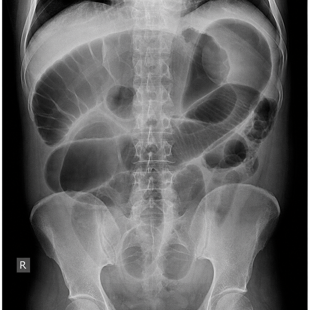
Most sensitive investigation for abdominal trauma in a hemodynamically stable patient is-
The "Target sign" ultrasonographically means:
In a patient with a tender and rigid abdomen, what is the expected finding on X-ray?
Identify the condition based on the non-contrast CT scan of a patient given below.

Gas absent from intestine (gasless abdomen) on x-ray is seen in which condition?
Gasless abdomen seen in-
What is the investigation of choice for an 8-year-old child presenting with an acute abdomen?
A patient presents to the emergency department with pain and distension of abdomen and absolute constipation. What is the investigation of choice ?
Explanation: ***Blunt abdominal trauma*** - **Focused Assessment with Sonography for Trauma (FAST) exam** is the initial imaging modality of choice for rapidly detecting **intra-abdominal free fluid** (hemoperitoneum) in hemodynamically unstable patients with blunt abdominal trauma due to its speed, portability, and non-invasiveness. - It helps guide the need for further imaging or surgical intervention, making it critical in the acute setting. *Somatostatinoma* - Diagnosed primarily through biochemical tests (elevated **somatostatin levels**) and imaging like **CT, MRI, or somatostatin receptor scintigraphy (SRS)**, which are superior for localizing these rare neuroendocrine tumors. - Although ultrasound can sometimes detect pancreatic masses, it is not the **investigation of choice** for definitive diagnosis or staging of somatostatinomas. *Intraductal Pancreatic calculi* - Often best visualized with **Endoscopic Retrograde Cholangiopancreatography (ERCP)** or **Magnetic Resonance Cholangiopancreatography (MRCP)**, which provide detailed imaging of the pancreatic and bile ducts. - While transabdominal ultrasound can sometimes detect dilated ducts or large calculi, **Endoscopic Ultrasound (EUS)** is more sensitive and specific for intraductal pathologies, making routine transabdominal ultrasound not the primary choice. *Urethral stricture* - The gold standard for diagnosing urethral strictures is **urethrography** (retrograde urethrogram), which directly visualizes the stricture and its extent. - While ultrasound can sometimes be used to assess the urethra, it is less effective than urethrography for defining the length and severity of a stricture.
Explanation: ***USG (FAST Exam)*** - In an **unstable patient** with blunt abdominal trauma, **Focused Assessment with Sonography for Trauma (FAST) exam** is the investigation of choice. - It is **rapid, non-invasive, and bedside**, allowing immediate detection of **free fluid** (blood) in the peritoneal cavity, pericardium, and pleural spaces without transporting the patient. - Guides immediate decision for **laparotomy** in hemodynamically unstable patients. - **Note:** In **stable patients**, **CT abdomen** is the gold standard as it provides detailed anatomical information, but it requires patient transport and time. *X-ray abdomen* - Provides limited information in blunt trauma, primarily showing **free air** (bowel perforation) or **bony fractures**. - **Not sensitive** for detecting intraperitoneal bleeding, which is the primary concern in unstable patients. *MRI* - Offers excellent soft tissue detail but is **time-consuming** and requires the patient to be **hemodynamically stable**. - **Impractical** for unstable trauma patients requiring rapid assessment and intervention. *Diagnostic Peritoneal Lavage (DPL)* - An **invasive procedure** that is sensitive for detecting intra-abdominal hemorrhage. - Has largely been **replaced by FAST exam** in most trauma centers due to FAST being non-invasive, rapid, and repeatable. - DPL has a **higher false-positive rate** and cannot identify the source of bleeding.
Explanation: ***CT Scan (Computed Tomography)*** - **CT scans** offer superior anatomical detail and can accurately detect organ damage, hemorrhage, and other injuries in **hemodynamically stable** patients with abdominal trauma. - It is considered the **most sensitive** and specific imaging modality for evaluating blunt and penetrating abdominal trauma when the patient can tolerate the study. *Ultrasonography (FAST)* - While effective for detecting **free fluid** (blood) in specific abdominal areas, **Focused Assessment with Sonography for Trauma (FAST)** has lower sensitivity for solid organ injuries or bowel perforations. - Its primary role is rapid assessment for **hemoperitoneum** to guide immediate management in unstable patients, not detailed injury characterization. *Diagnostic peritoneal lavage (DPL)* - **DPL** is an invasive procedure with high sensitivity for detecting **intraperitoneal bleeding**, but it does not identify specific organ injuries or retroperitoneal hemorrhage. - It is rarely used in hemodynamically stable patients due to its invasiveness and the availability of more detailed imaging techniques. *MRI (Magnetic Resonance Imaging)* - **MRI** provides excellent soft tissue contrast but is typically too **time-consuming** and less accessible in urgent trauma settings compared to CT. - It's generally not the first-line investigation for acute abdominal trauma due to motion artifacts and limited utility in detecting air or bone injuries.
Explanation: ***Intussusception*** - The **"target sign"** (also known as the **"donut sign"** or **"pseudokidney sign"**) on ultrasound is a classic finding for **intussusception**. - It represents concentric layers of bowel telescoping into an adjacent segment, creating a central hyperechoic core surrounded by hypoechoic rings. *Liver metastasis* - Liver metastases often appear as **hypoechoic, hyperechoic, or mixed echogenicity lesions** on ultrasound, and vary widely in appearance. - While some can have a "target-like" appearance with a hyperechoic rim, it's not the primary or most specific sign for liver metastasis and is less distinct than in intussusception. *Ectopic kidney* - An ectopic kidney is an anatomical variant where the kidney is located outside its normal position, most commonly in the **pelvis**. - On ultrasound, it would appear as a normally formed kidney in an atypical location, without the distinct concentric layers seen in the "target sign." *Ovarian carcinoma* - Ovarian carcinomas present with **complex masses** that can be solid, cystic, or mixed, often with septations, papillary projections, and areas of necrosis. - Their ultrasound appearance is highly variable but does not typically manifest as a "target sign" with concentric rings.
Explanation: ***Air under the diaphragm*** - The presence of **free air** (pneumoperitoneum) beneath the diaphragm on an upright abdominal X-ray is a classic sign of **visceral perforation**. - A **tender and rigid abdomen** (peritoneal signs) indicates irritation of the peritoneum, most commonly due to a ruptured hollow viscus. *Blood under the diaphragm* - While blood can accumulate under the diaphragm (e.g., from **trauma** or a ruptured ectopic pregnancy), it typically manifests as a **hemoperitoneum** on imaging. - Blood is **fluid** and would appear as a fluid collection, not free air, on X-ray. *Hazy lung fields* - **Hazy lung fields** suggest conditions like **pulmonary edema**, pneumonia, or acute respiratory distress syndrome (ARDS). - These findings are primarily associated with pulmonary pathology and are not directly indicative of an acute abdominal emergency like perforation. *Prominent vascular markings* - **Prominent vascular markings** often indicate increased blood flow to the lungs or **pulmonary hypertension**. - This finding is unrelated to acute abdominal pain or peritoneal irritation.
Explanation: ***Hydatid cyst*** - The image distinctly shows a **large, well-defined cyst with internal septations**, consistent with the daughter cysts and collapsed membranes within a hydatid cyst (the "**water lily sign**"). - The thick, often calcified wall surrounding the lesion is a characteristic feature often seen in **Echinococcus granulosa** infection. *Hepatocellular carcinoma* - **Hepatocellular carcinoma (HCC)** typically appears as a **solid, enhancing mass** (especially on contrast-enhanced CT) and does not usually present with clearly defined internal septations or "water lily" sign on non-contrast imaging. - While HCC can show necrosis, it does not form the characteristic cystic structure seen here. *Liver abscess* - A **liver abscess** would typically appear as a ill-defined, fluid-filled lesion that may have a rim of enhancement on contrast CT, but it generally lacks the **distinct internal septations** or daughter cysts characteristic of a hydatid cyst. - Abscesses are often associated with signs of infection like fever and elevated inflammatory markers. *Fibronodular hyperplasia* - **Focal nodular hyperplasia (FNH)** is a benign liver lesion characterized by a central scar and is typically **isodense or slightly hypodense** to the liver parenchyma on non-contrast CT. - It does not present as a cystic lesion with internal daughter cysts or calcified walls.
Explanation: ***Acute pancreatitis*** - In **severe acute pancreatitis**, a **gasless or relatively gasless abdomen** may be seen due to profound **ileus** with fluid accumulation displacing intestinal gas. - The marked inflammatory process can lead to complete loss of intestinal motility and fluid sequestration (third-spacing), resulting in minimal visible bowel gas on X-ray. - **Note**: Classic signs include **sentinel loop sign** (dilated jejunal loop) or **colon cut-off sign**, but in severe cases with massive ascites or fluid collections, a gasless pattern may occur. *Ulcerative colitis* - Typically presents with **dilated loops of large bowel** with visible gas and **toxic megacolon** in severe cases. - Inflammatory changes cause bowel wall thickening, but gas is usually **present and often increased**. *Intussusception* - May show a **target sign** or **meniscus sign** on imaging, with bowel loops dilated proximal to the obstruction. - Gas is typically **present** within the bowel or proximal to the invagination, not absent from the entire abdomen. *Necrotizing enterocolitis* - Characterized by **pneumatosis intestinalis** (gas in the bowel wall) and **portal venous gas**, features directly contradicting a gasless abdomen. - Shows dilated loops with gas and evidence of bowel wall necrosis - **gas is prominently present**.
Explanation: ***Acute pancreatitis*** - A **gasless abdomen** on X-ray can be a finding in severe **acute pancreatitis** due to **ileus** (paralytic ileus secondary to inflammation), and the presence of significant peritoneal fluid or inflammation may obscure gas. - The inflammatory process can lead to **adynamic ileus** affecting segments of the bowel, limiting gas accumulation. *Ulcerative colitis* - Often presents with **toxic megacolon** in severe cases, which would show a **dilated, gas-filled colon**, not a gasless abdomen. - Inflammation is typically confined to the colon and does not usually lead to a widespread adynamic ileus that would result in a gasless appearance. *Intussusception* - While it can manifest as an **abdominal mass** and **bowel obstruction**, gas is typically present proximal to the obstruction. - The classic X-ray finding is a **target sign** (telescoping bowel) or signs of **bowel obstruction**, which includes dilated loops of bowel with air-fluid levels. *Necrotising enterocolitis* - Characterized by **pneumatosis intestinalis** (gas in the bowel wall) and sometimes **portal venous gas**, indicating the presence of gas, not its absence. - A sign of advanced disease, this condition involves gas within the bowel layers rather than a gasless abdomen.
Explanation: ***USG*** - An **ultrasound (USG)** is the preferred initial imaging modality in pediatric acute abdomen due to its **lack of ionizing radiation**, ease of use, and ability to visualize common causes like appendicitis and intussusception. - It is particularly useful for assessing **fluid collections**, inflammation, and obstruction in a non-invasive manner suitable for children. *CT Scan* - While it offers detailed anatomical views, **CT scans** involve significant **ionizing radiation**, which is a concern in children due to increased lifetime cancer risk. - It is typically reserved for cases where **USG is inconclusive** or if there is a high suspicion of conditions not well visualized by ultrasound. *X-ray* - **X-rays** provide limited information for soft tissue pathologies and are primarily useful for detecting **bowel obstruction (air-fluid levels)** or **free air** (perforation). - They lack the resolution to diagnose many common causes of acute abdomen in children, such as appendicitis or intussusception. *MRI* - **MRI** provides excellent soft tissue contrast without ionizing radiation but often requires **sedation** in young children due to the long scan times and need for stillness. - It is less readily available and more expensive than USG, making it a less practical first-line investigation for an acute presentation.
Explanation: ***Plain X-ray abdomen (Erect)*** - An erect plain X-ray of the abdomen is the initial and often diagnostic investigation for **bowel obstruction**, revealing **dilated bowel loops** and **air-fluid levels**. - It helps confirm the presence of obstruction and can sometimes indicate its location and severity, though it does not provide information about the cause. *Ultrasonography* - While ultrasound can detect **bowel dilation** and **peristalsis**, it is limited in visualizing the entire bowel and cannot reliably differentiate between various causes of obstruction. - It is more useful for assessing **extraluminal pathology** or **fluid collections** but less effective as a primary diagnostic tool for bowel obstruction. *Barium meal follow-through* - This study involves oral **barium administration** and serial X-rays to visualize the small bowel, but it is **contraindicated** in suspected bowel obstruction due to the risk of exacerbating the obstruction or causing **barium impaction**. - Its primary role is in evaluating chronic or partial obstructions, or malabsorption, not acute presentations with complete obstruction. *Colonoscopy* - **Colonoscopy** is an invasive procedure primarily used for diagnosis and treatment of **colonic pathology**, such as polyps, strictures, or bleeding. - It is **contraindicated** in acute, complete bowel obstruction due to the risk of **perforation** and is not the initial diagnostic choice for acute abdominal pain and absolute constipation.
Get full access to all questions, explanations, and performance tracking.
Start For Free