HR-180, BP-60/40, temp-39.5°C, ETCO2-65 post induction. Most likely diagnosis:
Which does not cause malignant hyperthermia –
Malignant hyperthermia is
A person working in a hot environment who consumes more water without salt is likely to develop a condition called
Which does not cause malignant hyperthermia
Which of the following measurement sites most closely reflects core body temperature?
A cold exposure which is expected to bring the body temperature from 37°C to 20°C, actually brings it down to only 36.5°C. Calculate the 'Gain' of the thermoregulatory system.
Regarding haemorrhagic shock, which one of the following statements is correct?
All the following mediate their action using cAMP as second messenger except:
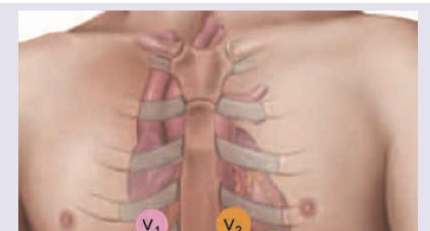
The lead of ECG marked as $X$ is called:

Explanation: ***Malignant hyperthermia*** - The rapid onset of **tachycardia (HR-180)**, **hyperthermia (temp-39.5°C)**, and profoundly elevated **ETCO2 (65 mmHg)** immediately following anesthetic induction is the classic presentation of malignant hyperthermia. - This condition is a pharmacogenetic disorder, triggered by volatile anesthetics (e.g., isoflurane) and succinylcholine, leading to uncontrolled skeletal muscle metabolism and hypercapnia. *Thyroid storm* - While thyroid storm can cause **tachycardia** and **hyperthermia**, the sudden and dramatic rise in **ETCO2** is not a characteristic feature. - Onset is typically less abrupt and often associated with pre-existing hyperthyroidism or a precipitating event like surgery or infection, rather than immediate post-induction. *Anaphylaxis* - Anaphylaxis typically presents with **hypotension (BP-60/40)**, **tachycardia**, and often features like **bronchospasm**, **rash**, or **angioedema**. - Although it can cause **bronchospasm** leading to increased ETCO2, the extreme elevation to 65 mmHg is less typical, and **profound hyperthermia** is not a primary symptom. *Septic shock* - **Septic shock** is characterized by **hypotension** and **tachycardia**, often accompanied by **fever**, but its onset is usually prolonged over hours to days. - A sudden increase in **ETCO2** to 65 mmHg immediately post-induction is uncharacteristic for sepsis, which relates to an exaggerated, systemic inflammatory response to infection.
Explanation: ***N2O*** - **Nitrous oxide** is a weak inhaled anesthetic and does not trigger **malignant hyperthermia** (MH). - Its mechanism of action does not involve the **ryanodine receptor** or calcium release, which are central to MH pathophysiology. *Enflurane* - **Enflurane** is a volatile inhaled anesthetic that is a known trigger for **malignant hyperthermia**. - It induces uncontrolled **intracellular calcium release** in skeletal muscle, leading to hypermetabolism. *Desflurane* - **Desflurane** is another potent volatile inhaled anesthetic and a classic trigger agent for **malignant hyperthermia**. - Its use can result in a rapid onset of MH symptoms due to its quick pharmacokinetics. *Isoflurane* - **Isoflurane** is also a volatile inhaled anesthetic and is well-established as a trigger for **malignant hyperthermia**. - Like other volatile agents, it can bind to the **ryanodine receptor** (RyR1), causing excessive calcium efflux.
Explanation: ***Succinylcholine is a triggering agent*** - **Succinylcholine**, a depolarizing neuromuscular blocker, is a potent trigger for malignant hyperthermia due to its interaction with the **ryanodine receptor**. - Other common triggering agents include **volatile anesthetic agents** such as halothane, isoflurane, and sevoflurane, all leading to uncontrolled calcium release. *Metabolic alkalosis and hypokalemia* - Malignant hyperthermia typically presents with a rapidly developing severe **metabolic acidosis**, not alkalosis, due to increased lactic acid production from muscle hypermetabolism. - Patients usually experience **hyperkalemia** due to muscle cell breakdown and potassium release, which can lead to cardiac arrhythmias. *Calcium infusion is used for treatment.* - The immediate treatment for malignant hyperthermia is **dantrolene sodium**, which acts by blocking calcium release from the sarcoplasmic reticulum in muscle cells. - **Calcium channel blockers** are generally contraindicated in malignant hyperthermia, especially when dantrolene has been administered, due to the risk of exacerbating hyperkalemia. *Autosomal recessive pharmacogenetic disease* - Malignant hyperthermia is inherited as an **autosomal dominant** pharmacogenetic disorder, primarily linked to mutations in the **RYR1 gene** (ryanodine receptor 1 gene). - This dominant inheritance pattern means that only one copy of the mutated gene is sufficient to predispose an individual to the condition.
Explanation: ***Heat cramps*** - This condition occurs due to **excessive sweating** in a hot environment, leading to significant **electrolyte (salt) loss**, particularly sodium. - Consuming large amounts of **plain water without electrolyte replacement** further dilutes the remaining electrolytes in the body, exacerbating hyponatremia and increasing the likelihood of painful muscle cramps. *Heat stroke* - **Heat stroke** is a life-threatening condition characterized by a **core body temperature >104°F (40°C)** and central nervous system dysfunction (e.g., altered mental status). - While fluid and electrolyte imbalances can contribute, its defining feature is the severe **thermoregulatory failure** leading to organ damage, which is distinct from simple muscle cramps. *Heat hyperpyrexia* - This term refers to an **extremely high body temperature** (often above 106°F or 41.1°C) but is not a specific diagnosis in the context of heat-related illness. - It is more of a symptom that could be present in severe heatstroke, not a primary condition resulting from excessive plain water intake. *Heat exhaustion* - **Heat exhaustion** presents with symptoms like fatigue, dizziness, nausea, and profuse sweating, but without significant central nervous system dysfunction or extremely high core body temperature. - While it involves fluid and electrolyte loss, the specific scenario of drinking plain water without salt primarily leads to muscle cramps due to electrolyte dilution, rather than the broader symptoms of heat exhaustion.
Explanation: **N2O** - **Nitrous oxide (N2O)**, or laughing gas, is an inhaled anesthetic that does not trigger **malignant hyperthermia (MH)**. It is considered a safe anesthetic agent for MH-susceptible patients. - MH is a genetic disorder of skeletal muscle characterized by uncontrolled **calcium release** from the **sarcoplasmic reticulum**, leading to a hypermetabolic state. *Enflurane* - **Enflurane** is a **volatile inhaled anesthetic** that can trigger malignant hyperthermia. Volatile anesthetics are a primary class of agents known to induce MH in susceptible individuals. - It works by affecting the **ryanodine receptor (RyR1)** in muscle cells, leading to excessive calcium release. *Isoflurane* - **Isoflurane** is also a **volatile inhaled anesthetic** and is a known trigger for malignant hyperthermia. - Like other volatile agents, it causes a rapid increase in **intracellular calcium**, muscle rigidity, and a systemic hypermetabolic response. *Desflurane* - **Desflurane** is another **volatile inhaled anesthetic** that is a potent trigger of malignant hyperthermia. - Its rapid onset and offset properties do not prevent it from causing the rapid **calcium release** characteristic of MH.
Explanation: ***Rectal*** - **Rectal temperature** is considered the most accurate non-invasive measure of **core body temperature** because of its proximity to the body's internal organs and consistent blood supply. - It is typically about **0.5-0.7°C higher than oral temperature** and reflects the true core warmth of the body. *Axillary* - **Axillary temperature** is taken in the armpit and is generally the **least accurate** and lowest reading. - It often **underestimates core body temperature** by 1°C or more due to exposure to ambient air. *Oral* - **Oral temperature** is a common and convenient site for temperature measurement but can be influenced by recent ingestion of hot or cold foods/liquids, or breathing through the mouth. - While generally reliable, it typically measures about **0.5°C lower than rectal temperature**. *Surface* - **Surface temperature**, such as that taken on the skin (e.g., forehead thermometer), is highly variable and easily affected by environmental factors like ambient temperature, airflow, and sweating. - It provides a less accurate reflection of the **body's internal core temperature** compared to deeper measurements.
Explanation: ***33*** - The **error signal** (or uncorrected temperature drop) is the difference between the actual drop and the expected drop without regulation. Here, the expected drop is 37°C - 20°C = 17°C. The actual drop is 37°C - 36.5°C = 0.5°C. So, the error signal caused by the regulatory system's action is 36.5°C - 20°C = 16.5°C. Alternatively calculated as 17°C (expected) - 0.5°C (actual) = 16.5°C. - The **gain** of a thermoregulatory system is calculated as the expected change in temperature (without regulation) divided by the actual observed change in temperature after regulation when the body resists the change. Here, the body would have cooled by 17°C (37°C - 20°C) without compensation, but it only cooled by 0.5°C (37°C - 36.5°C). The gain is therefore 16.5 / 0.5 = 33. *34* - This answer likely arises from a miscalculation of the error signal or the expected temperature drop. - The key is to correctly identify the **change that would have occurred without regulation** and the **change that actually occurred.** *16.5* - This value represents the **change in temperature that was prevented by the thermoregulatory system** (17°C expected drop minus 0.5°C actual drop), but it is not the gain. - The gain is a ratio, not an absolute temperature difference. *66* - This value would result from an incorrect calculation, possibly by inverting the gain formula or multiplying by an incorrect factor. - The gain is specifically the ratio of the "corrected" change to the "uncorrected" error.
Explanation: ***Loss of 40% of circulating volume is life threatening*** - A loss of **40% or more** of circulating blood volume corresponds to **Class IV haemorrhagic shock**, which is a severe, life-threatening condition requiring immediate and aggressive resuscitation. - At this stage, the body's compensatory mechanisms are overwhelmed, leading to profound systemic hypoperfusion, **organ dysfunction**, and a high risk of mortality. *Tachycardia presents in 100% of hypovolemic patients* - While **tachycardia** is a common compensatory mechanism in hypovolemia, it is not present in 100% of patients due to factors such as **beta-blocker use** or **pacemaker rhythm**. - In some early stages of blood loss, especially in young, healthy individuals, sufficient compensatory mechanisms may delay the onset of significant tachycardia. *Clinically manifested when > 10% of loss of total blood volume occurs* - Haemorrhagic shock is typically **clinically manifest** when there is a blood loss greater than **15%** (Class I shock), which represents approximately 750 mL in an average adult. - A loss of **less than 10%** often does not produce overt clinical signs as the body's compensatory mechanisms can effectively maintain vital signs within normal ranges. *In acute stage of shock, systemic vasodilation becomes evident* - In the acute stage of hemorrhagic shock, the body's primary compensatory mechanism is **systemic vasoconstriction**, not vasodilation, to maintain central blood pressure and perfuse vital organs. - **Vasodilation** can occur in the later, decompensated stages of shock, particularly in instances of **septic or neurogenic shock**, leading to a further drop in blood pressure.
Explanation: ***Vasopressin (ADH)*** - Vasopressin has **dual signaling mechanisms** depending on receptor type: - **V2 receptors** (kidney collecting duct): Use **Gs-protein → cAMP pathway** for water reabsorption via aquaporin-2 insertion - **V1 receptors** (vascular smooth muscle): Use **Gq-protein → IP3/DAG pathway** for vasoconstriction - In the context of this question, vasopressin is considered the exception because it has **significant non-cAMP mediated actions** through V1 receptors, unlike the other hormones listed which **predominantly or exclusively** use cAMP - **Note**: This is a teaching point about receptor subtypes; vasopressin DOES use cAMP at V2 receptors *Glucagon* - **Exclusively uses cAMP pathway** in hepatocytes and adipocytes - Binds to **glucagon receptor** (GPCR) → **Gs-protein** → adenylyl cyclase activation → **increased cAMP** → PKA activation - Promotes glycogenolysis, gluconeogenesis, and lipolysis *Dopamine* - **D1 and D5 receptors** are **Gs-coupled** → **stimulate adenylyl cyclase** → **increase cAMP** - Important for neurotransmission (motor control, reward) and renal vasodilation - D2-family receptors (D2, D3, D4) inhibit cAMP but D1-family predominates in many physiological contexts *Corticotropin (ACTH)* - Binds to **melanocortin-2 receptor (MC2R)** on adrenal cortex - **Gs-protein coupled** → adenylyl cyclase activation → **increased cAMP** → PKA activation - Stimulates steroidogenesis and cortisol secretion - **Exclusively cAMP-dependent mechanism**
Explanation: ***Lewis lead*** - This image displays the placement of electrodes for a **Lewis lead** ECG, used to enhance the detection of **atrial activity**, particularly for P waves. - The Lewis lead involves placing the right arm electrode (usually from a standard ECG setup) at the **right sternal border in the second intercostal space**, and the left arm electrode at the **right parasternal border in the fourth intercostal space**. *V4R* - **V4R** is a right-sided precordial lead used to detect **right ventricular infarction** and is placed in the fifth intercostal space at the right midclavicular line. - The electrode placement shown in the image is not consistent with V4R. *aVR* - **aVR** is an augmented unipolar limb lead that records electrical activity from the **right arm** relative to the average of the left arm and left leg electrodes. - It is not a chest lead placement, and therefore does not correspond to the image. *V_{a}* - **V_{a}** is not a standard or recognized ECG lead designation in clinical practice. - The commonly used precordial leads are denoted as V1 through V6.
Get full access to all questions, explanations, and performance tracking.
Start For Free