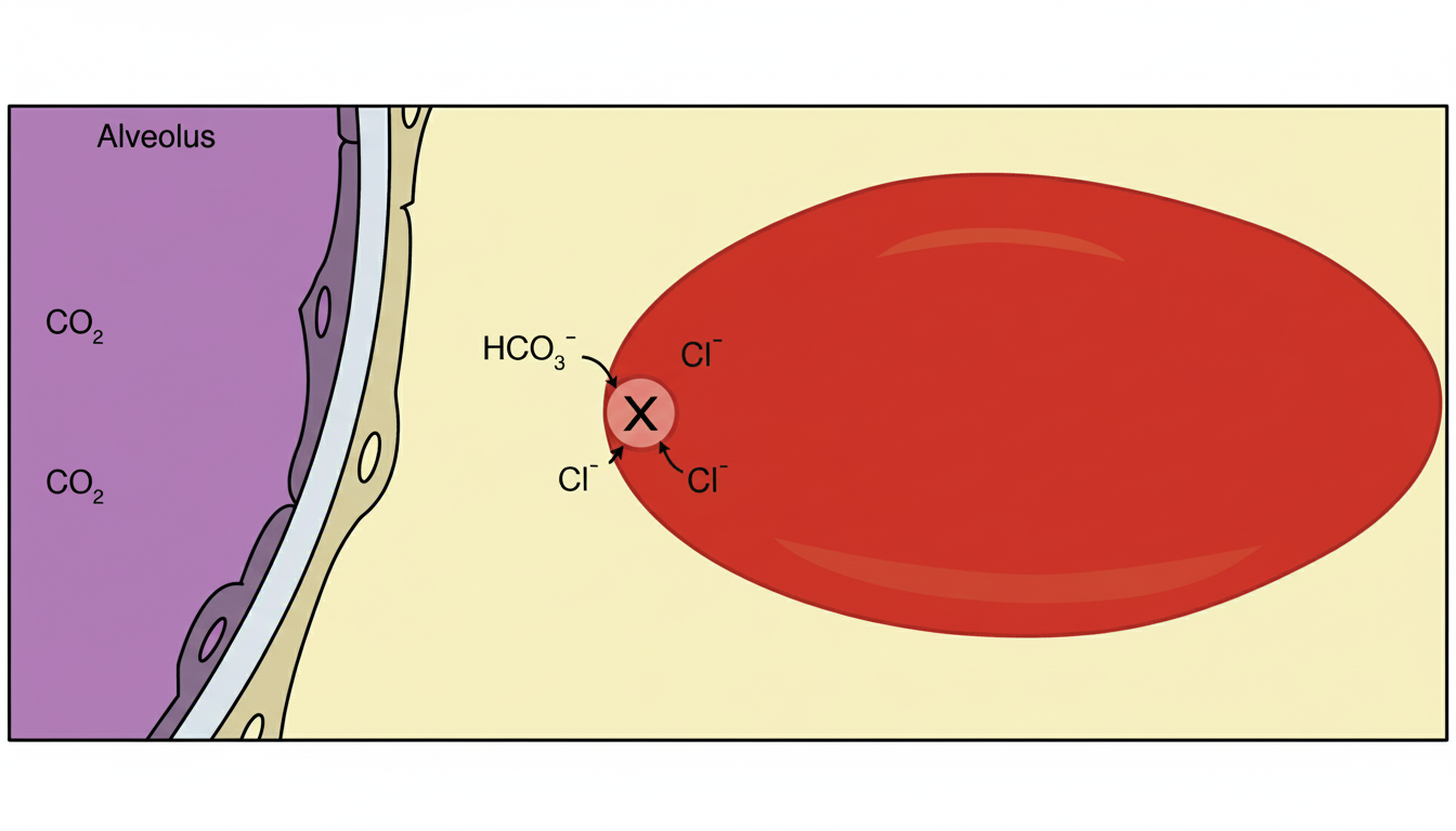
Which protein is responsible for the effect shown in RBC marked as $X$ ?

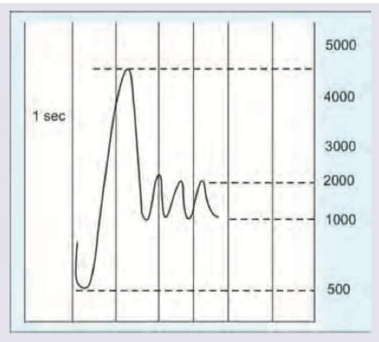
Calculate the FEV1/FVC ratio from the spirometry reading shown below.

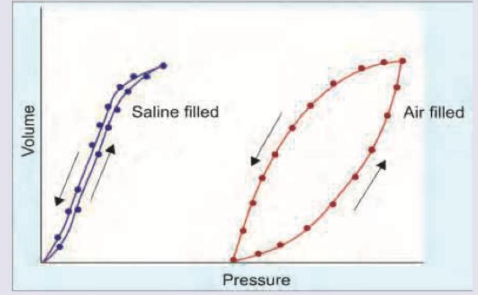
Hysteresis is observed between the deflation and inflation curves in an isolated lung compliance diagram. What is the best description for the same?

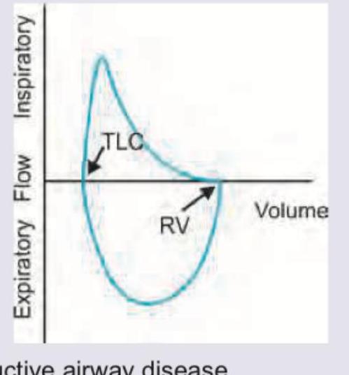
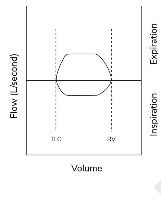
Which of the following pattern is reflected in the flow-volume curve?

Consider the following statements regarding respiratory function in old age: I. There is increasing ventilation-perfusion mismatch II. There is increased ventilatory response to hypoxia and hypercapnia III. There is a decline in maximum oxygen uptake leading to reduction in cardiorespiratory reserve IV. There is decline in the Forced Expiratory Volume to Forced Vital Capacity ratio (FEV1/FVC) by around 0.2% per year after the forties Which of the statements given above are correct?
Which of the following are the functions of larynx ? 1. Fixation of the chest 2. Aids in swallowing of food 3. Phonation 4. Respiration Select the correct answer using the code given below :
O2 (Oxygen) dissociation curve is shifted to right in the following except
What does zero pressure indicate in the pressure-volume curve?
Identify the pathology from the given flow-volume loop:

Which among the following has the highest airway resistance?
Explanation: ***Band 3 protein*** - The image indicates that **X** facilitates the exchange of **bicarbonate (HCO₃⁻)** out of the red blood cell and **chloride (Cl⁻)** into the red blood cell, a process known as the **chloride shift** or **Hamburger phenomenon**. - This specific transporter is the **Band 3 anion exchanger 1 (AE1)**, also known as **Band 3 protein**, which is crucial for maintaining electrical neutrality during CO₂ transport. - **Band 3 protein** is the most abundant membrane protein in RBCs and accounts for approximately **25% of RBC membrane protein**. *Spectrin* - Spectrin is a **cytoskeletal protein** that forms a structural network on the inner surface of the RBC membrane. - It provides **mechanical stability and flexibility** to RBCs but does not participate in anion exchange. - Defects in spectrin cause hereditary spherocytosis and elliptocytosis. *Glycophorin* - Glycophorins are **surface glycoproteins** that carry many of the carbohydrate groups on the RBC surface. - They play roles in **membrane stability and blood group antigens** (MN blood group system). - They do not function as anion exchangers or transporters for bicarbonate/chloride. *Ankyrin* - Ankyrin is an **anchoring protein** that links the cytoskeletal protein spectrin to Band 3 protein. - It provides **structural support** and maintains membrane integrity but does not transport ions. - Ankyrin deficiency can cause hereditary spherocytosis.
Explanation: ***80-89 %*** - **Normal FEV1/FVC ratio is >70% in adults, with healthy individuals typically showing 80-90%.** - From the spirometry graph, the total vital capacity (FVC) after full exhalation is approximately **4500 mL**. The volume exhaled in the first second (FEV1) is approximately **4000 mL**. - Therefore, FEV1/FVC = (4000 mL / 4500 mL) × 100% = **88.8%**. This falls within the 80-89% range, indicating **normal lung function**. *60-69 %* - This percentage indicates **severe airflow obstruction**, where the FEV1 is significantly reduced relative to the FVC, which is not supported by the graph's values of **FEV1 ~4000 mL** and **FVC ~4500 mL**. - A ratio of 60-69% is seen in **moderate to severe obstructive lung disease** (COPD, severe asthma). *70-79 %* - This range suggests **mild airflow obstruction**, corresponding to an **FEV1/FVC ratio** that is borderline or slightly reduced (below the normal 80% threshold but above the diagnostic cutoff for obstruction at 70%). - While less severe than 60-69%, it still implies some degree of airway limitation, which is not the case with the calculated ratio of 88.8%. *90-99 %* - This percentage implies an **FEV1/FVC ratio** of 0.9 or higher, meaning that nearly all of the vital capacity is exhaled in the first second. While **88.8%** is close to this range, it does not fall within it. - A ratio this high might be seen in individuals with **excellent lung function** or paradoxically in some cases of **restrictive lung disease** where both FEV1 and FVC are proportionally reduced, but the exact calculated value from the graph is 88.8%, which falls just below 90%.
Explanation: ***Variation in surface tension forces at air-liquid interface*** - The phenomenon of **hysteresis** in lung compliance, particularly the larger loop seen with air-filled lungs compared to saline-filled lungs, is primarily attributable to the **dynamic changes in surface tension** at the air-liquid interface within the alveoli. - During inflation, more energy is required to overcome the opening forces of collapsed alveoli and recruit new ones, leading to a lower volume for a given pressure, while during deflation, previously opened alveoli remain open or close at lower pressures, contributing to the observed difference. *Stretching of elastic elements of lung parenchyma* - While the **elastic elements** of the lung parenchyma contribute to lung compliance, their contribution to hysteresis is relatively minor and would be observed even in saline-filled lungs to a lesser extent. - The difference in hysteresis between air-filled and saline-filled lungs strongly suggests that factors beyond the tissue elasticity are predominantly responsible for the larger hysteresis with air. *Decrease in surface tension in air-water interface at higher lung volumes* - This statement is partially correct regarding surfactant's action. **Surfactant** does reduce surface tension, especially at lower lung volumes, and prevents alveolar collapse. - However, the overall *variation* in surface tension forces throughout the breathing cycle, not just a decrease at higher volumes, is what creates the inspiratory and expiratory limbs of the pressure-volume curve. *Hering Breuer reflex is operational at higher lung volumes* - The **Hering-Breuer reflex** is a protective neurological reflex that terminates inspiration and initiates expiration when the lungs are overinflated. - This reflex is a **neurophysiological control mechanism** for breathing and does not directly explain the physical properties of the lung that contribute to the pressure-volume hysteresis loop.
Explanation: ***Restrictive airway disease*** - The flow-volume loop for **restrictive lung disease** shows decreased volumes (both **TLC** and **RV** are reduced) while maintaining a relatively normal expiratory flow rate. - The loop appears 'narrower' along the volume axis compared to a normal loop, indicating a **reduction in lung capacity**. *Obstructive airway disease* - An **obstructive pattern** would exhibit a "scooped out" appearance on the expiratory limb of the curve, with reduced peak expiratory flow and **increased residual volume (RV)**. - The **total lung capacity (TLC)** might be normal or increased. *Mixed pattern airway disease* - A **mixed pattern** would show features of both obstruction and restriction, characterized by a decrease in absolute lung volumes (like restriction) and a reduction in expiratory flow rates (like obstruction). - This typically results in a small, narrowed loop with a **concave expiratory limb**. *Central hypoventilation syndrome* - **Central hypoventilation syndrome** primarily affects the **respiratory drive**, leading to underventilation, especially during sleep, and does not produce a characteristic shape on a flow-volume loop. - It would be diagnosed based on **blood gas abnormalities** (e.g., hypercapnia) and polysomnography, not specific flow-volume loop patterns.
Explanation: ***I, III and IV*** - With aging, there is a **loss of elastic recoil** in the lungs and a structural decrease in **alveolar surface area**, leading to increased **ventilation-perfusion (V/Q) mismatch** as gravity-dependent areas collapse. - The **maximum oxygen uptake (VO2 max)** declines with age due to reduced cardiac output and skeletal muscle mass, thus decreasing **cardiorespiratory reserve**. The **FEV1/FVC ratio** also decreases by approximately **0.2% per year** after age 40 because of reduced elastic recoil and increased airway collapsibility. *I, II and IV* - While statement I and IV are correct, statement II is incorrect because the **ventilatory response to hypoxia and hypercapnia** actually **decreases** with age. - Older adults have a blunted response to changes in oxygen and carbon dioxide levels, making them more susceptible to respiratory compromise. *II, III and IV* - Statement II is incorrect as the **ventilatory response to hypoxia and hypercapnia decreases** with age, not increases. - Statements III and IV accurately describe the decline in **maximum oxygen uptake** and the **FEV1/FVC ratio** with aging. *I, II and III* - Statement II is incorrect; the **ventilatory response to hypoxia and hypercapnia is diminished** in older adults. - Statements I and III correctly identify increased **ventilation-perfusion mismatch** and decreased **maximum oxygen uptake** as age-related changes in respiratory function.
Explanation: ***All of the above (1, 2, 3 and 4)*** - The larynx performs **all four functions** listed in the question. - **Respiration**: The larynx serves as a vital conduit for airflow. The posterior cricoarytenoid muscles actively abduct the vocal cords during inspiration, and the larynx regulates airflow through glottic opening and closure. - **Phonation**: The vocal cords housed within the larynx vibrate to produce sound, making this the primary organ of voice production. - **Aids in swallowing**: During deglutition, the larynx elevates and the epiglottis closes the laryngeal inlet to prevent aspiration of food into the trachea. - **Chest fixation**: The larynx closes the glottis during the Valsalva maneuver, creating a closed air column that stabilizes the chest for activities like lifting, coughing, defecation, and parturition. *1, 2 and 3* - This option incorrectly excludes **respiration**, which is a fundamental function of the larynx as part of the conducting airways. - The larynx is not merely a passive tube but actively regulates airflow through intrinsic muscle activity. *2, 3 and 4* - While these are all valid laryngeal functions, this option incorrectly excludes **chest fixation**, which is accomplished through glottic closure during the Valsalva maneuver. *1, 3 and 4* - This option incorrectly excludes the larynx's role in **aiding swallowing** through laryngeal elevation and airway protection during deglutition.
Explanation: ***Metabolic alkalosis*** - A shift to the **right** on the oxygen dissociation curve indicates **decreased affinity** for oxygen, promoting oxygen release to tissues. - In **metabolic alkalosis**, the blood pH is elevated, which **increases hemoglobin's affinity for oxygen**, leading to a **left shift** in the curve. *Hypercapnia* - **Hypercapnia** (increased PCO2) decreases blood pH, reducing hemoglobin's affinity for oxygen via the **Bohr effect**, resulting in a **right shift**. - This facilitates oxygen release to tissues where CO2 production is high. *Rise in temperature* - An increase in **body temperature** weakens the binding of oxygen to hemoglobin, causing a **right shift** in the oxygen dissociation curve. - This is beneficial during exercise, when active tissues generate heat and require more oxygen. *Raised 2, 3 DPG level* - **2,3-bisphosphoglycerate (2,3-BPG)** binds to deoxygenated hemoglobin, stabilizing its T-state and **reducing its affinity for oxygen**, causing a **right shift**. - This is a key adaptation to chronic hypoxia, enhancing oxygen delivery to tissues.
Explanation: ***Functional residual capacity*** - This is the lung volume at which the **elastic recoil of the lungs** exactly balances the **elastic recoil of the chest wall**, resulting in zero net pressure across the respiratory system. - At **functional residual capacity (FRC)**, there is no airflow, and the **alveolar pressure equals atmospheric pressure (zero)**, indicating the equilibrium point. - Note: The **transpulmonary pressure remains positive** at FRC (approximately +5 cm H₂O), which keeps the lungs inflated against their elastic recoil. *Inspiratory reserve volume* - This is the **extra volume of air** that can be forcibly inhaled after a normal inspiration. - It involves active inspiration and therefore is associated with a **negative intrathoracic pressure**, not zero pressure. *Tidal volume* - This is the **volume of air inhaled and exhaled** during a normal quiet breathing cycle. - While breathing, pressures fluctuate, and the respiratory system is not at an equilibrium point of **zero pressure** throughout the tidal breath. *Residual volume* - This is the **volume of air remaining in the lungs** after a maximal exhalation. - The chest wall's outward recoil is greater than the lung's inward recoil at this point, resulting in a **negative intrapleural pressure** to keep the lungs from collapsing.
Explanation: ***Fixed central airway obstruction*** - This flow-volume loop shows **flattening of both the inspiratory and expiratory limbs**, creating a characteristic "box" or "square" shape. - This pattern indicates that airflow is limited equally during both inspiration and expiration, regardless of lung volume changes, which is characteristic of a **fixed central airway obstruction**. - Examples include **tracheal stenosis, tracheal tumors, or fixed goiters** compressing the trachea. *Variable extrathoracic obstruction* - Characterized by flattening of the **inspiratory limb only**, as negative intrathoracic pressure during inspiration exacerbates the obstruction. - The expiratory limb typically remains normal as positive intrathoracic pressure tends to open the airway. - Examples include **vocal cord paralysis or extrathoracic tracheal tumors**. *Variable intrathoracic obstruction* - Characterized by flattening of the **expiratory limb only**, as positive intrathoracic pressure during forced expiration collapses the airway. - The inspiratory limb usually remains normal as negative pressure helps maintain airway patency. - Examples include **intrathoracic tracheal tumors or tracheomalacia**. *Fixed distal airway obstruction* - Fixed obstructions producing the characteristic "box" pattern are typically **central (proximal) lesions in large airways**, not distal. - Distal airway obstructions (like **COPD or asthma**) produce a different flow-volume loop pattern characterized by **decreased peak expiratory flow** and "scooping" or "concave" appearance of the expiratory limb, not the flat bilateral pattern seen here.
Explanation: ***Bronchi (Medium-sized bronchi)*** - The **medium-sized bronchi** (approximately 4th-8th generation airways) contribute the **highest proportion to total airway resistance** in the tracheobronchial tree. - At this level, airways are still relatively **narrow** but arranged more in **series** rather than parallel, concentrating resistance. - This is the point of **maximum resistance** before the extensive branching of smaller airways creates parallel pathways. - Accounts for approximately **40-50% of total airway resistance** during normal breathing. *Small bronchioles* - While individual small bronchioles (<1 mm diameter) have narrow lumens, they branch extensively into **thousands of parallel airways**. - This creates an **enormous total cross-sectional area** (up to 20x larger than trachea), which dramatically **reduces total resistance**. - According to principles of parallel resistance, total resistance decreases as more parallel pathways are added: 1/R_total = 1/R₁ + 1/R₂ + ... + 1/Rₙ - Despite small individual diameter, collective parallel arrangement makes them **low resistance** pathways. *Alveolar ducts* - Have the **largest cumulative cross-sectional area** in the entire respiratory system. - Airflow velocity is minimal and flow is entirely **laminar**, offering negligible resistance. - These are part of the respiratory zone where gas exchange occurs primarily by diffusion. *Respiratory bronchioles* - Part of the **transitional/respiratory zone** with extensive branching and large total cross-sectional area. - Offer very low resistance due to their **parallel arrangement** and slow airflow velocity. - Contribute minimally to total airway resistance.
Mechanics of Breathing
Practice Questions
Pulmonary Ventilation
Practice Questions
Pulmonary Circulation
Practice Questions
Gas Exchange in the Lungs
Practice Questions
Oxygen and Carbon Dioxide Transport
Practice Questions
Control of Breathing
Practice Questions
Respiratory Adjustments in Health and Disease
Practice Questions
High Altitude Physiology
Practice Questions
Diving Physiology
Practice Questions
Respiratory Function Tests
Practice Questions
Get full access to all questions, explanations, and performance tracking.
Start For Free