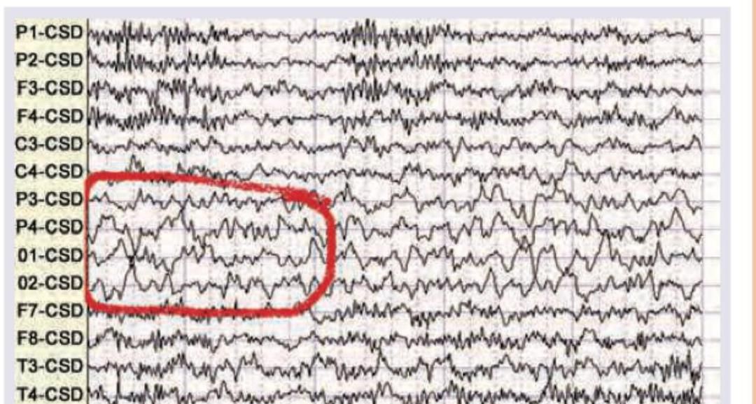
Which wave is seen in the given EEG recording?

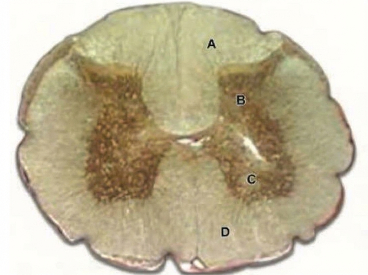
In the image shown below, which of the marked area is involved in relieving pain in response to massage?

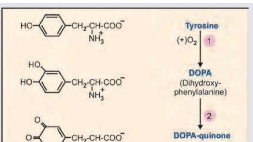
Name the product marked as X in the image shown below:

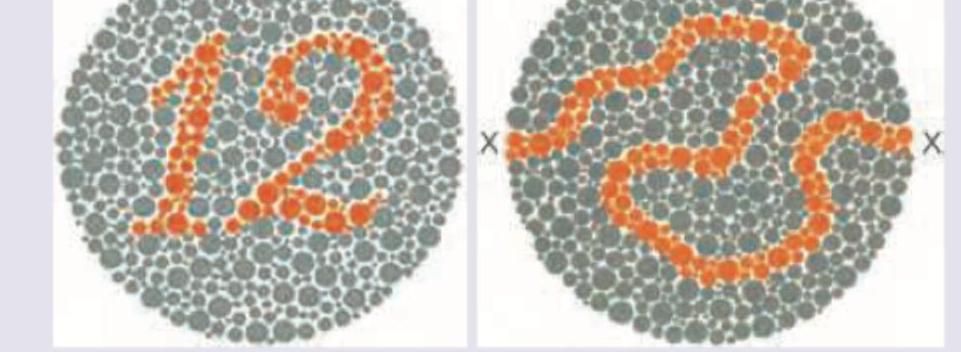
Which of the following cells in the brain are responsible for handling information regarding ability to read the slide below? (Recent NEET Pattern 2016-17)

A patient is experiencing phantom limb pain after the amputation of the right limb. What is observed on a PET scan in a patient with phantom limb pain?
What are the effects of a lesion in Brodmann area 22?
A woman with right-sided loss of sensations of both the upper and lower limb complains of shooting pain from her fingers to the right shoulder and a burning sensation when touching cold water. Motor functions are normal. Which of the following structures is likely to be involved?
What is the primary mechanism for maintaining constant cerebral blood flow despite changes in systemic blood pressure?
Which change in CSF production most directly affects intracranial pressure?
Which receptor type mediates the slow phase of synaptic transmission in autonomic ganglia?
Explanation: ***Delta waves*** - The highlighted EEG activity shows **large amplitude, low-frequency waves** (typically 0.5-4 Hz), which are characteristic of delta waves. - Delta waves are normally associated with **deep sleep** (NREM stage 3) or **pathological conditions** in awake adults, indicating significant brain dysfunction. *Alpha waves* - Alpha waves have a frequency range of **8-13 Hz** and typically appear when an individual is in a relaxed, awake state with eyes closed. - The waves in the image are much slower and higher in amplitude than typical alpha waves. *Beta waves* - Beta waves are characterized by a higher frequency range of **14-30 Hz** and are associated with active thinking, alertness, and concentration. - The observed activity is significantly slower and higher in amplitude than beta waves. *Epsilon wave* - The term "epsilon wave" is not a standard classification for EEG brain waves in the context of normal or common pathological activity, unlike alpha, beta, theta, and delta waves. - In cardiology, "epsilon wave" refers to a specific finding on an ECG in **Arrhythmogenic Right Ventricular Cardiomyopathy (ARVC)**, not an EEG.
Explanation: ***A*** - Area A points to the **dorsal columns** (specifically the fasciculus gracilis and cuneatus) of the spinal cord, which carry **mechanoreceptive** and proprioceptive information. - Massage activates large diameter afferent fibers which transmit signals via the dorsal columns, inhibiting pain transmission through the **gate control theory of pain**. *B* - Area B points to the **dorsal horn** (specifically lamina I, II, and V), which is primarily involved in receiving and processing **nociceptive input**. - While integration of pain occurs here, it is not the primary pathway for the **modulatory effects of touch/pressure** from massage for pain relief. *C* - Area C indicates the **ventral horn** of the spinal cord, which contains **motor neurons** that control skeletal muscle activity. - It is involved in motor output, not directly in the sensory processing or modulation of pain signals from massage. *D* - Area D points to the **lateral white matter**, which contains various ascending and descending tracts, including the **spinothalamic tract** (primary pain pathway) and corticospinal tract. - While the spinothalamic tract carries pain signals, the direct pain-relieving effect of massage primarily involves activation of mechanoreceptors whose signals ascend via the dorsal columns.
Explanation: ***Indolequinone*** - The image depicts the **melanin biosynthesis pathway** starting from **tyrosine**. - Tyrosine is converted to **L-DOPA** by tyrosinase, which is then oxidized to **dopaquinone** (also called DOPA-quinone). - Dopaquinone undergoes intramolecular cyclization to form **leucodopachrome**, which is subsequently oxidized to **dopachrome**. - Dopachrome is then converted through a series of steps to **5,6-dihydroxyindole**, which is finally oxidized to **indole-5,6-quinone** (indolequinone). - **Indolequinone** is a key intermediate that polymerizes to form **melanin**, the pigment responsible for coloration in skin, hair, and eyes. - Based on the pathway shown, X represents indolequinone, an oxidized indole derivative in melanin synthesis. *Tetrabenazine* - **Tetrabenazine** is a pharmaceutical drug that inhibits **vesicular monoamine transporter 2 (VMAT2)**. - It is used therapeutically to treat hyperkinetic movement disorders such as **Huntington's disease** and tardive dyskinesia. - It is not a natural metabolic intermediate in the tyrosine-to-melanin biosynthetic pathway. *Homovanillic acid* - **Homovanillic acid (HVA)** is a major end metabolite of **dopamine** in the catecholamine degradation pathway. - It is formed by the sequential actions of **monoamine oxidase (MAO)** and **catechol-O-methyltransferase (COMT)** on dopamine. - This represents a completely different branch of tyrosine metabolism (catecholamine pathway), not the melanin synthesis pathway. *Kynurenine* - **Kynurenine** is an intermediate metabolite in the **kynurenine pathway**, which is the major route of **tryptophan** degradation. - The kynurenine pathway leads to the formation of NAD+ and various neuroactive metabolites. - This pathway is completely separate from tyrosine metabolism and is unrelated to the melanin synthesis pathway shown in the image.
Explanation: ***Parvocellular cells*** - **Parvocellular cells** (P-cells) are responsible for processing detailed visual information, including **color**, **form**, and fine **texture**. Reading the Ishihara test requires the ability to distinguish specific colors and fine patterns. - They have **small receptive fields** and transmit information about high spatial resolution and chromatic detail, crucial for tasks such as reading and recognizing fine visual cues. *Magnocellular cells* - **Magnocellular cells** (M-cells) are primarily involved in detecting **motion** and processing **low-spatial frequency information**, such as global shape and location. - They have **large receptive fields** and are not primarily responsible for detailed color or pattern discrimination needed for reading. *Purkinje cells* - **Purkinje cells** are a type of large, extensively branched neuron located in the **cerebellar cortex**. - Their primary function is motor coordination, balance, and motor learning, not visual processing or reading. *Pyramidal cells* - **Pyramidal cells** are excitatory neurons found in the cerebral cortex and hippocampus, characterized by their pyramidal-shaped cell bodies. - While they are involved in complex cognitive functions, including parts of visual perception, they are not the specific cells in the primary visual pathway responsible for initial processing of fine details and color as required for reading this type of visual test.
Explanation: ***Neighboring cortical areas extending into the hand representation area*** - Phantom limb pain is associated with **cortical remapping** in the somatosensory cortex, where neighboring body part representations (face, upper arm) **invade the deafferented cortex zone** previously occupied by the amputated limb. - This reorganization is observed on PET scans as **increased metabolic activity in areas adjacent to the hand representation**, extending into the hand area that lost its sensory input. - This cortical reorganization correlates with the **intensity of phantom limb pain** and is a well-established finding in neuroimaging studies. *General expansion of left somatosensory cortex* - While the left somatosensory cortex (contralateral to the right amputated limb) does undergo changes, the key finding is **not a general expansion** of the entire cortex. - The characteristic observation is **specific reorganization** where neighboring representations invade the deafferented zone, rather than a diffuse enlargement. *Expansion of right somatosensory cortex* - Since the **right limb was amputated**, the **left somatosensory cortex** (which processes right-sided body sensations) is where reorganization occurs. - The right somatosensory cortex processes the left (intact) side and would not show the characteristic remapping associated with phantom limb pain. *Expansion of hand representation in the left somatosensory cortex into neighboring areas* - This describes the **opposite direction** of cortical remapping. - In phantom limb pain, **neighboring areas (face, upper arm) expand INTO the hand area**, not the hand area expanding outward. - The hand representation has lost its peripheral input due to amputation and is invaded by adjacent cortical representations.
Explanation: ***Receptive aphasia*** - A lesion in **Brodmann area 22**, specifically in **Wernicke's area**, leads to **receptive aphasia** (Wernicke's aphasia). - This condition is characterized by **impaired comprehension** of spoken and written language, **fluent but paraphasic speech**, and **poor repetition**. - This is the most comprehensive answer as it describes the entire clinical syndrome. *Expressive aphasia* - **Brodmann areas 44 and 45** (Broca's area) in the frontal lobe are associated with expressive aphasia (Broca's aphasia). - Patients have good comprehension but struggle to produce fluent speech, with effortful, telegraphic output. *Poor repetition of language* - While poor repetition is indeed a feature of Wernicke's aphasia, this option describes only one component of the syndrome rather than the complete clinical picture. - **Conduction aphasia** (from arcuate fasciculus lesions) is characterized by poor repetition with **relatively preserved** comprehension and fluent speech, distinguishing it from Wernicke's aphasia. - "Receptive aphasia" is the more complete answer. *Poor naming* - Difficulty with naming, or **anomia**, is a common feature across various types of aphasia, including both receptive and expressive aphasia. - It reflects disruption in language networks involving the **temporal and parietal lobes** but is not specific to Brodmann area 22 lesions.
Explanation: ***Lateral spinothalamic tract*** - The symptoms described, such as **loss of sensations**, **shooting pain** (neuropathic pain), and **burning sensation** with cold water (dysesthesia/allodynia), are characteristic of damage to the **spinothalamic tract**, which carries **pain and temperature** sensations. - Involvement of the **right-sided upper and lower limb** indicates a lesion affecting sensory pathways on the ipsilateral side of the body before decussation, or more commonly a contralateral lesion above the level of decussation for the specific tract. Given the symptoms affecting pain and temperature, the lateral spinothalamic tract is the primary candidate. *Anterior spinothalamic tract* - This tract primarily transmits **crude touch** and **pressure** sensations. - While loss of sensation is present, the prominent **shooting pain** and **burning sensation with cold water** are not typically associated with isolated anterior spinothalamic tract lesions. *Spinocerebellar tract* - This tract is responsible for transmitting **proprioceptive information** to the cerebellum for motor coordination. - Damage to the spinocerebellar tracts would manifest as **ataxia** and **incoordination**, not pain or loss of touch/temperature sensation, and motor functions are stated as normal in the patient. *Posterior column* - The posterior column (dorsal column-medial lemniscus pathway) transmits **fine touch**, **vibration**, and **proprioception**. - While a loss of sensation is present, the specific complaints of **shooting pain** and **burning sensation to cold water** are not characteristic of posterior column damage, which would typically present with deficits in discriminative touch, vibratory sense, and position sense.
Explanation: ***Myogenic autoregulation*** - **Myogenic autoregulation** is the intrinsic ability of vascular smooth muscle to contract when stretched by increased blood pressure, thereby maintaining a constant cerebral blood flow. - This mechanism operates within a specific range of mean arterial pressures (typically **60-150 mmHg**) to prevent both hypoperfusion and hyperperfusion of the brain. *Endothelial factors* - Endothelial cells release various vasoactive substances like **nitric oxide** and **endothelin**, which influence vascular tone. - While important for local blood flow regulation, these factors play a secondary role to myogenic autoregulation in maintaining constant cerebral blood flow against systemic pressure changes. *Baroreceptor reflex* - The **baroreceptor reflex** primarily controls systemic blood pressure by regulating heart rate and peripheral vascular resistance. - It does not directly regulate cerebral blood flow stability in response to systemic pressure changes; its main role is to stabilize the overall systemic arterial pressure. *Metabolic control* - **Metabolic control** regulates cerebral blood flow in response to the brain's metabolic demands, primarily by sensing local concentrations of **CO2**, **pH**, and **oxygen**. - While essential for matching blood supply to neuronal activity, it is not the primary mechanism for maintaining cerebral blood flow despite changes in systemic blood pressure.
Explanation: ***Decreased carbonic anhydrase activity*** - The **choroid plexus** produces CSF primarily through an active secretion process involving carbonic anhydrase. - Decreased activity of this enzyme directly reduces the formation of **bicarbonate ions** and **protons (H+)**, which are crucial for the active transport of Na+ and Cl- into the CSF, thereby lowering CSF production and subsequently **intracranial pressure**. *Decreased arachnoid granulation function* - This change would lead to a **decreased reabsorption** of CSF, which would *increase* intracranial pressure, not directly affect production to lower it. - Arachnoid granulations are responsible for the **resorption of CSF** into the venous system. *Increased choroid plexus blood flow* - While increased blood flow could potentially increase the delivery of substrates for CSF production, it is **not the most direct or primary determinant** of CSF production rate. - CSF production is predominantly an **active secretory process**, not a passive filtration process dependent solely on blood flow. *Increased osmotic gradient* - An increased osmotic gradient, if referring to a higher osmolality in the CSF compared to plasma, would tend to **draw water into the CSF**, potentially *increasing* CSF volume and intracranial pressure. - If referring to a gradient drawing water *out* of the CSF, it would *decrease* intracranial pressure but is not a primary mechanism of CSF production regulation.
Explanation: ***Muscarinic (M1)*** - **M1 receptors** are **Gq-protein coupled receptors** that activate phospholipase C, leading to increased intracellular calcium and diacylglycerol, which mediates the slow excitatory postsynaptic potential in autonomic ganglia. - This activation results in a **slow depolarization** that prolongs the excitability of ganglionic neurons after the initial fast synaptic transmission. *Muscarinic (M3)* - **M3 receptors** are primarily found on **smooth muscle**, glands, and endothelium, mediating contraction, secretion, and vasodilation, respectively. - While also **Gq-protein coupled**, their role in autonomic ganglia is not the main mediator of the slow phase of synaptic transmission. *Muscarinic (M2)* - **M2 receptors** are **Gi-protein coupled receptors** mainly found in the heart, mediating decreased heart rate and contractility. - In autonomic ganglia, M2 receptors could have a modulatory role, but they are not responsible for the slow excitatory phase of synaptic transmission. *Nicotinic (N2)* - **Nicotinic N2 receptors** (also known as **NN or neuronal nicotinic receptors**) mediate the **fast excitatory postsynaptic potential** (EPSP) in autonomic ganglia by opening ion channels. - This leads to rapid depolarization and action potential generation, which is distinct from the **slower, prolonged phase** of transmission.
Neurons and Glial Cells
Practice Questions
Synaptic Transmission
Practice Questions
Sensory Processing
Practice Questions
Motor Control Systems
Practice Questions
Autonomic Nervous System
Practice Questions
Hypothalamus and Limbic System
Practice Questions
Cerebral Cortex Functions
Practice Questions
Electroencephalography
Practice Questions
Neuroplasticity
Practice Questions
Sleep and Wakefulness
Practice Questions
Get full access to all questions, explanations, and performance tracking.
Start For Free