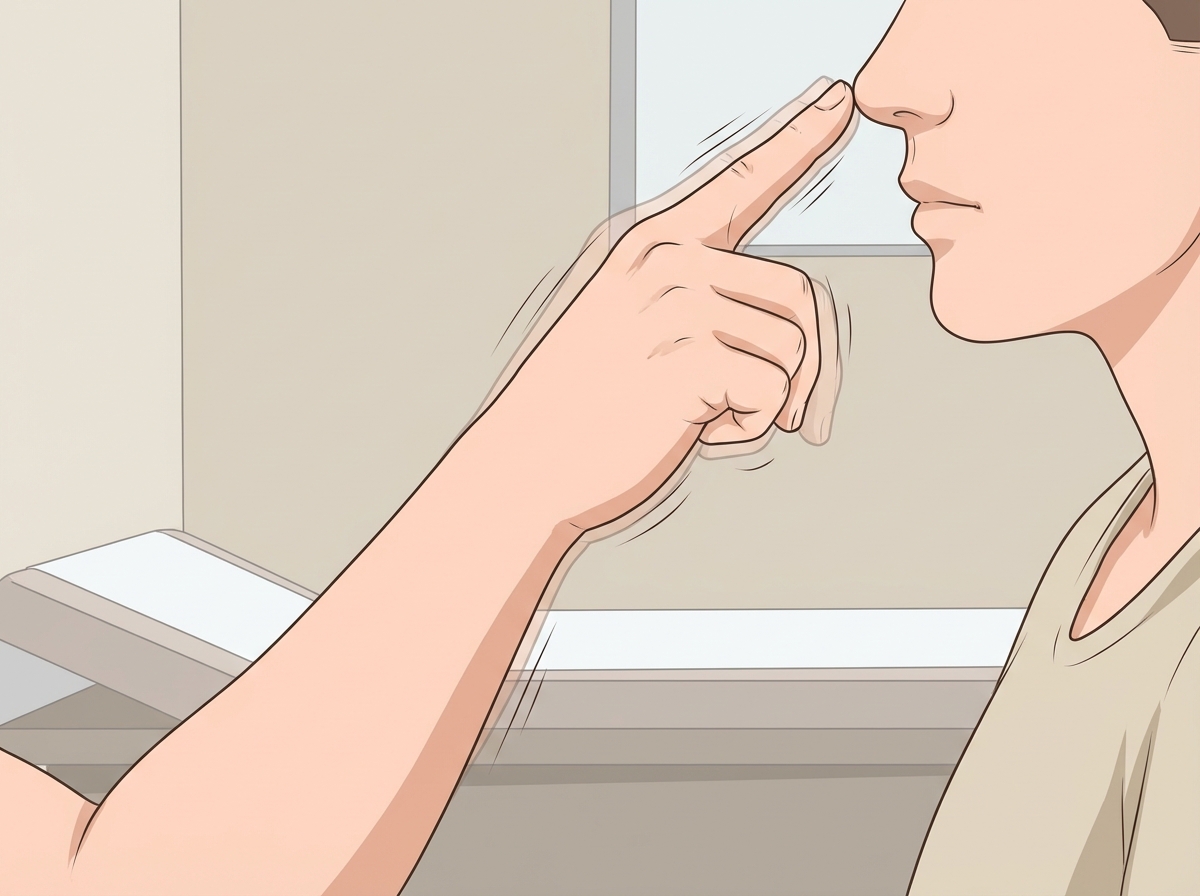
Based on the clinical sign demonstrated in the image, identify the most likely site of damage:

"Reward pathway" is associated with which structure?
Which of the following is an excitatory neurotransmitter?
What are postganglionic sympathetic fibres?
In decerebrate animals, which reflex is lost?
Which EEG rhythm is recorded from the surface of the scalp during REM sleep?
What does long-term potentiation refer to?
The rubrospinal tract primarily influences which of the following?
In peripheral tissues, which of the following contains substance P?
All except one are true about the spinothalamic tract?
Explanation: ***Neocerebellum*** - **Intention tremor** and **dysmetria** during finger-nose test are pathognomonic signs of **neocerebellum (lateral cerebellar hemisphere)** damage. - The neocerebellum controls **coordinated voluntary movements** and **motor planning**, causing tremor that worsens as the finger approaches the target. *Substantia Nigra* - Damage causes **resting tremor** that decreases with voluntary movement, opposite to the intention tremor shown. - Associated with **bradykinesia** and **rigidity** characteristic of **Parkinson's disease**, not cerebellar signs. *Archicerebellum* - Primarily affects **balance** and **posture**, causing **truncal ataxia** and **gait disturbances**. - Would not typically present with **intention tremor** during precise finger movements but rather broad-based gait abnormalities. *Ventromedial nucleus thalamus* - Damage causes **sensory deficits** and **thalamic pain syndrome**, not motor coordination problems. - May cause **thalamic tremor** but this is typically **mixed type** (resting + postural), not pure intention tremor during goal-directed movements.
Explanation: **Explanation:** The **Reward Pathway** (Mesolimbic pathway) is the primary dopaminergic circuit in the brain responsible for incentive salience, reinforcement, and pleasure. **1. Why Nucleus Accumbens is correct:** The Nucleus Accumbens (NAc), located in the ventral striatum, is the central "hub" of the reward system. It receives dopaminergic projections from the **Ventral Tegmental Area (VTA)**. When a rewarding stimulus is encountered, dopamine levels spike in the NAc, reinforcing the behavior. This pathway is heavily implicated in addiction and the "high" associated with drugs of abuse. **2. Why other options are incorrect:** * **Nucleus Ambiguous:** This is a motor nucleus in the medulla that gives rise to the efferent fibers of the Vagus (CN X) and Glossopharyngeal (CN IX) nerves, controlling muscles of the pharynx and larynx (swallowing and phonation). * **Dentate Nucleus:** This is the largest of the deep cerebellar nuclei. It is involved in the planning, initiation, and control of voluntary movements, not emotional reward. * **Substantia Nigra:** While it is a dopaminergic center, its **Pars Compacta** primarily projects to the dorsal striatum (Nigrostriatal pathway) to regulate motor control. Degeneration here leads to Parkinson’s disease. **High-Yield Clinical Pearls for NEET-PG:** * **The Mesolimbic Pathway:** VTA → Nucleus Accumbens (Reward/Addiction). * **The Mesocortical Pathway:** VTA → Prefrontal Cortex (Cognition/Executive function; dysfunction leads to negative symptoms of Schizophrenia). * **Neurotransmitter:** **Dopamine** is the key neurotransmitter of the reward system. * **Addiction:** Almost all addictive drugs (cocaine, amphetamines, nicotine) act by increasing dopamine release in the Nucleus Accumbens.
Explanation: ### Explanation Neurotransmitters are chemical messengers categorized as either excitatory (promoting action potentials) or inhibitory (hyperpolarizing the post-synaptic membrane). **1. Why Glutamate (Glutamine) is Correct:** Glutamate is the **primary excitatory neurotransmitter** in the central nervous system (CNS). It acts on ionotropic receptors (NMDA, AMPA, and Kainate) to increase sodium and calcium conductance, leading to depolarization. *Note on terminology:* In many competitive exams, "Glutamine" is occasionally used interchangeably with "Glutamate" in options, though physiologically, Glutamate is the active neurotransmitter and Glutamine is its inactive precursor/metabolite in the glutamate-glutamine cycle. **2. Why the Other Options are Incorrect:** * **GABA (Gamma-Aminobutyric Acid):** This is the **major inhibitory neurotransmitter** in the brain. It acts on $GABA_A$ (ligand-gated $Cl^-$ channels) and $GABA_B$ (G-protein coupled) receptors to cause hyperpolarization. * **Glycine:** This is the **major inhibitory neurotransmitter** in the spinal cord and brainstem. It increases chloride conductance. (Interestingly, it acts as a co-agonist for NMDA receptors, but its primary classification is inhibitory). * **Acetylcholine:** While acetylcholine is excitatory at the neuromuscular junction and in autonomic ganglia (Nicotinic receptors), it can be inhibitory in other areas (like the heart via $M_2$ Muscarinic receptors). In the context of CNS neurotransmitter classification, Glutamate is the definitive excitatory answer. **3. High-Yield Clinical Pearls for NEET-PG:** * **Most common inhibitory NT in Brain:** GABA. * **Most common inhibitory NT in Spinal Cord:** Glycine. * **Excitotoxicity:** Excessive glutamate release is linked to neuronal death in stroke and ALS. * **Strychnine Poisoning:** Acts by inhibiting Glycine receptors, leading to unchecked muscle contractions (opisthotonus). * **Benzodiazepines/Barbiturates:** Work by potentiating the action of GABA at the $GABA_A$ receptor.
Explanation: ### Explanation The classification of nerve fibres is a high-yield topic in neurophysiology, primarily based on the **Erlanger-Gasser classification**, which categorizes fibres according to their diameter, myelination, and conduction velocity. **Why C fibres are the correct answer:** Postganglionic sympathetic fibres are unique because they are **unmyelinated**. According to the Erlanger-Gasser classification, **Type C fibres** are the only unmyelinated fibres. They have the smallest diameter (0.4–1.2 μm) and the slowest conduction velocity (0.5–2.0 m/s). These fibres carry postganglionic autonomic signals and slow pain (prolonged, burning sensation). **Analysis of Incorrect Options:** * **Option A (A-alpha):** These are the largest, most heavily myelinated fibres. They function as somatic motor fibres (to extrafusal muscle fibres) and carry proprioception (from muscle spindles and Golgi tendon organs). * **Option C (B fibres):** These are small, lightly myelinated fibres. Crucially, they represent **preganglionic** autonomic fibres (both sympathetic and parasympathetic). * **Option D (A-gamma):** These are myelinated fibres that supply the intrafusal muscle fibres of the muscle spindle, regulating muscle tone. **NEET-PG High-Yield Pearls:** 1. **The "B" vs. "C" Rule:** Always remember: **B** comes before **C** in the alphabet, just as **Preganglionic (B)** comes before **Postganglionic (C)** in the autonomic pathway. 2. **Susceptibility:** * **Local Anesthetics:** Block **C fibres** first (smallest diameter). * **Pressure:** Affects **A fibres** first (largest diameter). * **Hypoxia:** Affects **B fibres** first. 3. **Fast vs. Slow Pain:** Fast pain is carried by **A-delta** fibres, while slow, chronic pain is carried by **C fibres**.
Explanation: **Explanation:** **Concept:** Decerebration involves a transection of the brainstem between the **superior and inferior colliculi** (midbrain level). This procedure disconnects the cerebral cortex and basal ganglia from the lower brainstem and spinal cord. The **hopping and placing reflexes** are **cortical reflexes**; they require an intact cerebral cortex and corticospinal tracts to process sensory input and coordinate motor output. Since decerebration removes cortical influence, these reflexes are permanently lost. **Analysis of Options:** * **A. Hopping and placing reflex (Correct):** These are complex postural reactions that depend on the **cerebral cortex**. In decerebrate animals, the connection to the cortex is severed, leading to the loss of these reflexes. * **B. Tonic neck reflex:** These are integrated at the level of the **medulla**. Since the medulla remains intact in a decerebrate preparation, these reflexes are preserved (and often exaggerated). * **C. Tonic labyrinthine reflex:** These are integrated in the **medulla/pons** (vestibular nuclei). They remain present in decerebrate animals. * **D. Stretch reflex:** This is a **monosynaptic spinal reflex**. In decerebration, the stretch reflex is actually **markedly enhanced** due to the removal of inhibitory cortical input and the facilitation of the lateral reticulospinal and vestibulospinal tracts, leading to "decerebrate rigidity." **High-Yield Facts for NEET-PG:** * **Decerebrate Rigidity:** Characterized by extension of all four limbs (gamma-motor neuron hyperactivity). It occurs due to the facilitation of the **Pontine Reticular Formation** and **Vestibular Nuclei** when inhibitory inputs from the Red Nucleus and Cortex are removed. * **Decorticate Posturing:** Lesion above the Red Nucleus (superior to colliculi). Presents with **flexion of upper limbs** (Red Nucleus intact) and extension of lower limbs. * **Integration Levels:** * Spinal Cord: Stretch reflex. * Medulla: Tonic neck/labyrinthine reflexes. * Midbrain: Righting reflexes. * Cortex: Hopping and placing reflexes.
Explanation: **Explanation:** The correct answer is **Alpha (Option A)**. During **REM (Rapid Eye Movement) sleep**, the brain is highly active, a state often referred to as "paradoxical sleep." The EEG pattern during REM sleep is characterized by low-voltage, high-frequency activity that closely resembles the **Alpha rhythm** (8–13 Hz) seen in an awake, relaxed state with eyes closed. In some classifications, this is also described as "desynchronized" or "sawtooth" waves. **Analysis of Options:** * **Alpha (Correct):** Predominates during REM sleep and relaxed wakefulness. It signifies a state of cortical activation despite the body being in a state of muscle atonia. * **Beta:** These are high-frequency waves (>13 Hz) associated with active thinking, intense mental concentration, or the use of benzodiazepines. While REM is "Beta-like," the classic textbook description for REM EEG is the Alpha rhythm. * **Delta:** These are low-frequency (0.5–4 Hz), high-amplitude waves characteristic of **Stage N3 (Deep/Slow-wave sleep)**. Their presence in REM would be abnormal. * **Theta:** These waves (4–7 Hz) are typically seen in **Stage N1 (Light sleep)** and are the dominant rhythm in the hippocampus, but not the primary scalp recording for REM. **High-Yield Clinical Pearls for NEET-PG:** * **REM Sleep:** Characterized by PGO (Pontine-Geniculate-Occipital) spikes, muscle atonia (except extraocular muscles and diaphragm), and vivid dreaming. * **Sleep Spindles & K-complexes:** These are the hallmark of **Stage N2** sleep. * **Bruxism (Teeth grinding):** Occurs mostly in Stage N2. * **Somnambulism (Sleepwalking) & Night Terrors:** Occur during Stage N3 (Slow-wave sleep).
Explanation: **Explanation:** **Long-Term Potentiation (LTP)** is a persistent strengthening of synapses based on recent patterns of activity. It is the fundamental cellular mechanism underlying **synaptic plasticity**, learning, and memory formation, primarily occurring in the **hippocampus**. 1. **Why Option A is Correct:** LTP involves a long-lasting increase in **synaptic efficacy**. When a high-frequency stimulus (tetanus) is applied to a presynaptic neuron, it leads to an enhanced postsynaptic response. This "enhancement of signal transmission" occurs due to increased neurotransmitter release and increased sensitivity of the postsynaptic membrane, making the synapse more efficient at communicating. 2. **Why Other Options are Incorrect:** * **Option B:** While an increase in the number of **AMPA receptors** on the postsynaptic membrane is a *mechanism* that facilitates LTP, the term LTP itself refers to the functional outcome (the enhancement of the signal), not just the structural change. * **Option C:** LTP involves changes in existing synaptic strength, not the generation of new neurons (neurogenesis). * **Option D:** Muscle tone is a peripheral neuromuscular phenomenon; LTP is a central neurophysiological process related to memory. **High-Yield Facts for NEET-PG:** * **Key Neurotransmitter:** Glutamate. * **Key Receptors:** **NMDA receptors** (act as coincidence detectors) and **AMPA receptors** (responsible for increased sodium influx). * **Ion involved:** **Calcium ($Ca^{2+}$)** entry through NMDA receptors is the critical trigger for inducing LTP. * **Location:** Most extensively studied in the **Schaffer collaterals** and **Dentate gyrus** of the hippocampus. * **Opposite Process:** Long-Term Depression (LTD), which involves a long-lasting decrease in synaptic strength.
Explanation: **Explanation:** The **rubrospinal tract** is a key component of the **lateral descending system** of the spinal cord. It originates in the **red nucleus** of the midbrain, decussates immediately in the ventral tegmental decussation, and descends in the lateral funiculus. 1. **Why B is correct:** The rubrospinal tract primarily facilitates the activity of **flexor motor neurons** and inhibits extensor motor neurons, particularly in the distal parts of the limbs. In humans, while the corticospinal tract is the dominant pathway for fine motor skills, the rubrospinal tract acts as a significant non-pyramidal backup for **voluntary motor activity**, specifically assisting in large-muscle movement and flexor tone. 2. **Why other options are wrong:** * **Option A (Posture and balance):** These are primarily mediated by the **medial descending systems**, including the vestibulospinal, reticulospinal, and tectospinal tracts, which influence axial and proximal limb muscles. * **Option C (Vestibuloocular reflexes):** This reflex is mediated by the **medial longitudinal fasciculus (MLF)**, connecting the vestibular nuclei with the cranial nerve nuclei (III, IV, and VI) controlling eye movements, not the rubrospinal tract. **High-Yield Clinical Pearls for NEET-PG:** * **Somatotopy:** The red nucleus receives significant input from the motor cortex and the cerebellum (interposed nucleus). * **Decerebrate vs. Decorticate Rigidity:** The rubrospinal tract is the key differentiator. In **decorticate posturing** (lesion above the red nucleus), the rubrospinal tract is intact, leading to **flexion of the upper limbs**. In **decerebrate posturing** (lesion below the red nucleus), the rubrospinal influence is lost, leaving the excitatory vestibulospinal tract unopposed, resulting in **extension of all four limbs**.
Explanation: **Explanation:** **Substance P** is an 11-amino acid neuropeptide belonging to the tachykinin family. It is primarily synthesized in the cell bodies of **first-order sensory neurons** (located in the dorsal root ganglia) and is transported to both central and peripheral nerve terminals. 1. **Why Nerve Terminals are Correct:** In the periphery, Substance P is stored in the vesicles of **non-myelinated C-fibers** (nociceptors). Upon stimulation, it is released from these nerve terminals, where it mediates **neurogenic inflammation** by causing potent vasodilation and increasing capillary permeability. 2. **Why other options are incorrect:** * **Plasma cells:** These are differentiated B-lymphocytes responsible for antibody (immunoglobulin) production, not neuropeptide storage. * **Mast cells:** While mast cells do not contain Substance P, they possess receptors for it (NK1 receptors). When Substance P is released from nerve terminals, it triggers mast cell **degranulation**, releasing histamine. * **Vascular endothelium:** The endothelium responds to Substance P (via nitric oxide release), but it does not synthesize or store it. **High-Yield Clinical Pearls for NEET-PG:** * **Pain Transmission:** In the spinal cord (dorsal horn), Substance P acts as a major neurotransmitter for slow, chronic pain. * **Triple Response of Lewis:** Substance P release from peripheral nerve terminals is a key mediator of the "flare" component. * **Capsaicin:** Found in chili peppers, it works by initially stimulating and then **depleting Substance P** from peripheral nerve terminals, used clinically for pain relief in post-herpetic neuralgia. * **Antagonist:** Aprepitant is an NK1 receptor antagonist used as an anti-emetic in chemotherapy.
Explanation: ### Explanation The **Spinothalamic Tract (STT)** is the primary pathway for the anterolateral system, responsible for transmitting exteroceptive sensations. **Why Option C is the Correct Answer (False Statement):** While both the lateral spinothalamic tract and the medial lemniscus (from the dorsal column pathway) terminate in the same thalamic nucleus, they remain **anatomically distinct** throughout the brainstem. The lateral STT, along with the anterior STT and spinotectal tracts, merges to form the **Spinal Lemniscus**. It does not "join" or merge into the Medial Lemniscus; they ascend as separate bundles. **Analysis of Other Options:** * **Option A (True):** All sensory information from the body (excluding the face) carried by the STT relays in the **Ventral Posterolateral (VPL) nucleus** of the thalamus before reaching the somatosensory cortex. * **Option B (True):** In the medulla, the fibers of the anterior and lateral spinothalamic tracts unite with the spinotectal tract to form a single bundle known as the **Spinal Lemniscus**. * **Option D (True):** This is the classic functional division: the **Lateral STT** carries pain and temperature, while the **Anterior STT** carries crude touch, pressure, and itch/tickle sensations. **High-Yield NEET-PG Pearls:** * **Decussation:** STT fibers cross the midline in the **anterior white commissure** within 1–2 spinal segments of entry. * **Clinical Correlation:** A lesion of the STT (e.g., in **Brown-Séquard Syndrome**) results in **contralateral** loss of pain and temperature sensation beginning 1–2 segments below the level of the lesion. * **Somatotopic Organization:** In the STT, fibers are arranged such that sacral fibers are most lateral and cervical fibers are most medial.
Neurons and Glial Cells
Practice Questions
Synaptic Transmission
Practice Questions
Sensory Processing
Practice Questions
Motor Control Systems
Practice Questions
Autonomic Nervous System
Practice Questions
Hypothalamus and Limbic System
Practice Questions
Cerebral Cortex Functions
Practice Questions
Electroencephalography
Practice Questions
Neuroplasticity
Practice Questions
Sleep and Wakefulness
Practice Questions
Get full access to all questions, explanations, and performance tracking.
Start For Free