
Neurophysiology — MCQs

On this page
Which of the following sensations is carried by pathways that relay through the thalamus via the spinothalamic tract?
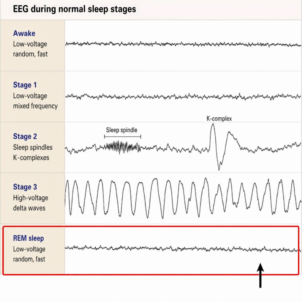
Identify the sleep wave marked in the EEG image during the sleep-wake cycle.
Which of the following is referred to as the "Window of the limbic system"?
Type of sensation lost on same side of Brown Sequard syndrome?
Gamma waves of REM sleep are associated with?
Which of the following statements about sleep is incorrect?
What is one of the specific functions of the primary motor cortex located on the anterior edge of the pre-central gyrus?
Which of the following has direct innervation from sympathetic system but no parasympathetic supply?
Which of the following neurons in the cerebellar cortex is primarily excitatory?
Which part of the sympathetic nervous system is responsible for secreting catecholamines?
Practice by Chapter
Neurons and Glial Cells
Practice Questions
Synaptic Transmission
Practice Questions
Sensory Processing
Practice Questions
Motor Control Systems
Practice Questions
Autonomic Nervous System
Practice Questions
Hypothalamus and Limbic System
Practice Questions
Cerebral Cortex Functions
Practice Questions
Electroencephalography
Practice Questions
Neuroplasticity
Practice Questions
Sleep and Wakefulness
Practice Questions
Want unlimited practice?
Get full access to all questions, explanations, and performance tracking.
Scan to download app