
Neurophysiology — MCQs

On this page
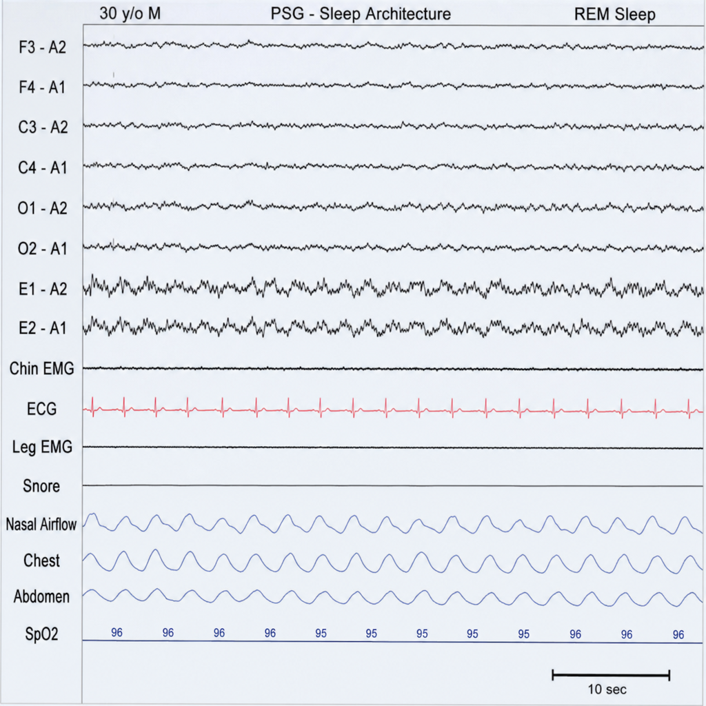
Identify the sleep stage marked in the EEG during the sleep-wake cycle?
True about decorticate rigidity:
What is the rate of CSF formation in children?
Withdrawal reflex is an example of which of the following?
Intracranial pressure is not raised during
'C' fibers carry sensations through which pathway?
What is the maximum duration of time spent in the NREM sleep stage N2?
Which of the following is the primary excitatory neurotransmitter in the central nervous system?
Which of the following is a primary function of the cerebellum?
What is the total volume of cerebrospinal fluid (CSF) in an adult human?
Practice by Chapter
Neurons and Glial Cells
Practice Questions
Synaptic Transmission
Practice Questions
Sensory Processing
Practice Questions
Motor Control Systems
Practice Questions
Autonomic Nervous System
Practice Questions
Hypothalamus and Limbic System
Practice Questions
Cerebral Cortex Functions
Practice Questions
Electroencephalography
Practice Questions
Neuroplasticity
Practice Questions
Sleep and Wakefulness
Practice Questions
Want unlimited practice?
Get full access to all questions, explanations, and performance tracking.
Scan to download app