
Neurophysiology — MCQs

On this page
Which of the following is an amino neurotransmitter?
What is the lowest level of integration for the stretch reflex?
The tonic neck reflex is lost in a lesion of which part of the central nervous system?
Which of the following is the afferent limb of the corneal reflex?
In a normal awake person at rest with eyes closed, EEG waves that are reduced on opening the eyes:
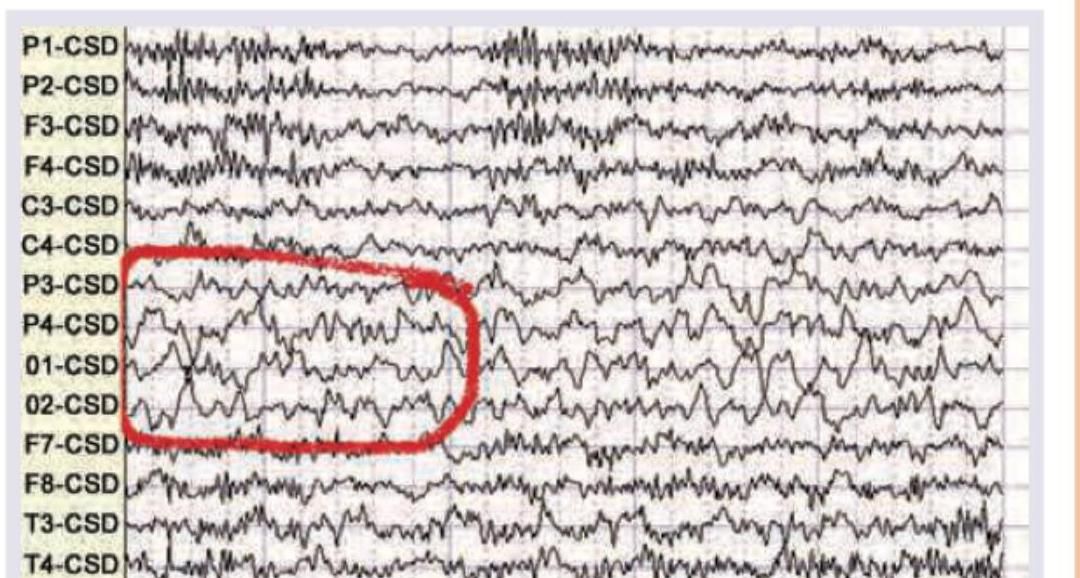
Which wave is seen in the given EEG recording?
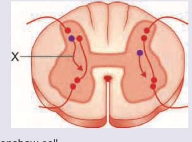
Name the interneuron marked X in colour purple involved in tetanus. (Recent NEET Pattern 2016-17)
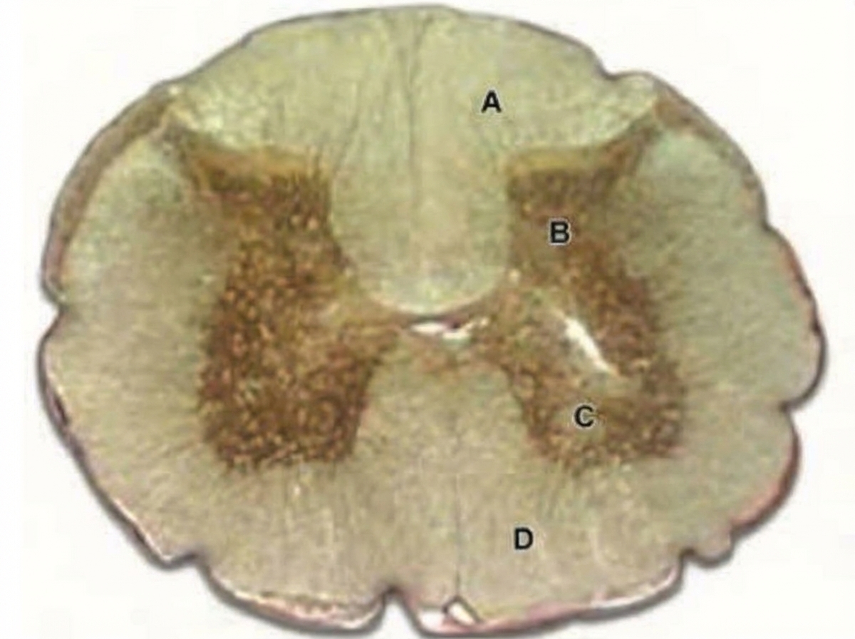
In the image shown below, which of the marked area is involved in relieving pain in response to massage?
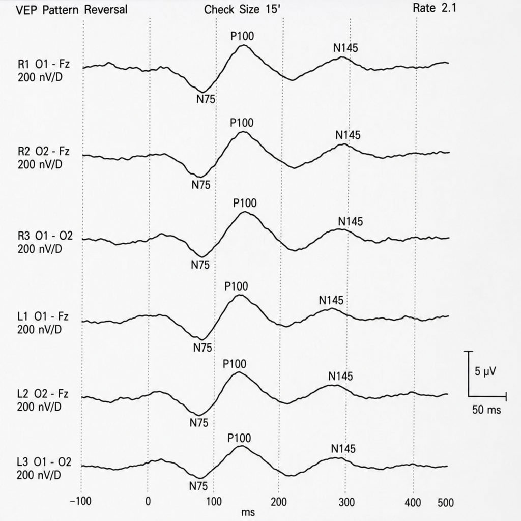
The following electrical recording is known as:
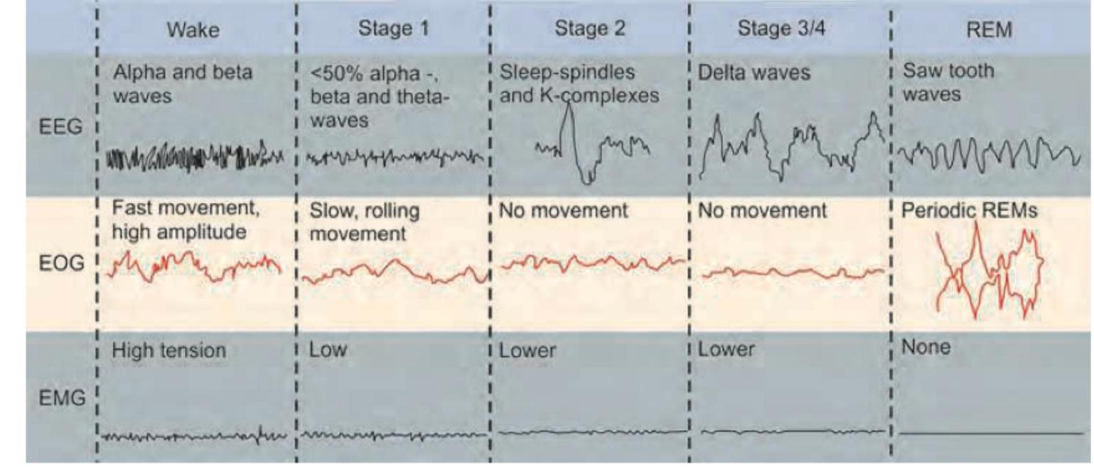
A polysomnography is performed on a patient. Based on the provided EEG, EOG, and EMG findings, which stage of sleep is indicated? (Image: img-182.jpeg)
Practice by Chapter
Neurons and Glial Cells
Practice Questions
Synaptic Transmission
Practice Questions
Sensory Processing
Practice Questions
Motor Control Systems
Practice Questions
Autonomic Nervous System
Practice Questions
Hypothalamus and Limbic System
Practice Questions
Cerebral Cortex Functions
Practice Questions
Electroencephalography
Practice Questions
Neuroplasticity
Practice Questions
Sleep and Wakefulness
Practice Questions
Want unlimited practice?
Get full access to all questions, explanations, and performance tracking.
Scan to download app