
Neurophysiology — MCQs

On this page
A transection made at the lower end of medulla through the pyramids would cause all of the following except:
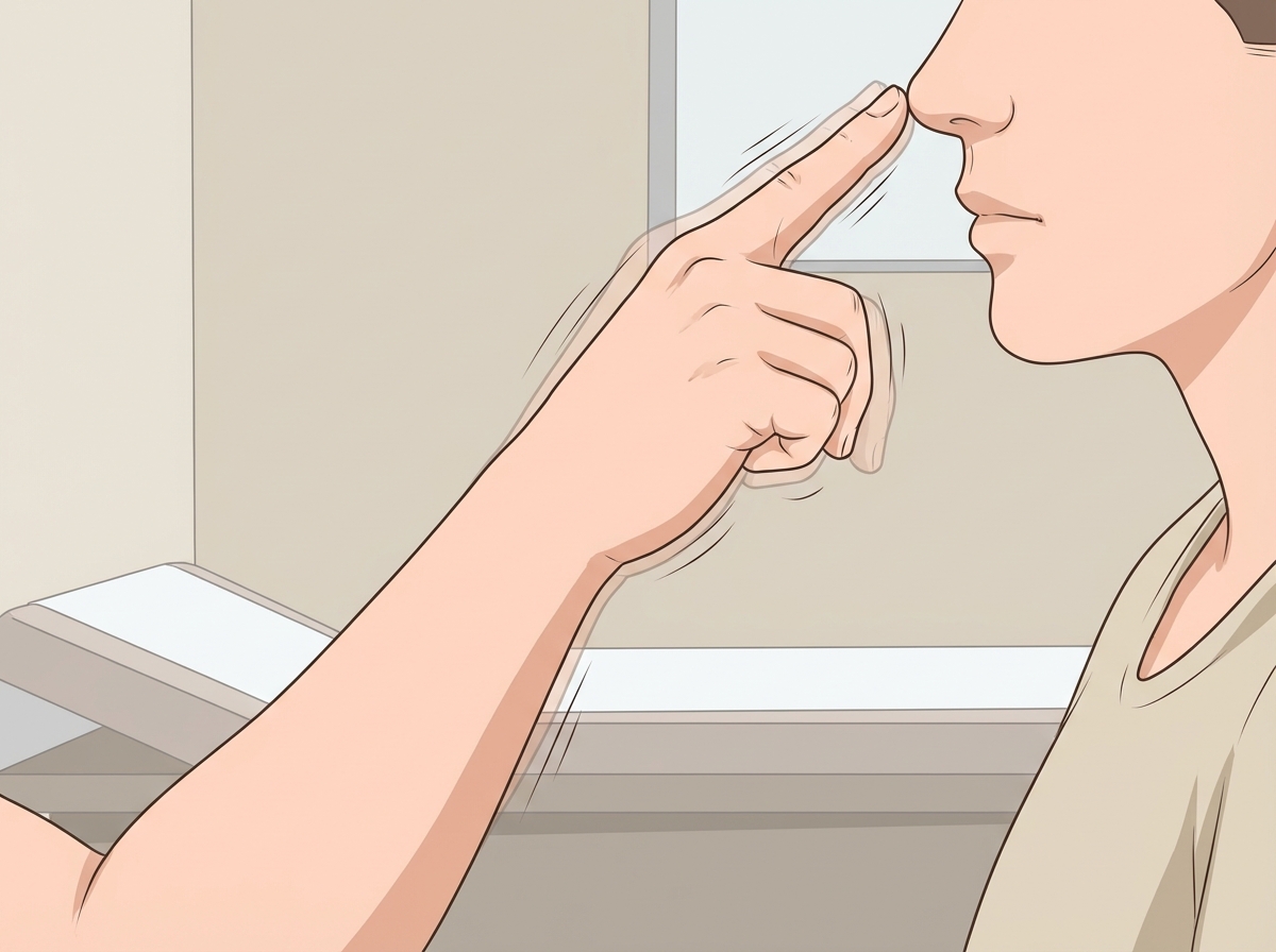
Based on the clinical sign demonstrated in the image, identify the most likely site of damage:
"Reward pathway" is associated with which structure?
In decerebrate animals, which reflex is lost?
What does long-term potentiation refer to?
The rubrospinal tract primarily influences which of the following?
In peripheral tissues, which of the following contains substance P?
Which Brodmann areas correspond to the motor cortex?
In Kluver-Bucy syndrome, which brain structure is typically lesioned?
The cyclical flexion and extension motions of a leg during walking result from activity at which level of the nervous system?
Practice by Chapter
Neurons and Glial Cells
Practice Questions
Synaptic Transmission
Practice Questions
Sensory Processing
Practice Questions
Motor Control Systems
Practice Questions
Autonomic Nervous System
Practice Questions
Hypothalamus and Limbic System
Practice Questions
Cerebral Cortex Functions
Practice Questions
Electroencephalography
Practice Questions
Neuroplasticity
Practice Questions
Sleep and Wakefulness
Practice Questions
Want unlimited practice?
Get full access to all questions, explanations, and performance tracking.
Scan to download app