Arrange the following nerve fibers sequentially in the descending order of nerve impulse transmission velocity: A. C fiber B. Aa fiber C. B fiber D. Ad fiber
In Myasthenia gravis, what is the fundamental defect at the neuromuscular junction?
Where does the action potential in a spinal motor neuron generate from?
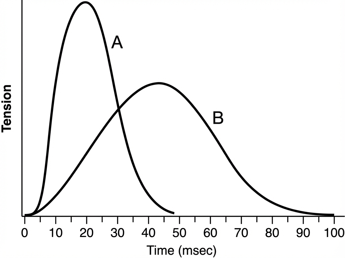
Which of the following best describes muscle B compared with muscle A?

Which of the following statements regarding intrafusal fibers is incorrect?
Post-tetanic facilitation is thought to be the result of which of the following?
Which of the following specifically blocks sodium channels?
The refractory period is minimum for which of the following nerve fibers?
Botulinum toxin blocks neuromuscular transmission by which of the following mechanisms?
Dynamic response is due to which type of muscle ending?
Explanation: **Explanation** The velocity of nerve impulse transmission is primarily determined by two factors: **fiber diameter** and the **presence of myelin**. According to the Erlanger-Gasser classification, thicker and more heavily myelinated fibers conduct impulses faster than thinner, unmyelinated ones. **The Correct Sequence (Descending Order):** 1. **Aα (Alpha) fiber:** The thickest (12–20 µm) and most heavily myelinated; conducts at 70–120 m/s. 2. **Aδ (Delta) fiber:** Thinner than Aα (2–5 µm) but still myelinated; conducts at 12–30 m/s. 3. **B fiber:** Small, preganglionic autonomic fibers ( <3 µm) with light myelination; conducts at 3–15 m/s. 4. **C fiber:** The thinnest (0.4–1.2 µm) and **unmyelinated**; conducts at the slowest speeds (0.5–2 m/s). Therefore, the descending order of velocity is **Aα > Aδ > B > C**, which corresponds to sequence **B-D-C-A**. **Analysis of Options:** * **Option B (Correct):** Correctly follows the hierarchy of diameter and myelination. * **Options A, C, and D:** These are incorrect because they place C fibers (slowest) or B fibers (intermediate) ahead of A-group fibers in velocity. **High-Yield NEET-PG Pearls:** * **Sensitivity to Local Anesthetics:** The order of blockade is **B > C > Aδ > Aγ > Aβ > Aα**. Note that B fibers are blocked first despite being myelinated because of their small diameter and anatomy. * **Sensitivity to Pressure:** A fibers are most sensitive (e.g., "limb falling asleep"). * **Sensitivity to Hypoxia:** B fibers are most sensitive. * **Function of C fibers:** They carry "slow pain" (dull, aching), temperature, and postganglionic sympathetic signals. Aδ fibers carry "fast pain" (sharp, localized).
Explanation: **Explanation:** **Myasthenia Gravis (MG)** is an autoimmune disorder characterized by the production of antibodies against the **nicotinic Acetylcholine receptors (nAChR)** located on the postsynaptic membrane of the neuromuscular junction (NMJ). 1. **Why Option C is Correct:** The fundamental defect is the **reduction in the number of functional postsynaptic receptors**. Autoantibodies (anti-AChR) bind to these receptors, leading to their destruction via complement-mediated lysis or internalization. Consequently, even though ACh is released normally, it cannot "attach" to enough receptors to generate a sufficient end-plate potential (EPP) to trigger a muscle contraction. This results in the hallmark symptom: fatiguable muscle weakness. 2. **Why Other Options are Incorrect:** * **Options A & B:** In MG, the synthesis and storage of Acetylcholine are normal. The defect is not a "deficiency" of the neurotransmitter itself, but a lack of available "docking sites" on the muscle side. * **Option D:** Impeded presynaptic release is characteristic of **Lambert-Eaton Myasthenic Syndrome (LEMS)**, where antibodies target the presynaptic voltage-gated calcium channels (VGCC), preventing ACh release. **High-Yield Clinical Pearls for NEET-PG:** * **Hallmark:** Fatiguability (weakness worsens with activity, improves with rest). * **Associated Pathology:** 75% of patients have **thymic hyperplasia**; 10% have a **thymoma**. * **Diagnosis:** Edrophonium (Tensilon) test (shows rapid improvement), repetitive nerve stimulation (shows **decremental response**), and anti-AChR antibody titers. * **Treatment:** Pyridostigmine (AChE inhibitor) is the first-line symptomatic treatment.
Explanation: ### Explanation The correct answer is **D. Initial segment**. **1. Why the Initial Segment is the Correct Answer:** In a spinal motor neuron, the action potential is generated at the **initial segment**, which is the unmyelinated portion of the axon just distal to the axon hillock. This region has the **lowest threshold** for excitation because it contains a significantly higher density of **voltage-gated sodium (Na+) channels** compared to the cell body or dendrites. When excitatory postsynaptic potentials (EPSPs) summate and reach the threshold (usually about -55mV), the high concentration of Na+ channels in the initial segment triggers the rapid depolarization necessary to fire an action potential. **2. Why Other Options are Incorrect:** * **A. Cell Body (Soma):** While the cell body integrates incoming signals, it has a relatively low density of voltage-gated Na+ channels, making its threshold for firing much higher than the initial segment. * **B. Dendrites:** Dendrites are primarily involved in receiving inputs (graded potentials). In most spinal motor neurons, they lack the necessary density of voltage-gated channels to generate a self-propagating action potential. * **C. Axon:** While the action potential *propagates* down the axon, it does not *originate* there. The axon serves as the transmission cable to the neuromuscular junction. **3. High-Yield Clinical Pearls for NEET-PG:** * **Axon Hillock vs. Initial Segment:** While often used interchangeably in casual discussion, the **initial segment** is the precise physiological "trigger zone." The axon hillock is the anatomical funnel-shaped region of the soma leading to the axon. * **Threshold Comparison:** The threshold at the initial segment is approximately **-45 to -55 mV**, whereas the cell body threshold is much higher (around -30 mV). * **Accommodation:** If a neuron is subjected to slow, constant depolarization, the threshold of the initial segment may rise; this is known as accommodation.
Explanation: ***Innervated by smaller nerve fibers*** - According to **Henneman's size principle**, slow-twitch (Type I) muscle fibers are innervated by **smaller motor neurons** with smaller axon diameters. - Type I fibers require **lower threshold stimulation** and are recruited first during muscle activation, consistent with smaller nerve fiber innervation. *Adapted for rapid contraction* - This describes **fast-twitch (Type II) fibers**, not slow-twitch fibers; Type I fibers contract **slowly** and are built for endurance. - Fast-twitch fibers have rapid **calcium release and reuptake** mechanisms, enabling quick contraction-relaxation cycles. *Composed of larger muscle fibers* - **Type II (fast-twitch) fibers** are typically larger in diameter to generate more force quickly. - Type I fibers are generally **smaller** as they prioritize sustained activity over peak force production. *Fewer mitochondria* - Type I fibers have **abundant mitochondria** for sustained **aerobic metabolism** and fatigue resistance. - Type II fibers rely more on **anaerobic glycolysis** and have fewer mitochondria compared to oxidative Type I fibers.
Explanation: **Explanation:** The muscle spindle is a complex sensory organ containing **intrafusal fibers**, which are responsible for detecting changes in muscle length and the rate of change. 1. **Why Option C is the "Incorrect Statement" (Correct Answer):** The question asks for the *incorrect* statement. Option C states that primary sensory endings are excited by both fibers—this is actually a **correct** physiological fact. Primary (Type Ia) afferent endings wrap around the central portions of **both** nuclear bag and nuclear chain fibers. Therefore, it cannot be the "incorrect" statement unless there is a nuance in the question's framing or a typo in the provided key. *Note: In standard physiology (Guyton/Ganong), Type Ia fibers supply both, while Type II fibers predominantly supply nuclear chain fibers.* 2. **Analysis of Other Options:** * **Option A (Correct Fact):** Nuclear chain fibers are indeed shorter and thinner, with nuclei arranged in a row (chain), whereas bag fibers are larger with nuclei clumped in a central "bag." * **Option B (Correct Fact):** In a typical spindle, there are usually 1–3 nuclear bag fibers and 3–9 nuclear chain fibers. Thus, bag fibers are fewer. * **Option D (Incorrect Fact):** Secondary (Type II) sensory endings primarily innervate **nuclear chain fibers**, but they can also have a minor contribution to static nuclear bag fibers. However, compared to primary endings, their association with chain fibers is their defining characteristic. **High-Yield NEET-PG Pearls:** * **Primary Endings (Ia):** Detect **dynamic** (velocity) and static changes. * **Secondary Endings (II):** Detect **static** (length) changes only. * **Gamma Motor Neurons:** Maintain spindle sensitivity during contraction. **Dynamic gamma** supplies bag fibers; **Static gamma** supplies chain fibers. * **Alpha-Gamma Co-activation:** Ensures the spindle does not go "slack" when the extrafusal muscle contracts.
Explanation: **Explanation:** **Post-tetanic facilitation (PTF)** is a phenomenon where a period of high-frequency (tetanic) stimulation is followed by an increased response to a subsequent single stimulus. **Why Option C is Correct:** The underlying mechanism is the **buildup of calcium ions ($Ca^{2+}$)** in the presynaptic terminal. During rapid, repetitive firing (tetanus), $Ca^{2+}$ enters the terminal through voltage-gated channels faster than the cell’s buffering systems (like the mitochondria and calcium pumps) can remove it. This "residual calcium" remains in the terminal after the tetanus ends. When a subsequent stimulus arrives, the newly entered $Ca^{2+}$ adds to this residual pool, leading to a massive release of neurotransmitters and a significantly larger post-synaptic potential. **Why the other options are incorrect:** * **Option A:** Voltage-gated sodium channels are responsible for the depolarization phase of the action potential, not the enhanced synaptic transmission seen in facilitation. * **Option B:** Opening of potassium channels leads to hyperpolarization and inhibition, which would decrease rather than facilitate neurotransmitter release. * **Option D:** Electrotonic conduction refers to the passive spread of electrical signals (like graded potentials) and does not account for the chemical enhancement seen at the synapse during PTF. **NEET-PG High-Yield Pearls:** * **Synaptic Plasticity:** PTF is a form of short-term synaptic plasticity. Long-term potentiation (LTP), involving NMDA receptors, is the long-term equivalent crucial for memory. * **Calcium is Key:** Neurotransmitter release is directly proportional to the **fourth power** of calcium concentration in the presynaptic terminal. * **Clinical Correlation:** PTF is classically demonstrated in the **Lambert-Eaton Myasthenic Syndrome (LEMS)**, where muscle strength improves after brief exercise (tetanic contraction) due to the buildup of calcium overcoming the deficiency of calcium channels.
Explanation: **Explanation:** The correct answer is **Tetrodotoxin (TTX)**. This potent neurotoxin, famously found in the liver and ovaries of the **Pufferfish (Fugu)**, acts by selectively and reversibly binding to the extracellular pores of **voltage-gated sodium channels (VGSCs)**. By blocking these channels, it prevents the influx of sodium ions required for the depolarization phase of an action potential, leading to muscle paralysis and respiratory failure. **Analysis of Options:** * **Nifedipine:** This is a **Dihydropyridine (DHP) Calcium Channel Blocker**. It primarily targets L-type calcium channels in vascular smooth muscle and the heart, used clinically for hypertension and angina. * **Tetraethylammonium (TEA):** This is a classic pharmacological tool used to block **Voltage-gated Potassium (K+) channels**. It inhibits the efflux of K+, thereby prolonging the repolarization phase of the action potential. * **Choline:** This is a precursor for the neurotransmitter **Acetylcholine**. It is not a channel blocker; rather, it is taken up by presynaptic neurons via a high-affinity transporter to synthesize new neurotransmitters. **High-Yield Facts for NEET-PG:** * **Saxitoxin:** Produced by red tide dinoflagellates; it has a mechanism identical to Tetrodotoxin (blocks Na+ channels). * **Batrachotoxin:** Found in poison dart frogs; it keeps Na+ channels **open**, preventing repolarization. * **Local Anesthetics (e.g., Lidocaine):** These also block Na+ channels but act on the **inner (cytoplasmic) side** of the channel, unlike TTX which acts on the outer surface. * **Dendrotoxin:** A snake toxin (Mamba) that blocks K+ channels.
Explanation: **Explanation:** The duration of the **refractory period** in a nerve fiber is inversely proportional to its **diameter** and **conduction velocity**. 1. **Why A alpha fibers are correct:** According to the Erlanger-Gasser classification, **A alpha (Aα) fibers** are the thickest (12–20 μm) and fastest (70–120 m/s) myelinated fibers. Because they have the largest diameter, they possess a shorter membrane time constant and a higher density of voltage-gated sodium channels at the Nodes of Ranvier. This allows them to recover from inactivation more rapidly, resulting in the **shortest absolute refractory period (ARP)**. A shorter refractory period allows these fibers to fire at higher frequencies. 2. **Why other options are incorrect:** * **A beta (Aβ) and A delta (Aδ) fibers:** These are progressively thinner and slower than A alpha fibers. As diameter decreases, the refractory period increases. * **C fibers:** These are the thinnest, unmyelinated fibers with the slowest conduction velocity (0.5–2 m/s). Consequently, they have the **longest refractory period** among all nerve fibers. **High-Yield Facts for NEET-PG:** * **Order of Refractory Period:** C > B > A (C fibers have the longest; A fibers have the shortest). * **Order of Velocity:** Aα > Aβ > Aγ > Aδ > B > C. * **Susceptibility to Blockade:** * **Hypoxia:** B fibers are most sensitive. * **Pressure:** A fibers are most sensitive (e.g., "Saturday Night Palsy"). * **Local Anesthetics:** C fibers are most sensitive (due to small diameter and lack of myelin).
Explanation: **Explanation:** **Mechanism of Action (The Correct Answer):** Botulinum toxin, produced by *Clostridium botulinum*, acts primarily at the **presynaptic terminal** of the neuromuscular junction (NMJ). Its primary mechanism involves the cleavage of **SNARE proteins** (such as synaptobrevin, SNAP-25, and syntaxin). These proteins are essential for docking and fusing acetylcholine (ACh) vesicles with the presynaptic membrane. By disrupting this process, the toxin effectively prevents the release of ACh into the synaptic cleft. In the context of ion channels, Botulinum toxin inhibits the **calcium-dependent exocytosis** of neurotransmitters, effectively mimicking a functional **closure or blockade of presynaptic Ca++ channels** (as Ca++ entry no longer triggers vesicle release). **Analysis of Incorrect Options:** * **Option B & D:** These refer to **postsynaptic** events. Botulinum toxin does not affect the postsynaptic membrane or its receptors (like the nicotinic ACh receptor). Postsynaptic blockade is characteristic of drugs like Curare (d-tubocurarine) or conditions like Myasthenia Gravis. * **Option C:** Opening presynaptic K+ channels would cause hyperpolarization and inhibit ACh release, but this is not the mechanism of Botulinum toxin. **High-Yield Clinical Pearls for NEET-PG:** * **Clinical Presentation:** Causes **flaccid paralysis** (descending) and "floppy baby syndrome" (infantile botulism via honey ingestion). * **Therapeutic Uses:** Used for focal dystonias, achalasia cardia, strabismus, and cosmetic reduction of wrinkles (Botox). * **Contrast with Tetanus Toxin:** While both cleave SNARE proteins, Tetanus toxin undergoes **retrograde axonal transport** to the CNS and inhibits GABA/Glycine release from Renshaw cells, causing **spastic paralysis**. * **Lambert-Eaton Syndrome:** Also affects the presynaptic terminal but specifically involves antibodies against voltage-gated Ca++ channels.
Explanation: **Explanation:** The muscle spindle is a complex sensory organ that monitors muscle length and the rate of change in length. It contains two types of intrafusal fibers: **Nuclear Bag fibers** and **Nuclear Chain fibers**. 1. **Why Primary Ending is Correct:** Primary sensory endings (also known as **Type Ia fibers**) wrap around the central portions of both nuclear bag and nuclear chain fibers. Because nuclear bag fibers are highly sensitive to the *velocity* of stretch, the primary endings provide a **dynamic response**. This means they fire rapidly during the actual movement or change in length, signaling how fast the muscle is being stretched. 2. **Why Other Options are Incorrect:** * **Secondary Ending (Type II fibers):** These are located primarily on nuclear chain fibers. They are responsible for the **static response**, meaning they fire at a constant rate proportional to the *absolute length* of the muscle, rather than the speed of change. * **Tertiary Ending:** This term is not standard in muscle spindle physiology. Sensory innervation is categorized into Primary (Ia) and Secondary (II) endings. * **All of the above:** Incorrect because dynamic and static responses are mediated by distinct fiber types with different physiological properties. **High-Yield Clinical Pearls for NEET-PG:** * **Primary Endings (Ia):** Responsible for the **monosynaptic stretch reflex** (e.g., Knee jerk). They are "velocity sensors." * **Secondary Endings (II):** Responsible for "position sensors." * **Gamma Motor Neurons:** Maintain spindle sensitivity during muscle contraction. **Gamma-dynamic** neurons affect bag fibers (dynamic response), while **Gamma-static** neurons affect chain fibers (static response). * **Golgi Tendon Organ (Ib):** Unlike spindles, these sense **muscle tension** and are arranged in series with extrafusal fibers.
Resting Membrane Potential
Practice Questions
Action Potential Generation and Propagation
Practice Questions
Neuromuscular Junction
Practice Questions
Skeletal Muscle Contraction
Practice Questions
Smooth Muscle Physiology
Practice Questions
Cardiac Muscle Properties
Practice Questions
Muscle Metabolism and Fatigue
Practice Questions
Motor Unit Function
Practice Questions
Neurotransmitters and Receptors
Practice Questions
Electrophysiological Measurements
Practice Questions
Get full access to all questions, explanations, and performance tracking.
Start For Free