
Nerve and Muscle Physiology — MCQs

On this page
Most common type of calcium channels of skeletal muscles is?
What is the primary action observed in the withdrawal reflex?
ATPase activity is present in
Which of the following fiber types is classically categorized as Group B nerve fibers?
Which of the following statements is true about red muscle fibers?
A man slept with his head over his forearm. The next morning, he complains of tingling and numbness over the forearm. If this were primarily due to hypoxia affecting nerve fibers, which of the following statements about nerve fiber sensitivity to hypoxia is correct?
What is a key difference between smooth muscle and skeletal muscle physiology?
What is the fixed length of a myosin filament?
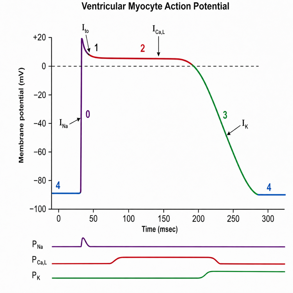
The plateau phase of this graph is due to:
Which of the following is NOT a location where multi-unit smooth muscle is present?
Practice by Chapter
Resting Membrane Potential
Practice Questions
Action Potential Generation and Propagation
Practice Questions
Neuromuscular Junction
Practice Questions
Skeletal Muscle Contraction
Practice Questions
Smooth Muscle Physiology
Practice Questions
Cardiac Muscle Properties
Practice Questions
Muscle Metabolism and Fatigue
Practice Questions
Motor Unit Function
Practice Questions
Neurotransmitters and Receptors
Practice Questions
Electrophysiological Measurements
Practice Questions
Want unlimited practice?
Get full access to all questions, explanations, and performance tracking.
Scan to download app