
Nerve and Muscle Physiology — MCQs

On this page
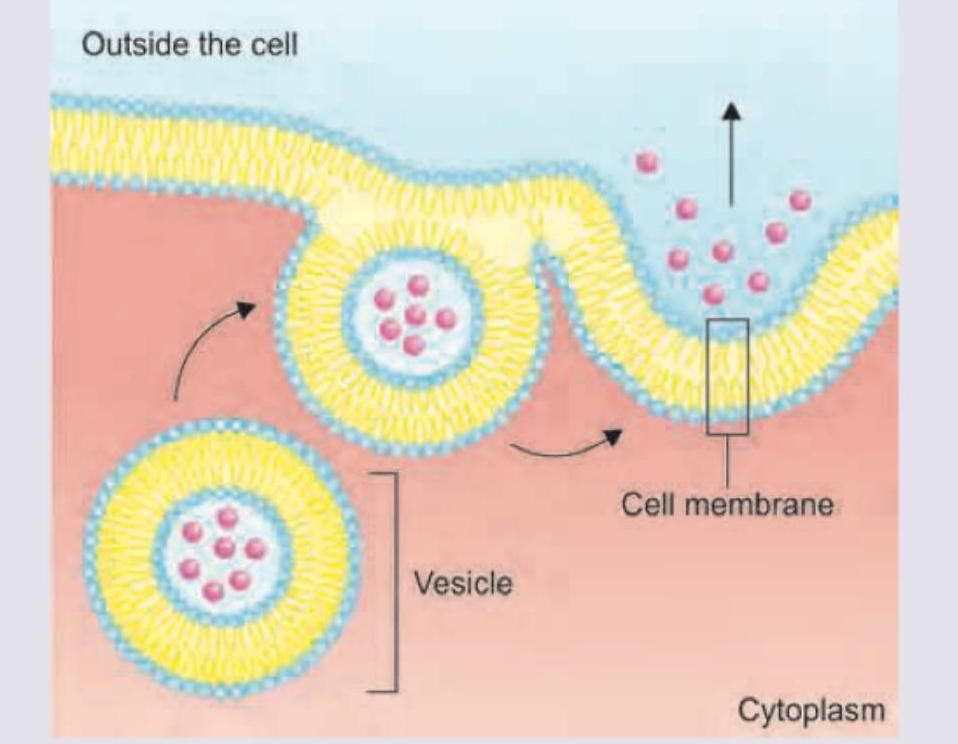
What best describes step 3 in the given diagram?
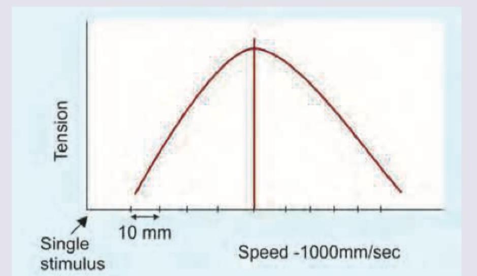
Which of the following changes occurs during muscle contraction while exercising, as shown in the image?
A single muscle twitch lasts 40 milliseconds. What is the minimum tetanization frequency required to produce a sustained (fused) contraction in this muscle?
The following skeletal muscle recording shows presence of: (Recent NEET Pattern 2016-17)
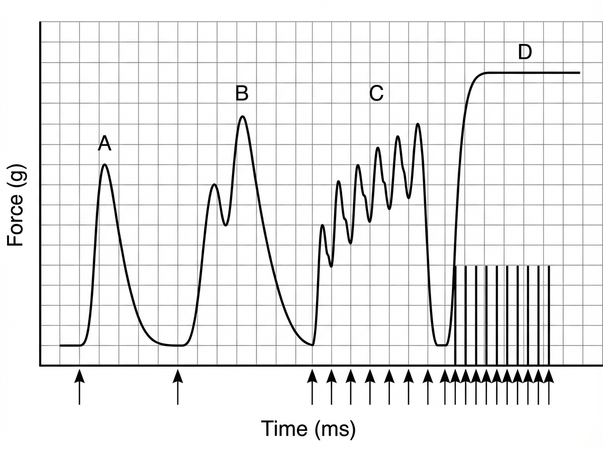
Calculate the tetanizing frequency based on the contraction dynamics of gastrocnemius muscle of frog shown in the image?
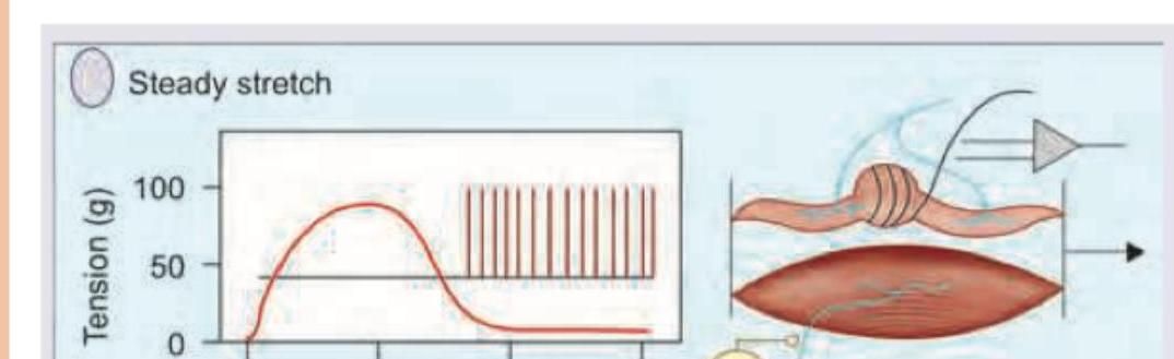
In the process shown below stretch stimulus is mediated by which of the following receptors?
Which ion plays a role in the process shown below? (Recent NEET Pattern 2018)
Sequence the events in neuromuscular action potential conduction: 1. Sodium channels open in the end plate 2. Calcium enters at the nerve terminal 3. Release of acetylcholine
Assertion: RMP depends on proteins and phosphate ions. Reason: Diffusion potential can be calculated using nernst equation. Choose the best statement regarding the assertion and reason.
Arrange the following parts of sarcomere from periphery to center. 1. Z line 2. M line 3. A band 4. H zone
Practice by Chapter
Resting Membrane Potential
Practice Questions
Action Potential Generation and Propagation
Practice Questions
Neuromuscular Junction
Practice Questions
Skeletal Muscle Contraction
Practice Questions
Smooth Muscle Physiology
Practice Questions
Cardiac Muscle Properties
Practice Questions
Muscle Metabolism and Fatigue
Practice Questions
Motor Unit Function
Practice Questions
Neurotransmitters and Receptors
Practice Questions
Electrophysiological Measurements
Practice Questions
Want unlimited practice?
Get full access to all questions, explanations, and performance tracking.
Scan to download app