Disruption of the myelin sheath without interruption in the axons is called:
Rigor mortis results after death is due to which of the following?
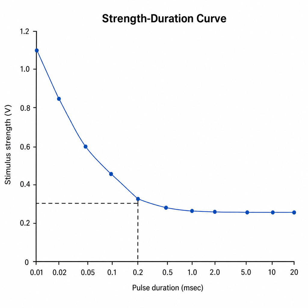
Conduction velocity of nerve is reduced in all of the following conditions, except?
Which of the following statements is TRUE regarding the excitation-contraction coupling mechanism in smooth muscles?
Wallerian degeneration is seen in which part of a nerve?
Which motor unit is recruited last?
Gamma neurons innervate which type of muscle fibers?
Nuclear bag fibers are related to which of the following?
Clasp knife rigidity is also known as:
Heart muscles act as a functional syncitium because:
Explanation: ### Explanation This question refers to the **Seddon Classification of Nerve Injury**, which categorizes nerve damage based on the severity of structural disruption. **1. Why Neuropraxia is correct:** Neuropraxia is the mildest form of nerve injury. It is characterized by a **localized conduction block** due to focal demyelination (disruption of the myelin sheath). Crucially, the **axon and the connective tissue sheaths (endoneurium, perineurium, and epineurium) remain intact**. Because the axon is not severed, Wallerian degeneration does not occur, and recovery is typically complete within days to weeks once the myelin repairs. **2. Why the other options are incorrect:** * **Axonotmesis:** This involves the **disruption of the axon** and its myelin sheath, but the supporting connective tissue framework (endoneurium) remains intact. Wallerian degeneration occurs distal to the injury. * **Neurotmesis:** This is the most severe grade, involving **complete transection** of both the axon and all surrounding connective tissue layers. Spontaneous recovery is impossible without surgical intervention. * **Transient ischemia:** While ischemia can cause temporary numbness (e.g., "Saturday Night Palsy"), it is a mechanism of injury rather than a structural classification of nerve damage. **3. High-Yield Clinical Pearls for NEET-PG:** * **Wallerian Degeneration:** Occurs in Axonotmesis and Neurotmesis, but **NOT** in Neuropraxia. * **Recovery Sequence:** Neuropraxia (Fastest/Complete) > Axonotmesis (Slow/Partial) > Neurotmesis (Requires Surgery). * **Tinel’s Sign:** Usually **absent** in Neuropraxia (since the axon is intact) but **present** in Axonotmesis as regenerating axonal sprouts advance. * **Sunderland Classification:** An expansion of Seddon’s; Neuropraxia corresponds to **Grade 1**, Axonotmesis to **Grades 2-4**, and Neurotmesis to **Grade 5**.
Explanation: **Explanation:** Rigor mortis is the post-mortem stiffening of muscles caused by the depletion of **Adenosine Triphosphate (ATP)**. **Why Option B is Correct:** In a living muscle, ATP is required for two critical steps in the cross-bridge cycle: 1. **Dissociation:** ATP binding to the myosin head is essential to break the bond between actin and myosin. 2. **Relaxation:** ATP powers the SERCA pump (Sarcoplasmic Endoplasmic Reticulum Calcium ATPase) to pump calcium back into the sarcoplasmic reticulum. After death, cellular respiration ceases, leading to a **failure of ATP supply**. Without ATP, the myosin heads remain permanently attached to actin filaments in a "latched" state, and calcium remains in the cytosol, maintaining a state of rigid contraction. **Why Other Options are Incorrect:** * **Option A:** Acetylcholine (ACh) is responsible for initiating depolarization at the neuromuscular junction. While ACh release stops after death, it is the lack of ATP for detachment, not the absence of ACh diffusion, that causes the physical rigidity. * **Option C:** This is a distractor. While calcium bridges (troponin-tropomyosin interactions) remain active because calcium cannot be sequestered, the fundamental "failure" is the lack of the energy molecule (ATP) required to break the actin-myosin cross-bridge. **High-Yield Clinical Pearls for NEET-PG:** * **Sequence:** Rigor mortis typically starts in the small muscles (eyelids, jaw) and follows a **proximal-to-distal** (Nysten’s Law) progression. * **Timeline:** It usually begins 2–3 hours after death, peaks at 12 hours, and disappears after 36–48 hours due to **autolysis** (proteolytic enzyme action). * **Temperature:** High ambient temperatures accelerate the onset of rigor mortis.
Explanation: ### Explanation The conduction velocity of a nerve is primarily determined by the **integrity of the myelin sheath**. According to the principles of saltatory conduction, myelin allows the action potential to "jump" between Nodes of Ranvier, significantly increasing speed. Therefore, **demyelinating disorders** cause a marked reduction in conduction velocity, whereas **pure axonal disorders** primarily affect the amplitude of the action potential while leaving the velocity relatively preserved. **1. Why AMAN is the correct answer:** Acute Motor Axonal Neuropathy (AMAN) is a variant of Guillain-Barré Syndrome (GBS) characterized by **primary axonal degeneration** rather than demyelination. In AMAN, the immune system targets the axolemma (nodes of Ranvier). Because the myelin remains largely intact, the conduction velocity remains normal or is only minimally reduced, despite a significant drop in the compound muscle action potential (CMAP) amplitude. **2. Analysis of Incorrect Options:** * **AIDP (Acute Inflammatory Demyelinating Polyradiculoneuropathy):** This is the most common form of GBS. It involves immune-mediated destruction of myelin, leading to significantly **prolonged latencies and reduced conduction velocities**. * **HSMN (Hereditary Sensory Motor Neuropathy):** Also known as Charcot-Marie-Tooth (CMT) disease. Type 1 (CMT1) is a classic **hypertrophic demyelinating neuropathy** where conduction velocities are uniformly and severely slowed (often <38 m/s). * **Multifocal Motor Neuropathy (MMN):** This is a chronic demyelinating condition characterized by **conduction blocks**. The underlying pathology involves focal demyelination, which slows or stops nerve impulse propagation. **Clinical Pearls for NEET-PG:** * **Conduction Velocity $\propto$ Nerve Diameter:** Larger fibers conduct faster. * **Temperature:** Cold temperatures reduce conduction velocity. * **Myelination:** The most critical factor for high-speed conduction. * **GBS Variants:** Remember that **AIDP = Demyelinating** (Slow velocity) while **AMAN/AMSAN = Axonal** (Normal velocity, low amplitude).
Explanation: ### Explanation **1. Why the Correct Answer is Right:** In smooth muscle, the rise in **intracellular calcium ([Ca²⁺]i)** is the fundamental trigger for contraction. Unlike skeletal muscle, smooth muscle lacks a well-developed sarcoplasmic reticulum and relies on both extracellular calcium (entering via voltage-gated channels) and intracellular stores. Once [Ca²⁺]i increases, it binds to **Calmodulin**. This Ca²⁺-Calmodulin complex activates **Myosin Light Chain Kinase (MLCK)**, which phosphorylates the myosin head, allowing it to bind to actin and initiate the cross-bridge cycle. **2. Why the Other Options are Wrong:** * **Option A:** Smooth muscle **lacks troponin**. Instead, it uses **Calmodulin** as the primary calcium-binding protein. Troponin is characteristic of skeletal and cardiac muscles. * **Option B:** Smooth muscle can maintain a **sustained contraction (Latch-bridge state)** with **low** energy consumption and **low** calcium levels. This allows organs like blood vessels to maintain tone without fatigue. * **Option C:** Contraction is triggered by the **phosphorylation of Myosin** (specifically the regulatory light chain), not actin. Actin in smooth muscle is always "ready," but myosin must be activated by MLCK. **3. High-Yield NEET-PG Pearls:** * **Caldesmon and Calponin:** These are proteins found in smooth muscle that inhibit the actin-myosin interaction; they are the functional equivalents of the troponin complex. * **Caveolae:** These are small invaginations of the sarcolemma in smooth muscle that act like the T-tubule system of skeletal muscle. * **Relaxation:** Requires **Myosin Light Chain Phosphatase (MLCP)** to dephosphorylate the myosin head. * **Multi-unit vs. Unitary:** Unitary (visceral) smooth muscle uses **gap junctions** to contract as a syncytium (e.g., GI tract, uterus).
Explanation: ### Explanation **Concept Overview:** Wallerian degeneration refers to the process of antegrade degeneration of an axon following a nerve injury. When an axon is transected, the segment distal to the cut is separated from the metabolic source—the **cell body (soma)**. Since the cell body provides essential proteins and nutrients via axonal transport, the distal segment undergoes enzymatic digestion and fragmentation. **Why Option C is Correct:** Wallerian degeneration occurs specifically in the **distal segment** (the part not attached to the cell body). Within 24–48 hours of injury, the cytoskeleton breaks down, the axonal membrane dissolves, and the myelin sheath undergoes phagocytosis by Schwann cells and macrophages. **Analysis of Incorrect Options:** * **Option A:** The part attached to the cell body is the **proximal segment**. It does not undergo Wallerian degeneration; instead, it may undergo limited "retrograde degeneration" up to the nearest Node of Ranvier. * **Options B & D:** Changes in the cell body are termed **Retrograde changes** or **Chromatolysis**. This involves the swelling of the cell body, displacement of the nucleus to the periphery, and disappearance of Nissl bodies (to prioritize protein synthesis for regeneration). **NEET-PG High-Yield Facts:** * **Chromatolysis:** The hallmark of the cell body's response to injury (Nissl substance disappears). * **Regeneration:** In the PNS, Schwann cells form **Bungner bands** (tubes) to guide the regenerating axon sprout toward the target organ. * **Rate of Growth:** Nerve fibers typically regenerate at a rate of **1–3 mm/day**. * **CNS vs. PNS:** Wallerian degeneration is much slower in the CNS because oligodendrocytes do not provide the same regenerative framework as Schwann cells and actually secrete inhibitory factors (e.g., Nogo protein).
Explanation: This question tests your understanding of the **Henneman Size Principle**, a fundamental concept in neurophysiology regarding motor unit recruitment. ### **Explanation of the Correct Answer** According to the **Henneman Size Principle**, motor units are recruited in order of increasing size of the alpha motor neuron. 1. **Small motor neurons** have a lower threshold for excitation and are recruited first. These innervate **Type I (Slow Oxidative)** fibers. 2. **Large motor neurons** have a higher threshold and are recruited only when high force is required. These innervate **Type IIb (Fast Fatigable)** fibers. Therefore, **Fast Fatigable (FF) units** are the last to be recruited because they require the highest level of neural drive. ### **Analysis of Incorrect Options** * **A. Slow oxidative motor unit:** These are Type I fibers. They are small, fatigue-resistant, and recruited **first** for low-intensity, aerobic activities (e.g., maintaining posture). * **C & D. Active phase/Relaxation:** These options describe the *timing* of a muscle twitch rather than the *type* of motor unit. Recruitment is a process of increasing the number of active motor units during the buildup of contractile force, not a specific phase of a single twitch. ### **High-Yield Facts for NEET-PG** * **Recruitment Order:** Type I (Slow) → Type IIa (Fast-Resistant) → Type IIb (Fast-Fatigable). * **De-recruitment:** This occurs in the exact **reverse order**; the largest units (FF) are the first to stop firing as force decreases. * **Physiological Basis:** Smaller neurons have a higher membrane resistance ($R$), so a given excitatory postsynaptic current ($I$) produces a larger voltage change ($V=IR$), reaching the threshold faster than in large neurons. * **Type IIb Fibers:** High glycogen content, low myoglobin (White muscle), and high ATPase activity.
Explanation: **Explanation:** The correct answer is **B. Intrafusal muscle fibers.** **1. Why it is correct:** The muscle spindle is a sensory receptor located within skeletal muscles that detects changes in muscle length. It consists of specialized fibers called **intrafusal fibers**. These fibers are innervated by **Gamma ($\gamma$) motor neurons**, which originate in the ventral horn of the spinal cord. When gamma neurons fire, they cause the poles (ends) of the intrafusal fibers to contract, which stretches the central sensory portion, thereby maintaining the sensitivity of the muscle spindle even when the muscle itself is shortened. **2. Why the other options are incorrect:** * **Option A:** **Extrafusal muscle fibers** are the standard contractile fibers responsible for muscle bulk and force generation. These are innervated by **Alpha ($\alpha$) motor neurons**, not gamma neurons. * **Option C:** The **primary motor innervation** to skeletal muscle is provided by Alpha motor neurons, which form the "final common pathway" for motor control. * **Option D:** Inhibition of antagonistic muscles is achieved via **Ia inhibitory interneurons** during the reciprocal inhibition reflex, not directly by gamma neurons. **Clinical Pearls & High-Yield Facts for NEET-PG:** * **Alpha-Gamma Co-activation:** During voluntary movement, both alpha and gamma neurons are activated simultaneously. This ensures the muscle spindle remains "taut" and sensitive to further changes in length during contraction. * **Gamma Loop:** This refers to the pathway: Gamma motor neuron $\rightarrow$ Intrafusal fiber contraction $\rightarrow$ Ia afferent stimulation $\rightarrow$ Alpha motor neuron activation $\rightarrow$ Extrafusal fiber contraction. * **Clinical Correlation:** Overactivity of gamma motor neurons is a primary contributor to **spasticity** and increased muscle tone in Upper Motor Neuron (UMN) lesions.
Explanation: **Explanation:** The muscle spindle is a complex sensory receptor responsible for detecting changes in muscle length. It consists of two types of intrafusal fibers: **Nuclear Bag fibers** and **Nuclear Chain fibers**. **Why the correct answer is right:** Nuclear bag fibers are further divided into **Dynamic bag fibers** and **Static bag fibers**. 1. **Dynamic bag fibers** are highly sensitive to the **rate of change** in muscle length (Velocity). This is known as the dynamic response. 2. **Static bag fibers** (along with nuclear chain fibers) detect the **actual change** in muscle length. This is known as the static response. Therefore, nuclear bag fibers collectively provide information regarding both the **length** and the **velocity** of muscle stretch. **Why incorrect options are wrong:** * **A. Force:** Force or tension is sensed by the **Golgi Tendon Organ (GTO)**, not the muscle spindle. * **B. Length:** While bag fibers do sense length, this option is incomplete because it ignores their unique role in sensing velocity (dynamic response). * **C. Tone:** Muscle tone is a clinical state maintained by the stretch reflex (monosynaptic reflex arc), but the primary physiological parameter sensed by the bag fiber itself is length/velocity. **High-Yield Clinical Pearls for NEET-PG:** * **Innervation:** Nuclear bag fibers are primarily supplied by **Primary (Type Ia) afferents**, which are responsible for the dynamic stretch reflex (e.g., Knee jerk). * **Motor Supply:** Dynamic bag fibers are innervated by **Dynamic Gamma Motor Neurons**, while static bag and chain fibers are innervated by **Static Gamma Motor Neurons**. * **Function:** Muscle spindles prevent overstretching (protection) and maintain posture through the stretch reflex.
Explanation: **Explanation:** **Clasp-knife rigidity** is a clinical sign seen in **Upper Motor Neuron (UMN) lesions** (spasticity). When a clinician attempts to passively stretch a spastic muscle, there is initial high resistance followed by a sudden "giving way" or relaxation, similar to the closing of a pocketknife. 1. **Why the correct answer is right:** The physiological basis for this phenomenon is the **Inverse Stretch Reflex** (also known as the **Autogenic Inhibition**). Unlike the standard stretch reflex (which causes contraction), the inverse stretch reflex is mediated by **Golgi Tendon Organs (GTOs)**. When a spastic muscle is forcefully stretched, the tension in the tendon increases significantly. This activates the GTOs, which send impulses via **Ib afferent fibers** to inhibitory interneurons in the spinal cord. These interneurons inhibit the alpha motor neurons of the same muscle, causing it to suddenly relax. 2. **Why the incorrect options are wrong:** * **Withdrawal reflex:** A polysynaptic nociceptive reflex where a limb is pulled away from a painful stimulus. * **Lengthening reaction:** While often used synonymously in older texts, in the context of NEET-PG, "Inverse Stretch Reflex" is the specific physiological mechanism. (Note: Some sources consider them identical, but the reflex mechanism is the primary academic answer). * **Crossed extensor reflex:** Occurs in conjunction with the withdrawal reflex; while one limb flexes to withdraw, the contralateral limb extends to maintain balance. **High-Yield Clinical Pearls for NEET-PG:** * **Receptor:** Golgi Tendon Organ (detects **tension**). * **Afferent Fiber:** Ib fibers. * **Lead-pipe vs. Cogwheel rigidity:** These are seen in **Extrapyramidal lesions** (e.g., Parkinson’s), whereas Clasp-knife is seen in **Pyramidal (UMN) lesions**. * **Gamma Motor Neurons:** Overactivity of these neurons is the primary cause of the initial spasticity (increased tone) in UMN lesions.
Explanation: **Explanation:** The heart functions as a **functional syncytium**, meaning that when a single atrial or ventricular cell is excited, the action potential spreads to all muscle cells, causing them to contract as a single unit. **1. Why Gap Junctions are correct:** The primary structural basis for this syncytial behavior is the **Gap Junction**. These are low-resistance protein channels (formed by connexins) located within the **intercalated discs**. They allow the rapid flow of ions and electrical impulses directly from the sarcoplasm of one cardiomyocyte to the next. This electrical coupling ensures synchronized contraction, which is essential for effective pumping. **2. Why other options are incorrect:** * **Striations (A):** While cardiac muscle is striated (like skeletal muscle) due to the organized arrangement of actin and myosin, striations relate to the mechanism of contraction, not the electrical connectivity between cells. * **Long Action Potential (C):** Cardiac muscle has a long action potential (due to the plateau phase/calcium influx), which prevents tetany and allows for filling time, but it does not facilitate the spread of impulses between cells. * **Voluntary (D):** Cardiac muscle is **involuntary** and regulated by the autonomic nervous system, unlike skeletal muscle. **High-Yield Facts for NEET-PG:** * **Two Syncytia:** The heart has two separate functional syncytia—the atrial and the ventricular—separated by the fibrous AV ring. * **Intercalated Discs:** These contain two types of junctions: **Gap junctions** (for electrical communication) and **Desmosomes** (mechanical junctions that hold cells together during contraction). * **Connexin 43:** This is the most abundant gap junction protein in the ventricles.
Resting Membrane Potential
Practice Questions
Action Potential Generation and Propagation
Practice Questions
Neuromuscular Junction
Practice Questions
Skeletal Muscle Contraction
Practice Questions
Smooth Muscle Physiology
Practice Questions
Cardiac Muscle Properties
Practice Questions
Muscle Metabolism and Fatigue
Practice Questions
Motor Unit Function
Practice Questions
Neurotransmitters and Receptors
Practice Questions
Electrophysiological Measurements
Practice Questions
Get full access to all questions, explanations, and performance tracking.
Start For Free