What is the primary function of a muscle spindle?
Which type of motor unit is of prime importance in generating the muscle power necessary for the maintenance of posture?
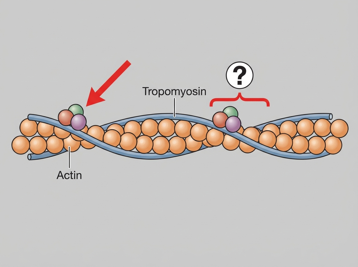
Unmyelinated nerve fibers differ from myelinated nerve fibers in that they:
What is the resting membrane potential of a neuron?
Which statement about rheobase is true?
Duchenne's Muscular Dystrophy is a disease of which of the following?
The spike in action potential of a nerve is due to which ion?
Absolute refractory period is due to which of the following?
What is true about the Golgi tendon organ?
Group A nerve fibers are most susceptible to which of the following?
Explanation: ### Explanation **1. Why Option C is Correct:** The muscle spindle is a specialized sensory receptor (proprioceptor) located within the belly of skeletal muscles. Its primary function is to detect changes in **muscle length** and the **rate of change in length**. When a muscle is stretched, the spindle sends afferent impulses (via Type Ia and II fibers) to the spinal cord. This triggers the **monosynaptic stretch reflex**, causing the muscle to contract to oppose the stretch. This feedback loop is essential for maintaining a constant muscle length and stabilizing posture. **2. Why Other Options are Incorrect:** * **Option A:** The withdrawal reflex (flexor reflex) is a polysynaptic reflex initiated by **nociceptors** (pain receptors) in response to a painful stimulus, not by muscle spindles. * **Option B:** While muscle spindles contribute to muscle tone via the stretch reflex, their *primary* physiological function is the sensing and regulation of length. Muscle tone is a broader clinical state maintained by multiple inputs, including the gamma motor system. * **Option C vs. D:** The **inverse stretch reflex** (autogenic inhibition) is mediated by the **Golgi Tendon Organ (GTO)**. While the spindle senses length, the GTO senses **muscle tension** and causes the muscle to relax to prevent injury. **3. NEET-PG High-Yield Pearls:** * **Innervation:** Muscle spindles are innervated by **Gamma ($\gamma$) motor neurons** (which maintain sensitivity during contraction) and **Type Ia (primary)** and **Type II (secondary)** sensory afferents. * **Nuclear Bag vs. Chain:** Nuclear bag fibers detect dynamic changes (velocity), while nuclear chain fibers detect static changes (length). * **Alpha-Gamma Co-activation:** This process ensures that muscle spindles remain sensitive to stretch even when the extrafusal fibers are contracting. * **Clinical Correlation:** The knee-jerk reflex (patellar reflex) is a direct clinical application of the muscle spindle-mediated stretch reflex.
Explanation: ### Explanation **1. Why Option A is Correct:** The maintenance of posture requires muscles to remain contracted for prolonged periods without tiring. This task is performed by **Type I (Slow-Twitch) muscle fibers**, which form **low-threshold, fatigue-resistant** motor units. * **Low Threshold:** According to **Henneman’s Size Principle**, smaller motor neurons (which innervate Type I fibers) have a lower threshold for excitation and are recruited first. * **Fatigue-Resistant:** These fibers are rich in mitochondria and myoglobin (Red muscle), relying on aerobic metabolism to provide a steady supply of ATP, making them ideal for sustained activities like standing or sitting. **2. Why Other Options are Incorrect:** * **Option B & D (High threshold, fatigable/high force):** These describe **Type IIb (Fast-Twitch)** motor units. They are recruited only when high force or rapid movement is needed (e.g., sprinting or jumping). They rely on anaerobic glycolysis and fatigue rapidly, making them unsuitable for postural maintenance. * **Option C (Intrafusal, gamma controlled):** Intrafusal fibers are part of the **muscle spindle** and are responsible for sensing muscle length (proprioception) rather than generating the contractile power needed to maintain posture. **3. High-Yield Facts for NEET-PG:** * **Henneman’s Size Principle:** Motor units are recruited in order of increasing size (Small/Type I → Large/Type II). * **Soleus vs. Gastrocnemius:** The Soleus is a classic "postural muscle" dominated by Type I fibers, whereas the Gastrocnemius contains more Type II fibers for explosive movements. * **Myoglobin Content:** Type I fibers are "Red" (high myoglobin/oxidative), while Type IIb fibers are "White" (low myoglobin/glycolytic). * **ATPase Activity:** Type II fibers have high myosin ATPase activity, leading to faster contraction speeds compared to Type I.
Explanation: ### Explanation **1. Why the Correct Answer (B) is Right:** The fundamental structural difference between myelinated and unmyelinated fibers lies in the arrangement of the myelin sheath. In **myelinated fibers**, the myelin sheath is interrupted at regular intervals (1–3 mm) by gaps called **Nodes of Ranvier**. These nodes contain a high density of voltage-gated sodium channels, allowing for **saltatory conduction** (jumping of the action potential). **Unmyelinated fibers** are enveloped by Schwann cell cytoplasm but lack the concentric, multi-layered wrapping of myelin; consequently, they do not possess these specialized gaps or nodes. **2. Why the Other Options are Wrong:** * **A. Have increased excitability:** Excitability is generally higher in myelinated fibers because the nodal membrane has a very low threshold for stimulation due to the high concentration of sodium channels. * **C. Have no power of regeneration:** Both myelinated and unmyelinated fibers in the Peripheral Nervous System (PNS) possess the power of regeneration, provided the cell body is intact and the Schwann cell column (neurilemma) guides the regrowth. * **D. Are not associated with Schwann cells:** This is a common misconception. In the PNS, **all** axons (myelinated and unmyelinated) are associated with Schwann cells. In unmyelinated fibers, several axons are simply tucked into simple invaginations of a single Schwann cell without the spiral wrapping. **3. High-Yield Facts for NEET-PG:** * **Conduction Velocity:** Myelinated fibers conduct impulses much faster (up to 120 m/s) via saltatory conduction, whereas unmyelinated fibers use slow **continuous conduction** (0.5–2 m/s). * **Energy Efficiency:** Myelinated fibers are more energy-efficient because depolarization occurs only at the nodes, requiring less ATP for the Na+-K+ pump to restore ionic gradients. * **Classification:** **Type C fibers** (postganglionic autonomics and slow pain) are the classic examples of unmyelinated fibers. * **Erlanger-Gasser Classification:** Remember that velocity is directly proportional to the diameter in myelinated fibers ($V \propto D$).
Explanation: ### Explanation **Concept Overview:** The Resting Membrane Potential (RMP) is the electrical potential difference across the plasma membrane of a cell when it is not excited. In neurons, this potential is primarily established by the unequal distribution of ions (Na⁺ and K⁺) and the selective permeability of the membrane. The RMP of a typical large mammalian neuron is **-70 mV**, meaning the inside of the cell is 70 millivolts more negative than the outside. **Why Option D is Correct:** The value of -70 mV is maintained by two main factors: 1. **K⁺ Leak Channels:** The membrane is significantly more permeable to Potassium (K⁺) than Sodium (Na⁺) at rest. K⁺ leaks out of the cell down its concentration gradient, leaving behind immobile negative anions (proteins). 2. **Na⁺/K⁺ ATPase Pump:** This electrogenic pump actively transports 3 Na⁺ out and 2 K⁺ in, contributing a small but essential negative charge to the interior. **Analysis of Incorrect Options:** * **Option A (-700 mV):** This value is physiologically impossible in biological systems; such high voltage would lead to dielectric breakdown of the cell membrane. * **Option B (-7 mV):** This is too close to zero. At this potential, the cell would be in a state of extreme depolarization, unable to generate an action potential. * **Option C (-170 mV):** This represents extreme hyperpolarization. Even the equilibrium potential of Potassium ($E_{K^+}$), which is the most negative ion potential, is only about -90 mV. **High-Yield NEET-PG Pearls:** * **Goldman-Hodgkin-Katz Equation:** Used to calculate the RMP by considering the permeability of all contributing ions. * **Nernst Equation:** Used to calculate the equilibrium potential for a *single* ion (e.g., $E_{Na^+}$ is +60 mV, $E_{K^+}$ is -90 mV). * **RMP Variations:** While neurons are -70 mV, **Skeletal muscle** is -90 mV, and the **Sinoatrial (SA) node** is -55 to -60 mV. * The **K⁺ gradient** is the most important determinant of the RMP. Hyperkalemia makes the RMP less negative (partial depolarization), increasing excitability initially but eventually leading to inactivation of Na⁺ channels.
Explanation: ### Explanation **Understanding Rheobase and Chronaxie** The excitability of a nerve or muscle is defined by the relationship between the intensity (strength) and the duration of a stimulus. This is graphically represented by the **Strength-Duration Curve**. * **Rheobase (Correct Option C):** This is defined as the **minimum intensity (strength)** of a constant current that, when applied for an adequate period of time, will produce a response (action potential). If the stimulus strength is below the rheobase, no excitation occurs, regardless of how long the stimulus is applied. * **Chronaxie:** This is the minimum **time** required to excite the tissue using a stimulus that is exactly **twice the intensity of the rheobase**. Chronaxie is a measure of excitability; the shorter the chronaxie, the more excitable the tissue (e.g., nerve fibers have a shorter chronaxie than skeletal muscle). **Analysis of Incorrect Options:** * **Options A & B:** These refer to "time." The time required for a stimulus to excite a tissue is termed **Utilization Time** (at rheobase strength) or **Chronaxie** (at double rheobase strength). Rheobase itself is a measure of voltage/current (strength), not time. * **Option D:** There is no "maximum strength" defined in this context, as any stimulus above the rheobase (suprathreshold) will trigger an action potential according to the All-or-None Law. **High-Yield Clinical Pearls for NEET-PG:** 1. **Excitability ∝ 1/Chronaxie:** A lower chronaxie means higher excitability. 2. **Order of Chronaxie:** Nerve < Skeletal Muscle < Cardiac Muscle < Smooth Muscle (Nerves are the most excitable). 3. **Clinical Use:** Strength-duration curves are used in physical medicine to assess nerve regeneration or denervation (a denervated muscle shows a shift of the curve to the right and upwards).
Explanation: **Explanation:** **Duchenne’s Muscular Dystrophy (DMD)** is an X-linked recessive disorder caused by a mutation in the **DMD gene**, which encodes the protein **Dystrophin**. 1. **Why Option C is Correct:** Dystrophin is a vital structural protein that links the intracellular cytoskeleton (actin) of a muscle fiber to the surrounding extracellular matrix through the cell membrane (sarcolemma). While it is technically a "cytoskeletal" protein, in the context of standard medical examinations like NEET-PG, it is categorized under **muscle contractile/structural proteins** because its absence leads to mechanical instability during contraction, causing membrane tears, calcium influx, and eventual muscle fiber necrosis. 2. **Why Other Options are Incorrect:** * **Option A:** Diseases of the neuromuscular junction include Myasthenia Gravis (post-synaptic) and Lambert-Eaton Syndrome (pre-synaptic), not DMD. * **Option B:** While dystrophin interacts with the sarcolemma, it is primarily an intracellular protein. "Sarcolemmal proteins" usually refers to the Dystrophin-Glycoprotein Complex (DGC) or ion channels. * **Option D:** DMD is a primary myopathy (genetic destruction of muscle), not disuse atrophy. Disuse atrophy occurs when a muscle is healthy but not being stimulated (e.g., limb in a cast). **High-Yield Clinical Pearls for NEET-PG:** * **Inheritance:** X-linked recessive (most common and severe muscular dystrophy). * **Clinical Signs:** **Gower’s sign** (using hands to "climb up" the body to stand) and **Pseudohypertrophy of calves** (muscle replaced by fat/fibrosis). * **Diagnosis:** Elevated Creatine Kinase (CK-MM) levels; Muscle biopsy shows variation in fiber size; Genetic testing is the gold standard. * **Becker’s MD:** A milder form due to *truncated* dystrophin (DMD is a total *absence*).
Explanation: **Explanation:** The "spike" of an action potential refers to the rapid **depolarization** phase. This occurs when a stimulus reaches the threshold potential (approx. -55mV), triggering the opening of **voltage-gated Sodium (Na+) channels**. 1. **Why Sodium (Na+) is correct:** According to the electrochemical gradient, Na+ ions are more concentrated outside the cell. When these channels open, there is a massive, rapid **influx of Na+** into the neuron. This influx reverses the membrane polarity from negative to positive (reaching up to +35mV), creating the characteristic "spike" on the oscilloscope. 2. **Why other options are incorrect:** * **Potassium (K+):** K+ is primarily responsible for **repolarization** and hyperpolarization. After the spike, Na+ channels close and voltage-gated K+ channels open, leading to an **efflux of K+** out of the cell to restore the resting membrane potential. * **Chloride (Cl-):** Cl- influx typically causes **hyperpolarization** (making the interior more negative), which inhibits the generation of an action potential (e.g., GABAergic inhibitory post-synaptic potentials). **High-Yield NEET-PG Pearls:** * **Tetrodotoxin (Pufferfish) & Saxitoxin:** Block voltage-gated Na+ channels, preventing the spike and causing paralysis. * **Local Anesthetics (Lidocaine):** Work by blocking these same Na+ channels from the internal side of the membrane. * **Overshoot:** The portion of the action potential where the membrane potential is positive (>0 mV). * **Resting Membrane Potential (RMP):** Primarily determined by **K+ permeability** via leak channels.
Explanation: The **Absolute Refractory Period (ARP)** is the interval during which a second action potential cannot be initiated, regardless of the strength of the stimulus. ### 1. Why Option D is Correct The molecular basis of the ARP lies in the state of the **Voltage-Gated Sodium Channels (VGSCs)**. These channels have two gates: an outer **activation (m) gate** and an inner **inactivation (h) gate**. * During depolarization, the activation gate opens. * At the peak of the action potential, the **inactivation (h) gate closes**. * As long as the inactivation gate is closed, the channel is in an "inactivated state" and cannot be reopened. The ARP lasts from the start of depolarization until the midpoint of repolarization, when the inactivation gates finally reset to their original "closed but resting" state. ### 2. Why Other Options are Incorrect * **Option A:** Calcium channels are primarily involved in the plateau phase of the cardiac action potential or neurotransmitter release, not the initiation of the neuronal ARP. * **Option B:** Potassium channels open during repolarization. Their closure marks the end of the Relative Refractory Period (RRP), not the ARP. * **Option C:** Closure of the active (m) gate occurs during the resting state. In the ARP, the channel is blocked by the *inactivation* gate, which is a distinct mechanism. ### 3. NEET-PG High-Yield Pearls * **ARP vs. RRP:** The ARP ensures one-way propagation of action potentials. The **Relative Refractory Period (RRP)** occurs during hyperpolarization when some Na+ channels have reset, but a stronger-than-normal stimulus is required due to open K+ channels. * **Cardiac Muscle:** The ARP in cardiac muscle is significantly longer (250ms) than in skeletal muscle (2ms), which prevents **tetanization** and allows for ventricular filling. * **Accommodation:** If a nerve is depolarized slowly, the inactivation gates close before an action potential can fire; this is known as accommodation.
Explanation: The **Golgi Tendon Organ (GTO)** is a high-threshold mechanoreceptor located in series with extrafusal muscle fibers at the muscle-tendon junction. ### **Explanation of the Correct Answer** **D. Senses muscle tension:** The primary function of the GTO is to monitor the **force or tension** generated within a muscle. When a muscle contracts (either isometrically or isotonically), the collagen fibers in the tendon are pulled taut, compressing the nerve endings of the **Ib afferent fibers**. This triggers the **inverse stretch reflex** (autogenic inhibition), which inhibits the agonist muscle to prevent damage from excessive tension. ### **Why Other Options are Incorrect** * **A. Senses dynamic length:** This is the function of the **Muscle Spindle** (specifically the nuclear bag fibers via Type Ia afferents). Spindles are arranged in *parallel* to sense length, while GTOs are in *series* to sense tension. * **B. Reciprocal innervation:** This term usually refers to the **Stretch Reflex**, where the agonist contracts and the antagonist is inhibited. The GTO mediates **autogenic inhibition**, where the muscle experiencing tension is inhibited. * **C. Stimulated by alpha motor neuron:** GTOs are **sensory receptors** (afferent). Alpha motor neurons are efferent fibers that stimulate extrafusal muscle contraction, which in turn *activates* the GTO, but they do not innervate the GTO itself. ### **High-Yield NEET-PG Pearls** * **Afferent Fiber:** GTO uses **Ib fibers** (fast-conducting, myelinated). * **Arrangement:** GTO is in **series**; Muscle Spindle is in **parallel**. * **Reflex Type:** GTO mediates the **Inverse Stretch Reflex** (disynaptic reflex). * **Function:** Acts as a protective mechanism to prevent avulsion or muscle tearing during heavy loading.
Explanation: The susceptibility of nerve fibers to different insults depends on their diameter and myelination. This concept is a high-yield topic based on the **Erlanger-Gasser classification**. ### **Explanation** **Group A nerve fibers** are the thickest, most heavily myelinated fibers. Because they have the largest diameter, they are physically more vulnerable to mechanical compression. When **pressure** is applied to a nerve trunk, these large-diameter fibers are the first to undergo mechanical deformation and ischemia of their vasa nervorum, leading to a rapid conduction block. ### **Analysis of Other Options** * **B. Hypoxia:** **Group B fibers** (preganglionic autonomic fibers) are the most susceptible to hypoxia. Group A fibers are intermediate, and Group C fibers are the most resistant. * **C. Local Anesthesia:** **Group C fibers** (small, unmyelinated pain fibers) are the most susceptible to local anesthetics. Because they lack myelin and have a small surface area, the anesthetic can easily penetrate and block the sodium channels. * **D. Temperature:** While extreme temperatures can affect nerve conduction, it is not the primary differentiating factor used in the Erlanger-Gasser susceptibility hierarchy. ### **High-Yield NEET-PG Pearls** To remember the order of susceptibility (from most to least sensitive), use the mnemonic **"PHL"**: 1. **P**ressure: **A** > B > C (Think: "A" is big and easily squashed) 2. **H**ypoxia: **B** > A > C 3. **L**ocal Anesthesia: **C** > B > A (Think: "C" is small and easily drugged) **Clinical Correlation:** "Saturday Night Palsy" (radial nerve compression) primarily affects Group A motor and sensory fibers first, causing motor weakness and loss of touch before affecting pain sensation.
Resting Membrane Potential
Practice Questions
Action Potential Generation and Propagation
Practice Questions
Neuromuscular Junction
Practice Questions
Skeletal Muscle Contraction
Practice Questions
Smooth Muscle Physiology
Practice Questions
Cardiac Muscle Properties
Practice Questions
Muscle Metabolism and Fatigue
Practice Questions
Motor Unit Function
Practice Questions
Neurotransmitters and Receptors
Practice Questions
Electrophysiological Measurements
Practice Questions
Get full access to all questions, explanations, and performance tracking.
Start For Free