Decreased basal metabolic rate is seen in which of the following conditions?
Which of the following hormones is orexigenic?
What is the physiological effect of fever associated with infection?
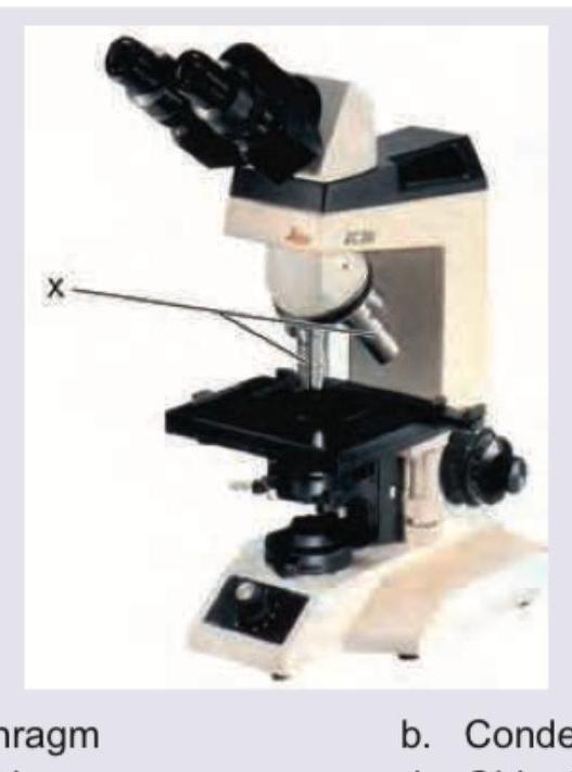
Name the part of the microscope marked as X.

Neural tube defects have which one of the following inheritance patterns ?
All are examples of negative feedback except
DEXA scan of lumbar vertebra and femoral neck are assessed for bone mineral density in menopausal women because of all except
BMR is increased in all except:
Which of the following is a height dependent index?
Which of the following is false regarding suspended animation
Explanation: **Explanation:** **Basal Metabolic Rate (BMR)** is the minimum energy expenditure required to maintain vital functions at rest. The correct answer is **Obesity** because BMR is primarily determined by **Lean Body Mass (LBM)**. In obese individuals, the ratio of adipose tissue to lean muscle is high. Since fat is metabolically less active than muscle, the overall BMR per unit of body weight is decreased compared to lean individuals. **Analysis of Options:** * **Hyperthyroidism (Incorrect):** Thyroid hormones ($T_3$ and $T_4$) are the primary regulators of BMR. In hyperthyroidism, increased $Na^+-K^+$ ATPase activity and cellular metabolism significantly **increase** BMR. * **Starvation (Incorrect):** While BMR does decrease during prolonged starvation as an adaptive mechanism to conserve energy, the question asks for a condition characterized by a lower baseline rate relative to body composition. (Note: In some contexts, starvation is a correct answer for decreased BMR, but in clinical physiology, the metabolic inefficiency of adipose tissue in obesity is a classic teaching point). * **Exercise (Incorrect):** Physical activity increases energy expenditure and oxygen consumption, leading to a significant **increase** in the metabolic rate above basal levels. **High-Yield Clinical Pearls for NEET-PG:** * **Surface Area Rule:** BMR is directly proportional to the surface area of the body. * **Age & Gender:** BMR is higher in children (due to growth) and males (due to higher testosterone and muscle mass). It declines with age. * **Specific Dynamic Action (SDA):** Protein has the highest SDA (30%), meaning it increases metabolic rate the most during digestion. * **Hormonal Influence:** Epinephrine and Testosterone increase BMR, while Hypothyroidism is the most common pathological cause of decreased BMR.
Explanation: **Explanation:** The regulation of food intake is controlled by the hypothalamus, specifically the balance between **orexigenic** (appetite-stimulating) and **anorexigenic** (appetite-suppressing) signals. **Correct Answer: B. Ghrelin** Ghrelin is the only major peripheral hormone that is **orexigenic**. It is secreted primarily by the P/D1 cells of the gastric fundus and epsilon cells of the pancreas. Ghrelin levels rise during fasting (the "hunger hormone") and stimulate the **NPY/AgRP neurons** in the Arcuate Nucleus of the hypothalamus to increase food intake and growth hormone secretion. **Analysis of Incorrect Options:** * **A. Serotonin (5-HT):** This is a potent **anorexigenic** neurotransmitter. It suppresses appetite by stimulating POMC/CART neurons and inhibiting NPY/AgRP neurons. Drugs like Lorcaserin (5-HT2C agonist) are used in obesity management for this reason. * **C & D (OCX and GA):** These are not standard physiological abbreviations for major metabolic hormones. In the context of appetite regulation, they serve as distractors. **High-Yield Clinical Pearls for NEET-PG:** * **Leptin:** Produced by adipocytes; it is the primary long-term **anorexigenic** hormone (satiety signal). It inhibits NPY and stimulates POMC. * **Prader-Willi Syndrome:** Characterized by hyperphagia and obesity due to pathologically high levels of Ghrelin. * **Vagus Nerve:** Ghrelin signals reach the brain via both the bloodstream and the vagus nerve. * **Sleep Deprivation:** Increases Ghrelin and decreases Leptin, leading to increased appetite and weight gain.
Explanation: **Explanation:** The correct answer is **D. Increases oxygen release to tissues.** Fever is a systemic response to infection characterized by an elevation in core body temperature. This physiological change has a direct impact on hemoglobin's affinity for oxygen. According to the **Bohr Effect** and the dynamics of the **Oxygen-Hemoglobin Dissociation Curve (ODC)**, an increase in temperature causes a **Right Shift** of the curve. A right shift indicates a decreased affinity of hemoglobin for oxygen, thereby facilitating the unloading (release) of oxygen into the peripheral tissues to meet the increased metabolic demands during an infection. **Analysis of Incorrect Options:** * **Option A:** Fever is actually a host defense mechanism. Elevated temperatures generally **inhibit** the replication of many bacteria and viruses while enhancing the phagocytic activity of leukocytes. * **Option B:** As stated, hyperthermia (fever) causes a **Right Shift**, not a left shift. A left shift (caused by hypothermia or alkalosis) would increase oxygen affinity and decrease tissue delivery. * **Option C:** While Interleukin-1 (IL-1) is a key endogenous pyrogen, it is primarily released by **macrophages and monocytes** (innate immunity) rather than CD4 T-helper cells, though T-cells can produce other cytokines like IL-6 and TNF-α. **High-Yield NEET-PG Pearls:** * **Right Shift Factors (Mnemonic: CADET, face Right!):** **C**O2 increase, **A**cidosis (H+), **D**PG (2,3-BPG) increase, **E**xercise, and **T**emperature increase. * **Pyrogen Pathway:** Exogenous pyrogens (endotoxins) → Macrophages release IL-1, IL-6, TNF → Hypothalamus → **PGE2** release (inhibited by NSAIDs/Antipyretics) → Set-point elevation → Fever. * **Benefit of Fever:** It enhances the "Respiratory Burst" in neutrophils, aiding bacterial killing.
Explanation: ***Condenser*** - The **condenser** is located beneath the stage and **focuses light** from the illuminator onto the specimen, enhancing image contrast and resolution. - The arrow 'X' points directly to this component, which typically contains lenses to gather and concentrate light. *Diaphragm* - The **diaphragm** is usually located within the condenser and controls the **amount of light** passing through the specimen. - While related to the condenser, the arrow 'X' points to the entire structure, not just the aperture within it. *Eye-piece* - The **eye-piece** (or ocular lens) is located at the top of the microscope, where the observer looks to view the magnified image. - The arrow 'X' is clearly pointing to a component below the stage, far from the eye-pieces. *Objective lens* - **Objective lenses** are mounted on the revolving nosepiece directly above the specimen and are responsible for the primary magnification of the specimen. - The arrow 'X' is pointing to a structure below the stage, not the objective lenses.
Explanation: ***Multi-factorial*** - Neural tube defects (NTDs) are considered **multi-factorial**, meaning they result from a complex interaction between multiple genetic predispositions and environmental factors. - While there are genetic components, no single gene mutation typically explains the recurrence risk, and external factors like **folic acid deficiency** play a significant role. *X-linked recessive* - This inheritance pattern typically affects males more severely and exclusively, with females often being carriers, which is not the primary pattern observed in NTDs. - Conditions like **Duchenne muscular dystrophy** exhibit X-linked recessive inheritance. *Autosomal dominant* - A single copy of an altered gene on a non-sex chromosome is sufficient to cause the condition, resulting in a 50% chance of transmission to offspring, which does not match the observed inheritance pattern for NTDs. - Examples include **Huntington's disease** and **Marfan syndrome**. *Autosomal recessive* - Both copies of a gene on a non-sex chromosome must be altered for the condition to manifest, meaning parents are often carriers but unaffected, which isn't the primary inheritance pattern for NTDs. - Conditions like **cystic fibrosis** and **sickle cell anemia** follow autosomal recessive inheritance.
Explanation: ***Coagulation of the blood*** - **Blood coagulation** is a classic example of **positive feedback**, where the initial clotting process amplifies itself until bleeding stops - Platelets aggregate and release factors that promote further platelet aggregation and activation of the clotting cascade, thereby **accelerating the response** rather than diminishing it - This is the exception among the options, as it represents positive feedback while all others are negative feedback *Regulation of blood CO2 level* - The regulation of **blood CO2 levels** is a vital example of **negative feedback**, where an increase in CO2 stimulates breathing to expel excess CO2 - This mechanism works to return the blood CO2 concentration to its homeostatic set point, thus **counteracting the initial stimulus** - Central and peripheral chemoreceptors detect elevated CO2 and trigger increased ventilation *Regulation of pituitary hormones* - The regulation of **pituitary hormones** involves **negative feedback loops**, where high levels of target gland hormones inhibit the release of stimulating hormones from the pituitary and hypothalamus - For example, high thyroid hormone levels inhibit TSH release from the pituitary and TRH from the hypothalamus - This effectively **reduces the initial stimulus** and maintains hormonal balance *Regulation of blood pressure* - The regulation of **blood pressure** is primarily controlled by **negative feedback mechanisms** involving baroreceptors, which detect changes in pressure - If blood pressure rises, baroreceptors in the carotid sinus and aortic arch signal the medulla to reduce heart rate and dilate blood vessels - This response **lowers the pressure back to the set point**, maintaining cardiovascular homeostasis
Explanation: ***Lumbar vertebra is 80% compact bone.*** - This statement is incorrect. The vertebral bodies of the lumbar spine are predominantly composed of **trabecular (spongy) bone**, accounting for approximately 75% of their mass, not compact bone. - **Compact bone** is dense and found mainly in the shafts of long bones and the outer layer of all bones, while **trabecular bone** is porous and found in the ends of long bones and inside vertebrae. *Trabecular bone has faster re-modelling rate.* - This is a correct statement and a reason why these sites are chosen for DEXA scans. **Trabecular bone** undergoes faster bone turnover compared to cortical bone, making it more sensitive to metabolic changes and bone loss. - Due to its higher metabolic activity, changes in bone density at these sites can be detected earlier in conditions like **osteoporosis**. *Early rapid bone loss can be determined by evaluation of this site with trabecular bone.* - This is a correct statement. Since the lumbar vertebrae and femoral neck contain a significant amount of **trabecular bone**, which has a higher remodeling rate, these sites are ideal for detecting **early and rapid bone loss**, especially in postmenopausal women. - The rapid turnover of trabecular bone means that changes in bone density due to estrogen deficiency or other causes will manifest here sooner than in predominantly cortical bone sites. *Lumbar vertebrae contain primarily trabecular bone.* - This is a correct statement and a reason for choosing this site. The **lumbar vertebral bodies are rich in trabecular bone**, which makes them highly susceptible to bone loss in conditions like osteoporosis. - The high proportion of **trabecular bone** in these areas allows for sensitive detection of density changes using a DEXA scan.
Explanation: ***Hypothyroidism*** - **Hypothyroidism** leads to a **decreased metabolic rate** due to insufficient thyroid hormone production, thus **reducing BMR**. - Symptoms like **weight gain**, **fatigue**, and **cold intolerance** are consistent with a lower metabolic rate. *Fever* - **Fever** causes an **increase in body temperature**, which directly elevates the **metabolic rate** as the body expends more energy to combat infection. - Each degree Celsius rise in body temperature increases BMR by approximately **13%**. *Exercise* - **Chronic exercise training** leads to an **increase in muscle mass**, which is metabolically more active than fat tissue. - This increased muscle mass contributes to a **higher resting metabolic rate (BMR)** over time, even when at rest. - The adaptive response to regular physical activity permanently elevates baseline energy expenditure. *Hyperthyroidism* - **Hyperthyroidism** results in an **overproduction of thyroid hormones**, significantly **increasing the metabolic rate**. - This leads to symptoms such as **weight loss**, **heat intolerance**, and **tachycardia**, all indicative of an elevated BMR.
Explanation: ***Ponderal*** - The **Ponderal Index** is calculated as **weight (kg) / height (m)³**, making it directly dependent on height. - It is the **standard height-dependent index** used in pediatrics and neonatal medicine to assess body proportionality. - Also known as the **Corpulence Index or Rohrer's Index**, it helps evaluate relative leanness or heaviness in relation to height. - The height³ in the denominator makes it highly sensitive to height changes. *Corpulence* - **Corpulence (Rohrer's Index)** is essentially the same as the Ponderal Index (weight/height³). - While technically also height-dependent, **Ponderal Index is the more widely recognized and standard term** in medical literature and examinations. *Lorent'z* - The **Lorentz formula** calculates ideal body weight using height. - Formula: IBW = Height (cm) - 100 - [(Height - 150)/k], where k varies by sex. - It's a **height-based calculation for ideal weight**, not a height-dependent proportionality index. *Brocas* - **Broca's Index** estimates ideal body weight using: Height (cm) - 100. - While it uses height, it's a **simple subtraction formula for target weight**, not a ratio/index that measures body proportions relative to height.
Explanation: ***Correct Answer: Suspended animation can be voluntarily induced*** **This statement is FALSE**, making it the correct answer to this question. - In accepted medical and scientific practice, suspended animation **cannot be voluntarily induced** by an individual - Suspended animation requires **medical interventions** such as therapeutic hypothermia, pharmacological agents, or extreme environmental conditions - It is not under voluntary control like breath-holding or meditation - While anecdotal claims exist about certain yogic practices, there is **no scientific evidence** supporting voluntary induction of true suspended animation in humans - This is a key distinction from other altered states that can be voluntarily achieved *Incorrect: Suspended animation is a state of apparent death* - This statement is **TRUE** - suspended animation is classically defined as a state of apparent death - The individual appears lifeless with dramatically reduced vital signs (heart rate, respiration, metabolism) - However, unlike actual death, this state is **reversible** - "Apparent death" is a legitimate medical term describing this condition *Incorrect: Patients in suspended animation can be resuscitated* - This statement is **TRUE** - reversibility is the defining characteristic of suspended animation - Successful resuscitation distinguishes suspended animation from actual death - The ability to restore normal physiological function is essential to the concept *Incorrect: Suspended animation typically lasts for 6 hours* - While this specific timeframe is imprecise, in **therapeutic hypothermia** and emergency preservation and resuscitation (EPR), durations can range from minutes to hours - The statement has some basis in clinical practice timeframes, though duration varies by method and patient - This is not definitively false compared to the voluntary induction claim
Cell Structure and Function
Practice Questions
Membrane Transport Mechanisms
Practice Questions
Bioelectric Phenomena
Practice Questions
Homeostasis and Feedback Mechanisms
Practice Questions
Body Fluid Compartments
Practice Questions
Signal Transduction Mechanisms
Practice Questions
Cell-to-Cell Communication
Practice Questions
Principles of Physiological Measurement
Practice Questions
Osmosis and Osmotic Pressure
Practice Questions
Physiological Adaptation Mechanisms
Practice Questions
Get full access to all questions, explanations, and performance tracking.
Start For Free