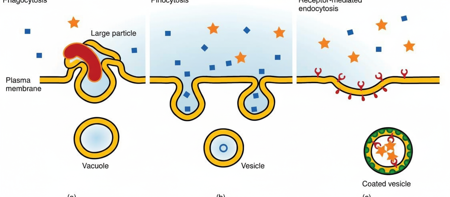
Which statement about the following cellular phenomenon is false?

All are seen more in ECF except?
Pacinian corpuscles transmit which sensation?
What is the approximate protein to lipid ratio in myelin?
What is the main constituent of the plasma membrane?
Inulin is used for the measurement of which body fluid compartment?
All of the following are stress responses except?
Gap junctions are present in?
Which is the efferent neuron for skeletal muscle?
Stem cells are found in which of the following locations?
Explanation: ***Clathrin-mediated endocytosis is involved in the uptake of dead tissues and bacteria.*** - **Clathrin-mediated endocytosis** is a form of **receptor-mediated endocytosis** that uses **clathrin-coated pits** to internalize specific molecules like **LDL cholesterol** and **transferrin**. - The uptake of **dead tissues** and **bacteria** is accomplished through **phagocytosis**, which involves **actin polymerization** and formation of **phagosomes**, not clathrin-coated vesicles. *This phenomenon includes phagocytosis and pinocytosis.* - **Endocytosis** encompasses multiple types including **phagocytosis** (cell eating), **pinocytosis** (cell drinking), and **receptor-mediated endocytosis**. - **Phagocytosis** involves uptake of large particles like bacteria, while **pinocytosis** involves uptake of fluid and dissolved substances. *It leads to endosome formation.* - **Endocytosis** results in the formation of **endosomes**, which are membrane-bound vesicles within the cytoplasm. - **Early endosomes** can mature into **late endosomes** and eventually fuse with **lysosomes** for degradation of internalized material. *None of the above.* - This option is incorrect because option B is indeed a **false statement** about endocytosis. - **Clathrin-mediated endocytosis** does not handle uptake of **dead tissues and bacteria**, making this the correct answer to identify the false statement.
Explanation: ### Explanation The composition of body fluids is a fundamental concept in General Physiology. The body is divided into the **Extracellular Fluid (ECF)** and **Intracellular Fluid (ICF)**, each maintained by specific transporters (like the Na+-K+ ATPase pump) to have distinct ionic profiles. **Why Magnesium (Mg++) is the Correct Answer:** Magnesium is primarily an **intracellular cation**. In fact, it is the **second most abundant intracellular cation** after Potassium (K+). Approximately 99% of total body magnesium is located within cells (mostly in bones and soft tissues), while only about 1% is present in the ECF. Therefore, its concentration is significantly higher inside the cell than outside. **Analysis of Incorrect Options (ECF Components):** * **Na+ (Sodium):** The predominant extracellular cation. It is vital for maintaining ECF volume and osmotic pressure. * **Cl- (Chloride):** The major extracellular anion. It usually follows sodium to maintain electrical neutrality. * **HCO3- (Bicarbonate):** A key extracellular buffer. While present in the ICF, its concentration is significantly higher in the ECF to manage systemic acid-base balance. **High-Yield NEET-PG Pearls:** * **Major Intracellular Cations:** Potassium (K+) > Magnesium (Mg++). * **Major Intracellular Anions:** Phosphates > Proteins. * **Major Extracellular Cations:** Sodium (Na+) > Calcium (Ca++). * **Major Extracellular Anions:** Chloride (Cl-) > Bicarbonate (HCO3-). * **Clinical Note:** Hypomagnesemia often coexists with hypokalemia and hypocalcemia; you cannot correct K+ levels effectively until Mg++ levels are replenished.
Explanation: **Explanation:** **1. Why Vibration is Correct:** Pacinian corpuscles are **rapidly adapting (phasic) mechanoreceptors** located in the deeper layers of the dermis, subcutaneous tissue, and mesentery. They are specifically designed to detect **high-frequency vibration** (250–350 Hz) and rapid changes in mechanical pressure. Their unique structure—consisting of a central nerve terminal surrounded by concentric layers of connective tissue (lamellae)—acts as a filter. This allows only rapid, transient stimuli to reach the nerve fiber, making them the most sensitive receptors for detecting "flutter" or vibratory sensations. **2. Why the Other Options are Incorrect:** * **Cold (Option A):** These sensations are primarily detected by **Krause end bulbs** and free nerve endings (specifically A-delta fibers). * **Heat (Option B):** Warmth is detected by **Ruffini endings** (which also sense skin stretch) and free nerve endings (C-fibers). * **Fine Touch (Option C):** This is the primary function of **Meissner’s corpuscles** (rapidly adapting, located in dermal papillae) and **Merkel discs** (slowly adapting, for pressure and texture). **3. High-Yield Clinical Pearls for NEET-PG:** * **Adaptation:** Pacinian corpuscles are the *fastest* adapting receptors in the body. * **Receptive Field:** They have **large receptive fields** with poorly defined borders, unlike Meissner’s corpuscles which have small, crisp fields. * **Pathway:** Vibratory sensation is carried via the **Dorsal Column-Medial Lemniscal (DCML) pathway**. * **Clinical Testing:** Vibration is often the first sensation lost in peripheral neuropathies (e.g., Diabetes Mellitus) and is tested clinically using a **128 Hz tuning fork**.
Explanation: **Explanation:** The composition of a cell membrane varies significantly depending on its physiological function. In most biological membranes (like the erythrocyte membrane), the protein-to-lipid ratio is approximately **1:1**. However, **myelin** is a unique exception. **Why 1:4 is correct:** Myelin serves as an electrical insulator for axons to facilitate saltatory conduction. To function as an effective insulator, it requires a high dielectric constant, which is provided by a high lipid content. Myelin is composed of approximately **75-80% lipids** and only **20-25% proteins**. This results in a protein-to-lipid ratio of roughly **1:4** (or 0.25). **Analysis of Incorrect Options:** * **A (1:1):** This is the standard ratio for a typical plasma membrane (e.g., Red Blood Cells), where proteins and lipids are present in roughly equal weights. * **B (2:1) & C (3:1):** These ratios represent protein-heavy membranes. For example, the **inner mitochondrial membrane** has a very high protein-to-lipid ratio (approx. 3:1) because it is packed with enzymes and carriers for the electron transport chain. **High-Yield Clinical Pearls for NEET-PG:** * **Primary Lipid in Myelin:** Cholesterol is the most common lipid, but **Galactocerebroside** is the characteristic glycolipid of myelin. * **Major Proteins:** Myelin Basic Protein (MBP) and Proteolipid Protein (PLP) in the CNS; Protein Zero (P0) in the PNS. * **Clinical Correlation:** In **Multiple Sclerosis**, the immune system attacks these myelin proteins, leading to demyelination and loss of the "insulation," which slows or blocks nerve impulses. * **Nodes of Ranvier:** These are the gaps in the myelin sheath where the protein density (specifically Na+ channels) is highest.
Explanation: **Explanation:** The plasma membrane is a selectively permeable lipid bilayer that defines the cell boundary. According to the **Fluid Mosaic Model** (Singer and Nicolson), the membrane is composed of proteins, lipids, and carbohydrates. **Why Protein is the Correct Answer:** While the "bilayer" is made of lipids, **proteins are the main constituent by weight** in most cell membranes. In a typical human cell membrane (like the erythrocyte membrane), the composition is approximately **52% Proteins**, 40% Lipids, and 8% Carbohydrates. Proteins are responsible for the membrane's functional diversity, acting as transporters, receptors, enzymes, and structural anchors. **Analysis of Incorrect Options:** * **B & C. Lipid/Phospholipid:** Although phospholipids form the structural backbone (the "matrix") of the membrane and are numerically more abundant, they account for only about 40% of the total mass. Phospholipids provide fluidity, but proteins provide the bulk of the weight. * **A. Carbohydrate:** These are the least abundant components (approx. 2-10%). They are found only on the outer surface, forming the **glycocalyx**, which is crucial for cell-to-cell recognition and immune response. **High-Yield NEET-PG Pearls:** * **Exception to the Rule:** The **Myelin sheath** is a notable exception where lipids (80%) outweigh proteins (20%) to provide electrical insulation. * **Mitochondrial Membrane:** The inner mitochondrial membrane has the highest protein-to-lipid ratio (approx. 3:1 or 75% protein) due to the presence of the electron transport chain. * **Cholesterol:** It is present in eukaryotic membranes to regulate fluidity; it is notably absent in prokaryotic membranes.
Explanation: **Explanation:** The measurement of body fluid compartments is based on the **Indicator Dilution Principle** ($Volume = Amount / Concentration$). To measure a specific compartment, the substance used must be able to distribute evenly within that compartment while being unable to cross the boundaries into others. **Why ECF is the correct answer:** **Inulin** is a polysaccharide that is freely filtered by the glomerulus but is neither secreted nor reabsorbed. Most importantly, it is a large molecule that **cannot cross the cell membrane**, meaning it stays outside the cells. However, it is small enough to pass through capillary pores into the interstitial space. Therefore, it distributes throughout the entire **Extracellular Fluid (ECF)**—which includes both plasma and interstitial fluid—making it the gold standard for ECF measurement. **Why other options are incorrect:** * **Intracellular Fluid (ICF):** There is no direct substance to measure ICF because no indicator distributes *only* inside cells. ICF is calculated indirectly: $ICF = Total Body Water (TBW) - ECF$. * **Plasma Volume:** Substances used for plasma must be too large to leave the capillaries. Examples include **Evans Blue dye** or **Radio-iodinated Serum Albumin (RISA)**. * **Blood Volume:** This is calculated using **Chromium-51 ($^{51}Cr$) labeled RBCs** or by using the formula: $Blood Volume = Plasma Volume / (1 - Hematocrit)$. **High-Yield Clinical Pearls for NEET-PG:** * **Total Body Water (TBW):** Measured using Tritiated water ($D_2O$), Deuterium oxide, or Aminopyrine. * **ECF Markers:** Inulin (Gold Standard), Mannitol, Sucrose, and Sodium Thiosulfate. * **Interstitial Fluid:** Calculated indirectly: $ECF - Plasma Volume$. * **Rule of thumb:** Inulin is also the gold standard for measuring **Glomerular Filtration Rate (GFR)**.
Explanation: The physiological response to stress (trauma, surgery, or sepsis) is a complex neuroendocrine reaction designed to maintain homeostasis and mobilize energy. **Why "Decrease in blood glucose" is the correct answer:** Stress triggers a state of **hyperglycemia**, not hypoglycemia. This occurs due to the surge of counter-regulatory hormones (cortisol, catecholamines, glucagon, and growth hormone) which promote gluconeogenesis and glycogenolysis in the liver. This "stress diabetes" ensures a steady supply of glucose for the brain and wound healing. **Explanation of other options:** * **A. Increased autonomic nervous system activity:** Stress activates the Sympathetic Nervous System (SNS), leading to the "fight or flight" response. This results in increased heart rate, blood pressure, and redirection of blood flow to vital organs. * **B. Increased peripheral insulin resistance:** High levels of cortisol and catecholamines interfere with insulin signaling and GLUT-4 translocation. This prevents peripheral tissues (like muscle) from consuming glucose, preserving it for the central nervous system. * **D. Loss of muscle protein:** Stress induces a **catabolic state**. Cortisol promotes proteolysis (breakdown of skeletal muscle) to provide amino acids for hepatic gluconeogenesis and the synthesis of acute-phase proteins. **High-Yield Clinical Pearls for NEET-PG:** * **The "ebb" and "flow" phases:** Stress response is divided into the Ebb phase (initial 24 hours, decreased BMR) and the Flow phase (hypermetabolism and catabolism). * **Negative Nitrogen Balance:** Due to massive protein breakdown and nitrogen excretion in urine, stress patients typically exhibit a negative nitrogen balance. * **Cytokine involvement:** IL-1, IL-6, and TNF-alpha are the primary inflammatory mediators driving the systemic metabolic response to stress.
Explanation: **Explanation:** **Gap junctions** (communicating junctions) are specialized intercellular connections composed of proteins called **connexins**. They allow the direct passage of ions and small molecules between adjacent cells, facilitating electrical and metabolic coupling. 1. **Why Smooth Muscle is Correct:** In **unitary (visceral) smooth muscle** (e.g., in the GI tract, uterus, and small blood vessels), gap junctions are essential for coordinated contraction. They allow action potentials to spread rapidly from one cell to another, enabling the tissue to function as a **functional syncytium**. Gap junctions are also prominently found in **cardiac muscle** (at the intercalated discs). 2. **Why Other Options are Incorrect:** * **Skeletal Muscle:** Unlike smooth or cardiac muscle, skeletal muscle fibers are electrically isolated from one another. Each fiber must be individually stimulated by a motor neuron at the neuromuscular junction; hence, gap junctions are absent. * **Choroid Plexus & Renal Tubular Epithelium:** These tissues are characterized by **Tight Junctions (Zonula occludens)**. Tight junctions are necessary to maintain a selective barrier (Blood-CSF barrier and tubular reabsorption barrier) and prevent the paracellular leakage of solutes, which is the opposite function of the "open" channels provided by gap junctions. **High-Yield NEET-PG Pearls:** * **Connexon:** A functional gap junction unit formed by 6 connexin subunits. * **Locations:** Gap junctions are found in the Heart, Smooth muscle, Osteocytes, and Electrical synapses (CNS). * **Clinical Correlation:** Mutations in connexin genes are linked to conditions like **Charcot-Marie-Tooth disease** (Cx32) and **congenital deafness** (Cx26). * **Regulation:** Gap junctions close in response to high intracellular $Ca^{2+}$ or low intracellular pH to prevent the spread of damage between cells.
Explanation: **Explanation:** The contraction of skeletal muscle is governed by the **Lower Motor Neurons (LMN)** located in the anterior horn of the spinal cord. 1. **Why Alpha (α) Motor Neurons are correct:** These are large, multipolar lower motor neurons that innervate the **extrafusal muscle fibers** (the regular muscle fibers responsible for force production and contraction). When an alpha motor neuron fires, it releases acetylcholine at the neuromuscular junction, leading to muscle shortening. This is the primary efferent pathway for voluntary and reflex skeletal muscle movement. 2. **Why the other options are incorrect:** * **Gamma (γ) motor neurons:** These are also efferent neurons, but they innervate **intrafusal fibers** within the muscle spindle. Their role is to regulate the sensitivity of the spindle to stretch, not to produce skeletal muscle contraction. * **Ia fibers:** These are **sensory (afferent)** fibers. They originate from the primary endings of the muscle spindle and carry information about the *rate of change* in muscle length to the CNS (responsible for the stretch reflex). * **Ib fibers:** These are **sensory (afferent)** fibers. They originate from the **Golgi Tendon Organ (GTO)** and carry information regarding *muscle tension* to prevent over-contraction and injury. **High-Yield Clinical Pearls for NEET-PG:** * **Alpha-Gamma Co-activation:** During voluntary movement, both alpha and gamma neurons fire simultaneously to ensure the muscle spindle remains sensitive even when the extrafusal muscle shortens. * **Size Principle (Henneman's):** Small alpha motor neurons are recruited first (for fine motor tasks), followed by larger ones (for explosive power). * **Lesion Sign:** Damage to alpha motor neurons results in **Lower Motor Neuron (LMN) signs**: flaccid paralysis, fasciculations, hypotonia, and areflexia.
Explanation: ### Explanation **Correct Option: C. Base of intestinal crypts** Adult stem cells (somatic stem cells) are undifferentiated cells found in various tissues that serve as an internal repair system. In the gastrointestinal tract, the epithelium undergoes rapid turnover (every 3–5 days). To maintain this, **intestinal stem cells (ISCs)** are located specifically at the **base of the Crypts of Lieberkühn**. These stem cells, often identified by the marker **Lgr5+**, divide to produce "transit-amplifying cells," which then migrate upward to differentiate into mature enterocytes, goblet cells, enteroendocrine cells, and Paneth cells (though Paneth cells remain at the base). **Why other options are incorrect:** * **A. Retina:** The adult human retina is considered a non-regenerative tissue. While some research explores Müller glia as potential progenitors, they do not function as active stem cell niches in the same way intestinal crypts do. * **B. Endometrium:** While the endometrium has high regenerative capacity, the question typically refers to the most classic, well-established physiological niche taught in standard medical textbooks (like Guyton or Ganong). *Note: While endometrial stem cells exist in the basalis layer, the "Base of intestinal crypts" is the gold-standard textbook example for epithelial stem cell niches.* **High-Yield Clinical Pearls for NEET-PG:** * **Marker for Intestinal Stem Cells:** Lgr5 (Leucine-rich repeat-containing G-protein coupled receptor 5). * **Paneth Cells:** Located at the base of the crypts, they provide the necessary "niche" signals (like Wnt) to maintain stem cell pluripotency. * **Hematopoietic Stem Cells (HSCs):** Found in the bone marrow; these are the most clinically utilized stem cells (CD34+ is the classic marker). * **Potency:** Adult stem cells are typically **multipotent** (can form multiple cell types of a specific lineage), whereas embryonic stem cells are **pluripotent**.
Cell Structure and Function
Practice Questions
Membrane Transport Mechanisms
Practice Questions
Bioelectric Phenomena
Practice Questions
Homeostasis and Feedback Mechanisms
Practice Questions
Body Fluid Compartments
Practice Questions
Signal Transduction Mechanisms
Practice Questions
Cell-to-Cell Communication
Practice Questions
Principles of Physiological Measurement
Practice Questions
Osmosis and Osmotic Pressure
Practice Questions
Physiological Adaptation Mechanisms
Practice Questions
Get full access to all questions, explanations, and performance tracking.
Start For Free