What is the primary function of ubiquitin?
All of the following intercellular communications occur via extracellular chemical messengers except?
What is the term for the smooth muscle cell membrane?
Which of the following statements is true regarding basal metabolic rate?
Total body water differences between males and females are not observed at which age range?
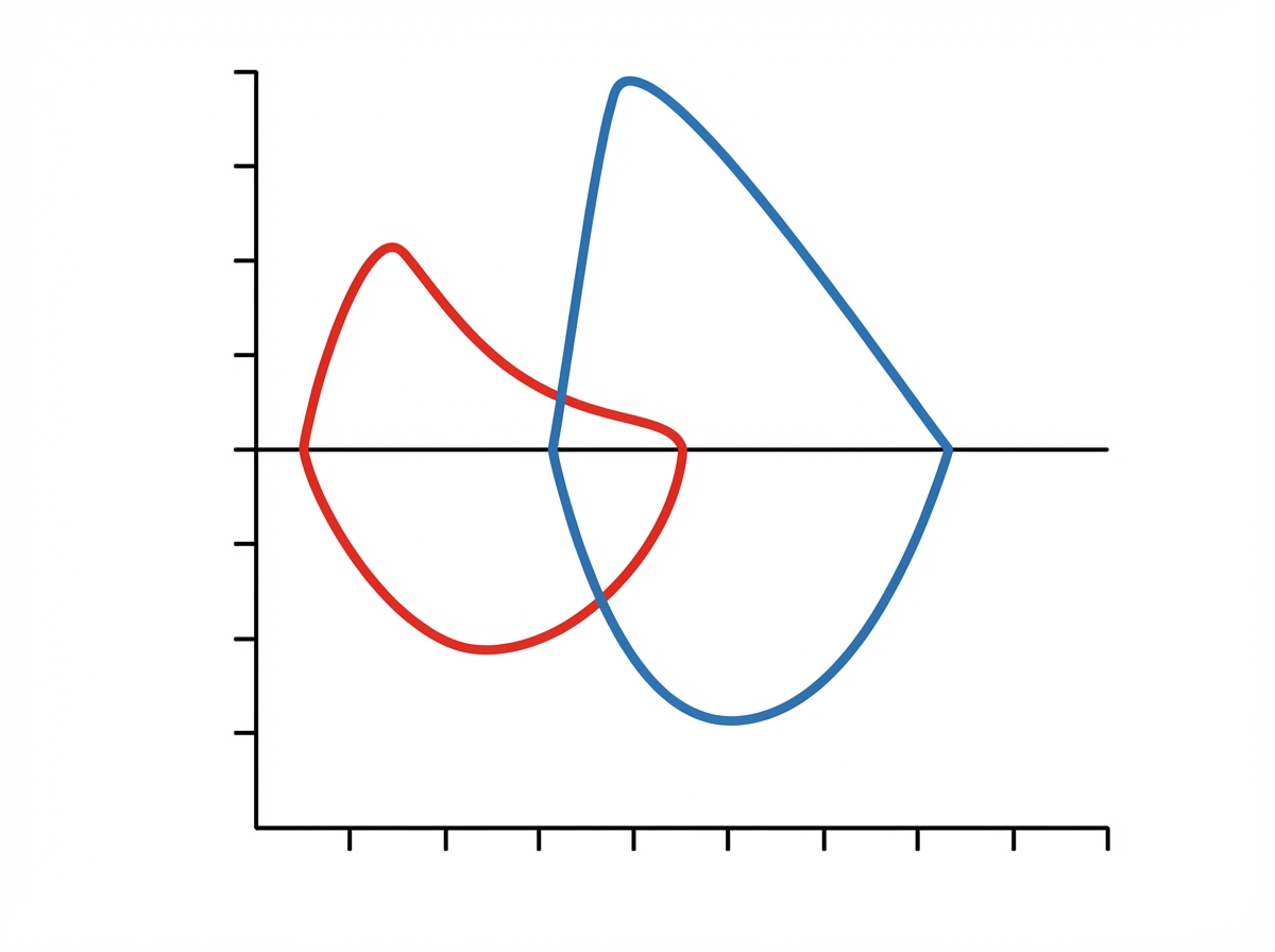
The given graph likely depicts which of the following conditions?

Within the endocrine system, specificity of communication is determined by what?
What is the reason for hyperosmolarity?
What are the functions of cytoplasmic enzymes in Red Blood Cells?
Which of the following processes requires a carrier protein?
Explanation: **Explanation:** The correct answer is **C. Degradation.** Ubiquitin is a small, highly conserved regulatory protein found in almost all eukaryotic tissues. Its primary function is to mark unwanted or damaged proteins for destruction via the **Ubiquitin-Proteasome Pathway (UPP)**. 1. **Why Degradation is Correct:** Proteins destined for degradation are tagged with a chain of ubiquitin molecules (polyubiquitination) through a series of enzymatic steps (E1, E2, and E3 enzymes). This "tag" acts as a molecular signal that directs the protein to the **26S Proteasome**, a barrel-shaped organelle that acts as the cell's "garbage disposal," breaking the protein down into small peptides. 2. **Why other options are incorrect:** * **Binding:** While ubiquitin does bind to proteins, this is a step in the process, not its ultimate physiological *function*. * **Transport:** Ubiquitin does not act as a primary carrier or transport protein (like albumin or hemoglobin). * **Assembly:** Ubiquitin is involved in disassembly (breakdown) rather than the synthesis or assembly of cellular structures. **High-Yield Clinical Pearls for NEET-PG:** * **ATP-Dependence:** The ubiquitin-proteasome pathway is an **ATP-dependent** process, distinguishing it from lysosomal degradation. * **Clinical Correlation:** Defects in the ubiquitin system are linked to neurodegenerative diseases like **Parkinson’s disease** (due to the accumulation of Lewy bodies) and **Alzheimer’s disease**. * **Bortezomib:** This is a proteasome inhibitor used clinically in the treatment of **Multiple Myeloma**, highlighting the importance of this pathway in cell cycle regulation. * **Angelman Syndrome:** Caused by a defect in the UBE3A gene, which encodes a ubiquitin ligase.
Explanation: ### Explanation Intercellular communication is broadly classified into two categories: **Direct communication** (physical contact) and **Indirect communication** (via chemical messengers). **Why Gap Junctions are the correct answer:** Gap junctions are specialized protein complexes (composed of **connexons**) that form direct physical bridges between the cytoplasm of adjacent cells. They allow the passage of ions and small molecules (like cAMP or $Ca^{2+}$) directly from one cell to another without the need for an extracellular chemical messenger. This allows for rapid electrical coupling and synchronized activity. **Analysis of Incorrect Options:** * **Paracrine (A):** Involves the release of chemical messengers into the interstitial fluid to act on neighboring cells (e.g., Histamine). * **Neurohormones (C):** These are chemicals released by neurons directly into the bloodstream to act on distant target cells (e.g., ADH/Oxytocin). * **Autocrine (D):** A cell secretes a chemical messenger that binds to receptors on its own surface, affecting its own function (e.g., Interleukin-1 in T-cells). All three (A, C, and D) rely on **extracellular chemical messengers** (ligands) to transmit signals. **High-Yield Facts for NEET-PG:** * **Connexon Structure:** Each gap junction is made of two connexons; each connexon is composed of **6 connexin** proteins. * **Location:** Gap junctions are vital in **cardiac muscle** (intercalated discs) and **smooth muscle** for functional syncytium. * **Juxtacrine Signaling:** Another form of direct communication where a surface protein on one cell binds to a receptor on an adjacent cell (e.g., Notch signaling). * **Speed:** Gap junctions provide the fastest mode of intercellular signal transmission.
Explanation: **Explanation:** The correct answer is **B. Sarcolemma**. In muscle physiology, specific terminology is used to describe the cellular components of muscle fibers (skeletal, cardiac, and smooth). The **sarcolemma** (derived from the Greek *sarx* meaning "flesh" and *lemma* meaning "sheath") is the specialized plasma membrane that surrounds a muscle cell. It acts as a barrier between the intracellular and extracellular compartments and plays a critical role in conducting action potentials that trigger contraction. **Analysis of Incorrect Options:** * **A. Sarcoplasm:** This refers to the **cytoplasm** of a muscle cell. It contains glycogen, myoglobin, and the organelles necessary for muscle metabolism. * **C. Sarcomere:** This is the **functional and structural unit** of a myofibril, defined as the segment between two successive Z-lines. Note: While skeletal and cardiac muscles have sarcomeres (striated), smooth muscle lacks organized sarcomeres, which is why it appears "smooth." * **D. Sarcoplasmic reticulum:** This is a specialized form of the **endoplasmic reticulum** that functions primarily as an intracellular storage site for calcium ions ($Ca^{2+}$). **High-Yield NEET-PG Pearls:** * **Caveolae:** Unlike skeletal muscle which has T-tubules, smooth muscle cells have small invaginations of the sarcolemma called **caveolae**, which help in transmitting signals and $Ca^{2+}$ entry. * **Dense Bodies:** In smooth muscle, actin filaments are attached to **dense bodies** (analogous to Z-lines in striated muscle), some of which are dispersed in the sarcoplasm and others attached to the sarcolemma. * **Gap Junctions:** The sarcolemma of "unitary" (visceral) smooth muscle contains numerous gap junctions to allow for coordinated, syncytial contraction.
Explanation: ### Explanation **Correct Answer: D. Decreased by 50% in starvation** **Understanding the Concept:** Basal Metabolic Rate (BMR) represents the minimum energy expenditure required to maintain vital functions (like breathing and circulation) at complete physical and mental rest. In states of **prolonged starvation**, the body initiates a survival mechanism to conserve energy. This leads to a significant reduction in BMR—potentially by as much as **50%**. This decrease is primarily mediated by a reduction in muscle mass (atrophy) and a decrease in the levels of circulating thyroid hormones (specifically the conversion of T4 to the more active T3) and catecholamines. **Analysis of Incorrect Options:** * **A. Increased in starvation:** This is incorrect. Starvation is a hypometabolic state. An increase in BMR is seen in conditions like hyperthyroidism, fever, and pregnancy. * **B. It is not influenced by hormonal changes:** This is false. BMR is heavily regulated by hormones. **Thyroid hormones (T3/T4)** are the primary determinants of BMR. Epinephrine, cortisol, and growth hormone also increase BMR. * **C. It is not affected by dietary changes:** This is false. Specific Dynamic Action (SDA) or the thermic effect of food increases metabolic rate after ingestion. Conversely, chronic calorie restriction leads to a compensatory drop in BMR. **High-Yield Clinical Pearls for NEET-PG:** * **Thyroid Status:** The most important factor affecting BMR. BMR increases by 50–100% in hyperthyroidism and decreases by 30–50% in hypothyroidism. * **Surface Area:** BMR is more closely related to surface area than body weight (Rubner’s Law). * **Gender & Age:** BMR is higher in males (due to higher muscle mass/testosterone) and decreases progressively with age. * **Climate:** BMR is higher in individuals living in cold climates to maintain body temperature.
Explanation: **Explanation:** The primary determinant of Total Body Water (TBW) differences between genders is the **ratio of adipose tissue to lean body mass**. Adipose tissue is hydrophobic and contains very little water (~10%), whereas muscle tissue is approximately 75% water. **Why 18-25 years is the correct answer:** In the context of standard physiological teaching (and specifically regarding this question's logic), the period of **young adulthood (18-25 years)** is often cited as the baseline where physiological parameters are most "standardized" before the significant divergence in body composition seen in later life. However, it is important to note that physiologically, the *least* difference is actually seen **before puberty** (infancy/childhood). In the context of the provided options, the question tests the understanding of when gender-based hormonal influences on fat distribution are most established versus when they begin to equalize or shift. **Analysis of Incorrect Options:** * **Above 60 years & 40-60 years:** As humans age, the percentage of TBW decreases in both genders due to a loss of muscle mass (sarcopenia) and an increase in fat. However, the gap between males and females persists or widens due to established post-menopausal changes in females. * **10-18 years:** This is the period of puberty. The surge in testosterone in males leads to increased muscle mass, while estrogen in females increases subcutaneous fat deposition. Therefore, significant differences in TBW begin to emerge during this stage. **High-Yield NEET-PG Pearls:** 1. **Average TBW:** Adult Male ≈ 60% of body weight; Adult Female ≈ 50% (due to higher subcutaneous fat). 2. **Infants:** Have the highest TBW (~75-80%), which is why they are highly susceptible to dehydration. There is **no gender difference** in TBW during infancy. 3. **Elderly:** Have the lowest TBW (~45-50%) due to increased fat and decreased muscle. 4. **Formula:** $TBW = 0.6 \times \text{Body Weight (kg)}$. 5. **Order of Water Content:** Heart/Muscle > Skin > Skeleton > Fat.
Explanation: ***Emphysema*** - Shows classic **obstructive pattern** with reduced **FEV1** and **FEV1/FVC ratio** (<70%), indicating airflow limitation. - The **scooped-out expiratory limb** on flow-volume loop is pathognomonic for emphysema due to **loss of elastic recoil** and **airway collapse**. *Interstitial lung disease* - Would show a **restrictive pattern** with reduced **total lung capacity (TLC)** and **vital capacity** but preserved FEV1/FVC ratio. - Flow-volume loop typically shows a **narrow, tall pattern** rather than the obstructive scooping seen here. *Normal study* - Normal spirometry shows **FEV1/FVC ratio >70%** with smooth, symmetric flow-volume loop curves. - **Peak expiratory flow** and lung volumes would be within normal predicted ranges for age, height, and gender. *Kyphosis* - Causes a **restrictive pattern** due to **chest wall deformity** limiting lung expansion, not airflow obstruction. - Would show reduced lung volumes but **preserved FEV1/FVC ratio**, unlike the obstructive pattern depicted.
Explanation: ### Explanation **1. Why the Correct Answer is Right:** In the endocrine system, hormones are secreted into the bloodstream and distributed throughout the entire body. Despite being exposed to numerous tissues, a hormone only elicits a response in specific **target cells**. This specificity is governed by the presence of **specific receptors** (proteins) that have a high affinity for that particular hormone. If a cell lacks the receptor, it remains "blind" to the hormone's signal, regardless of the hormone's concentration. This is the fundamental principle of ligand-receptor interaction in endocrinology. **2. Why the Other Options are Wrong:** * **Option A:** While the chemical nature (water-soluble vs. lipid-soluble) determines how a hormone travels in the blood and where its receptor is located (surface vs. intracellular), it does not determine which specific cell responds. * **Option B:** Unlike paracrine signaling, endocrine communication is independent of distance; hormones are designed to act on distant organs via the circulatory system. * **Option C:** This describes the **nervous system**, where specificity is determined by precise **anatomical "hard-wiring"** (synapses). In contrast, the endocrine system is "wireless" and relies on molecular recognition. **3. NEET-PG High-Yield Pearls:** * **Up-regulation vs. Down-regulation:** Target cells can modulate their sensitivity by changing the number of receptors (e.g., insulin resistance involves down-regulation of receptors). * **Receptor Locations:** * **Cell Surface:** For peptides (Insulin, GH) and catecholamines. * **Cytoplasmic:** For Steroids (Glucocorticoids). * **Nuclear:** For Thyroid hormones (T3/T4) and Retinoic acid. * **Spare Receptors:** Often, a maximal biological response is achieved when only a small fraction of receptors are occupied (e.g., LH receptors in Leydig cells).
Explanation: **Explanation:** The hyperosmolarity of the renal medullary interstitium is essential for the kidney's ability to concentrate urine. This gradient is primarily established by the **Countercurrent Multiplier system** in the Loop of Henle. **Why Option C is Correct:** The **Na+-K+-2Cl- cotransporter (NKCC2)**, located in the Thick Ascending Limb (TAL) of the Loop of Henle, is the "engine" of the countercurrent multiplier. It actively pumps sodium, potassium, and chloride ions from the tubular lumen into the medullary interstitium. Because the TAL is impermeable to water, these solutes accumulate in the interstitium without being followed by water, directly increasing medullary osmolarity (hyperosmolarity). Increased activity of this transporter enhances the corticomedullary gradient. **Analysis of Incorrect Options:** * **Option A:** Increased glomerular capillary pressure increases the Glomerular Filtration Rate (GFR) but does not directly contribute to the establishment of the medullary osmotic gradient. * **Option B:** Decreased Na+ absorption in the Proximal Convoluted Tubule (PCT) would lead to increased solute delivery to the loop, but it is the *active transport* in the loop itself that creates hyperosmolarity. * **Option D:** Decreased activity of Na+-K+ ATPase (which provides the primary active transport energy for NKCC2) would reduce solute deposition in the interstitium, leading to **hypoosmolarity** and a loss of concentrating ability. **High-Yield NEET-PG Pearls:** * **Loop Diuretics (Furosemide):** Act by inhibiting the NKCC2 transporter. This "washes out" the medullary gradient, preventing water reabsorption in the collecting duct. * **Urea Recycling:** Contributes nearly 50% of the medullary hyperosmolarity, especially during dehydration (mediated by ADH). * **Vasa Recta:** Acts as a **Countercurrent Exchanger**, maintaining the gradient by removing water and returning solutes to the interstitium.
Explanation: ### Explanation Mature Red Blood Cells (RBCs) lack a nucleus and mitochondria, making them entirely dependent on cytoplasmic enzymes for survival and function. These enzymes primarily drive **anaerobic glycolysis (Embden-Meyerhof pathway)** and the **Pentose Phosphate Pathway (PPP)**. **1. Why Option A is Correct:** The primary role of cytoplasmic enzymes is to generate **ATP** and **NADPH**. ATP is essential for fueling the Na⁺/K⁺-ATPase pumps, which regulate intracellular volume and osmotic pressure. By maintaining the correct ionic balance and preventing cell swelling, these enzymes ensure the RBC remains **pliable and deformable**. This flexibility is critical for the 8μm RBC to squeeze through 2-3μm splenic capillaries without undergoing hemolysis. **2. Analysis of Incorrect Options:** * **Option B:** Cytoplasmic enzymes do the opposite; they *facilitate* and power the transmembrane transport of ions (like Na⁺ and K⁺) to maintain homeostasis. * **Option C:** This is a conceptual reversal. Enzymes like **Methemoglobin Reductase** function to keep iron in the **Ferrous (Fe²⁺)** state. The Ferric (Fe³⁺) state (Methemoglobin) cannot bind oxygen. * **Option D:** Enzymes like **Glucose-6-Phosphate Dehydrogenase (G6PD)** produce NADPH to *prevent* the oxidation of proteins. Oxidation leads to the formation of Heinz bodies and membrane damage. **Clinical Pearls for NEET-PG:** * **G6PD Deficiency:** The most common enzyme deficiency; leads to oxidative stress, Heinz bodies, and "bite cells" in the peripheral smear. * **Pyruvate Kinase Deficiency:** The most common enzyme deficiency in the Embden-Meyerhof pathway, leading to decreased ATP and chronic hemolytic anemia. * **2,3-BPG:** A byproduct of RBC glycolysis (Rapoport-Luebering Shunt) that shifts the oxygen dissociation curve to the right, facilitating oxygen unloading to tissues.
Explanation: **Explanation:** The correct answer is **Facilitated diffusion**. This process is a type of carrier-mediated transport where substances move down their electrochemical gradient (from high to low concentration) without the expenditure of energy (ATP). Because the molecules involved (like glucose or amino acids) are often large or polar, they cannot cross the lipid bilayer alone and require specific **integral membrane proteins (carriers or channels)** to facilitate their passage. **Analysis of Options:** * **Exocytosis (A):** This is a form of vesicular transport (bulk transport) where substances are expelled from the cell via membrane-bound vesicles. It requires ATP and the rearrangement of the cytoskeleton, rather than a specific transmembrane carrier protein. * **Simple diffusion (C):** This involves the movement of small, non-polar, or lipid-soluble molecules (e.g., $O_2$, $CO_2$) directly through the phospholipid bilayer. No carrier protein is required. * **Osmosis (D):** This is the passive movement of water molecules across a semi-permeable membrane. While water can move through specialized channels called **aquaporins**, the classic definition of osmosis refers to the diffusion of solvent molecules, which does not inherently require a carrier protein. **High-Yield NEET-PG Pearls:** 1. **Kinetics:** Unlike simple diffusion, facilitated diffusion is **saturable**. It follows Michaelis-Menten kinetics, reaching a maximum transport rate ($V_{max}$) when all carrier binding sites are occupied. 2. **Specificity:** Carrier proteins are highly specific for their ligands (e.g., **GLUT transporters** for glucose). 3. **Competitive Inhibition:** Because carriers have binding sites, structurally similar molecules can compete for transport, a feature not seen in simple diffusion. 4. **Insulin Action:** The translocation of **GLUT-4** to the cell membrane in muscle and adipose tissue is a classic physiological example of regulated facilitated diffusion.
Cell Structure and Function
Practice Questions
Membrane Transport Mechanisms
Practice Questions
Bioelectric Phenomena
Practice Questions
Homeostasis and Feedback Mechanisms
Practice Questions
Body Fluid Compartments
Practice Questions
Signal Transduction Mechanisms
Practice Questions
Cell-to-Cell Communication
Practice Questions
Principles of Physiological Measurement
Practice Questions
Osmosis and Osmotic Pressure
Practice Questions
Physiological Adaptation Mechanisms
Practice Questions
Get full access to all questions, explanations, and performance tracking.
Start For Free