What is the diameter of Type C fibers?
The magnitude of the electrical potential difference that exists across a membrane can be determined by which of the following equations?
Which type of muscle exercise involves the muscle changing length against a constant load?
What does the Golgi tendon organ primarily detect?
Oxidative capacity is high in which type of skeletal muscle fiber?
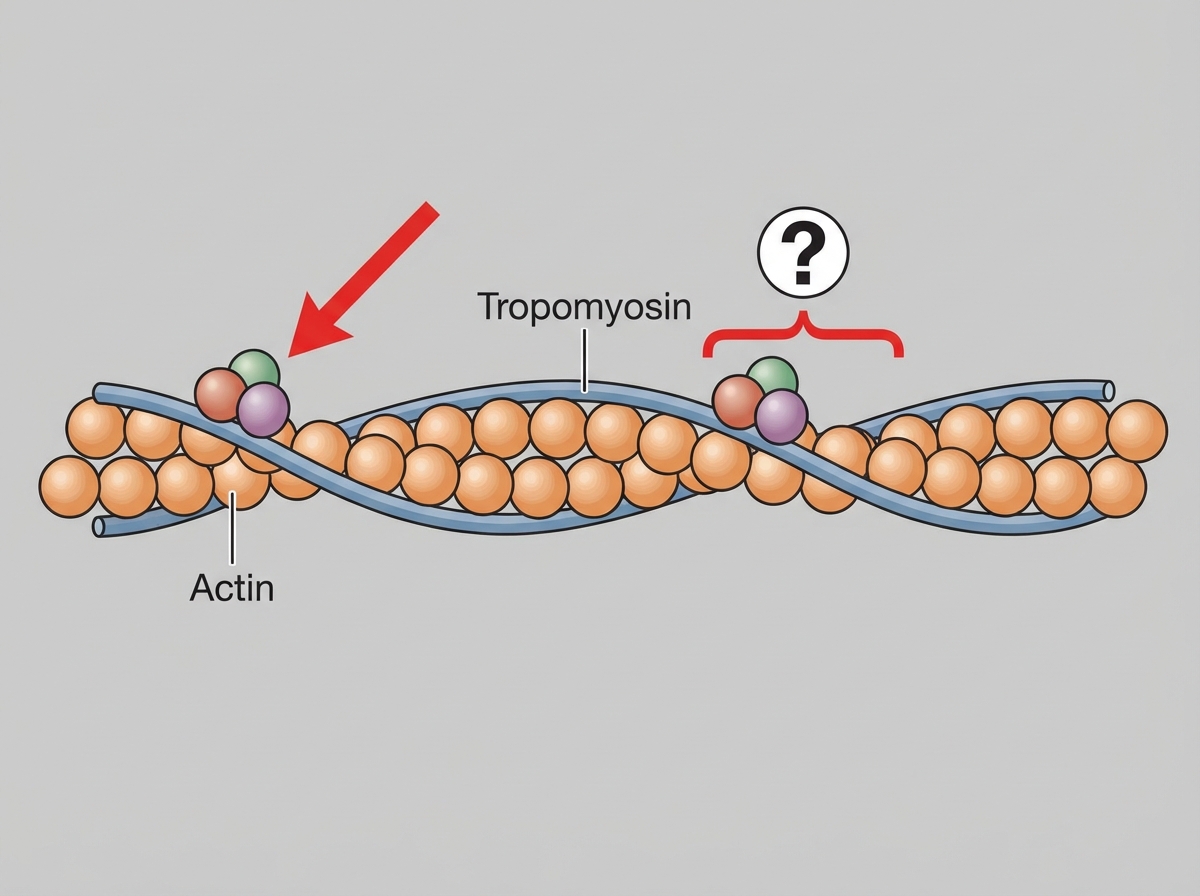
Which protein is shown in the thin filament as illustrated?

Positive feedback is seen in which of the following?
During skeletal muscle contraction, which bands shorten?
Which statement is true regarding Type-2 muscle fibers?
Which of the following statements is true about Nissl granules?
Explanation: **Explanation:** The classification of nerve fibers is based on the **Erlanger-Gasser classification**, which categorizes fibers according to their diameter, myelination, and conduction velocity. **Why Option D is Correct:** **Type C fibers** are the smallest nerve fibers in the human body. They are unique because they are **unmyelinated**, which results in a very small diameter (typically **0.5–2 micrometers**) and the slowest conduction velocity (0.5–2 m/s). These fibers primarily carry sensations of slow/chronic pain, temperature, and post-ganglionic autonomic signals. **Analysis of Incorrect Options:** * **Option A (13–20 µm):** This corresponds to **Type A-alpha (Aα)** fibers. These are the largest, most heavily myelinated fibers, responsible for proprioception and somatic motor function. * **Option B (4–13 µm):** This range corresponds to **Type A-beta (Aβ)** and **Type A-gamma (Aγ)** fibers, which mediate touch, pressure, and motor supply to muscle spindles. * **Option C (3–6 µm):** This range is characteristic of **Type B** fibers (pre-ganglionic autonomic fibers) or smaller **Type A-delta (Aδ)** fibers (fast pain and cold temperature). **High-Yield Clinical Pearls for NEET-PG:** * **Susceptibility to Blockade:** Type C fibers are the **most sensitive to local anesthetics** (due to lack of myelin) but the **least sensitive to hypoxia** and pressure. * **Pain Transmission:** Remember the "Double Pain" phenomenon: **Aδ fibers** carry "fast" (sharp/localized) pain, while **C fibers** carry "slow" (dull/aching/diffuse) pain. * **Rule of Thumb:** Conduction velocity (in m/s) for Type A fibers is roughly $6 \times \text{diameter (in µm)}$. This rule does not apply to Type C fibers as they lack myelin.
Explanation: ### Explanation **Correct Answer: C. Nernst equation** The **Nernst equation** is the fundamental formula used to calculate the **equilibrium potential** (electromotive force) for a single ion across a semi-permeable membrane. It determines the electrical potential difference required to exactly balance the concentration gradient of that ion, such that there is no net movement. * **Formula:** $E = \frac{RT}{zF} \ln \frac{[Ion]_{out}}{[Ion]_{in}}$ (Simplified at body temperature: $E = \pm 61 \log \frac{[Ion]_{out}}{[Ion]_{in}}$). * In physiology, it explains why the resting membrane potential is close to the equilibrium potential of Potassium ($K^+$), as the membrane is most permeable to it at rest. **Analysis of Incorrect Options:** * **A. Gibbs-Donnan equation:** Describes the behavior of charged particles near a semi-permeable membrane when one ion is non-diffusible (e.g., plasma proteins). It explains the resulting unequal distribution of diffusible ions, not the magnitude of the potential itself. * **B. Henderson-Hasselbalch equation:** Used in acid-base physiology to calculate the **pH** of a buffer solution or the ratio of bicarbonate to carbon dioxide in the blood. * **D. Fick’s law:** Relates to the **rate of diffusion** of a gas or solute across a membrane. It states that the flux is proportional to the concentration gradient and surface area. **High-Yield Clinical Pearls for NEET-PG:** * **Goldman-Hodgkin-Katz (GHK) Equation:** While Nernst is for a *single* ion, the GHK equation calculates the *resting membrane potential* by considering the permeability and concentration of *all* major ions ($Na^+, K^+, Cl^-$). * **Standard Potentials:** $E_{K^+} \approx -90\text{ mV}$, $E_{Na^+} \approx +60\text{ mV}$. * **Hypokalemia** makes the resting membrane potential more negative (hyperpolarization), making cells less excitable.
Explanation: **Explanation:** The correct answer is **Isotonic exercise**. This type of muscle contraction occurs when the **tension remains constant** while the **muscle length changes** (either shortening or lengthening) to move a load. The term is derived from the Greek words *"iso"* (same) and *"tonos"* (tension). Isotonic exercises are further divided into **concentric** (muscle shortens, e.g., upward phase of a bicep curl) and **eccentric** (muscle lengthens under tension, e.g., controlled lowering of a weight). **Why other options are incorrect:** * **Isometric exercise:** In this type, the **length of the muscle remains constant** while the **tension increases**. No external work is performed (Work = Force × Displacement; here displacement is zero). An example is pushing against a stationary wall. * **Aerobic exercise:** This refers to the metabolic pathway used (oxidative phosphorylation) rather than the mechanics of muscle length or tension. While many isotonic exercises are aerobic, the terms are not synonymous. **High-Yield NEET-PG Pearls:** 1. **Isokinetic exercise:** A type of exercise where the muscle contracts at a **constant velocity** throughout the entire range of motion (requires specialized equipment). 2. **Energy Expenditure:** Isotonic contractions perform external work and consume more energy compared to isometric contractions. 3. **Clinical Correlation:** Isometric exercises cause a more significant rise in **mean arterial pressure (MAP)** due to the compression of blood vessels within the contracting muscle, leading to increased peripheral resistance. 4. **Golgi Tendon Organs:** These are sensitive to changes in muscle **tension** (prominent in isometric), whereas **Muscle Spindles** are sensitive to changes in muscle **length** (prominent in isotonic).
Explanation: **Explanation:** The **Golgi Tendon Organ (GTO)** is a high-threshold mechanoreceptor located in the tendons of skeletal muscles, arranged in **series** with the muscle fibers. Its primary function is to monitor and respond to **muscle tension** (force of contraction). When a muscle contracts, it pulls on the tendon, compressing the nerve endings within the GTO. This triggers an inhibitory signal via **Ib afferent fibers** to the spinal cord, causing the muscle to relax (the **Inverse Stretch Reflex**). This mechanism protects the muscle and tendon from damage due to excessive force. **Analysis of Options:** * **Option A & D (Static and Dynamic Muscle Length):** These are detected by **Muscle Spindles**, which are arranged in **parallel** with muscle fibers. Nuclear chain fibers detect static length, while nuclear bag fibers detect dynamic changes in length (velocity). * **Option B (Muscle Action):** This is a general term. While GTOs are active during muscle action, their specific physiological stimulus is the resulting tension, not the action itself. **High-Yield Clinical Pearls for NEET-PG:** * **Location:** GTO is in **series**; Muscle Spindle is in **parallel**. * **Afferent Nerve:** GTO uses **Type Ib** fibers; Muscle Spindle uses **Type Ia** (primary) and **Type II** (secondary) fibers. * **Reflex:** GTO mediates the **Autogenic Inhibition** (Inverse Stretch Reflex), whereas the Muscle Spindle mediates the **Stretch Reflex** (DTRs). * **Function:** GTO prevents avulsion injuries; Muscle Spindles maintain muscle tone and posture.
Explanation: **Explanation:** Skeletal muscle fibers are classified based on their contraction speed and primary metabolic pathway. The correct answer is **Type I fiber** because these fibers are specifically designed for endurance and continuous aerobic activity. **1. Why Type I is correct:** Type I fibers, also known as **Slow-Twitch (Red) fibers**, have a high oxidative capacity. This is due to a high density of **mitochondria**, high concentrations of **myoglobin** (which stores oxygen and gives the muscle its red color), and a rich capillary supply. They rely on aerobic metabolism (oxidative phosphorylation) to produce ATP, making them highly resistant to fatigue. **2. Why the other options are incorrect:** * **Type IIA (Fast-Twitch Oxidative-Glycolytic):** These are intermediate fibers. While they possess some oxidative capacity, they also rely significantly on anaerobic glycolysis. Their oxidative capacity is lower than Type I but higher than Type IIB. * **Type IIB (Fast-Twitch Glycolytic):** These are "White fibers" with **low oxidative capacity**. They have few mitochondria and low myoglobin. They rely almost exclusively on anaerobic glycolysis for rapid, powerful bursts of activity but fatigue very quickly. **High-Yield NEET-PG Clinical Pearls:** * **Postural Muscles:** Muscles like the *soleus* are predominantly Type I (endurance). * **Sprint Muscles:** Muscles like the *extraocular muscles* or *gastrocnemius* have a higher proportion of Type II fibers (speed). * **Mnemonic:** **"One Slow Red Ox"** (Type **I**, **Slow**-twitch, **Red** color, **Ox**idative). * **ATPase Activity:** Type II fibers have high myosin ATPase activity (fast contraction), whereas Type I has low activity.
Explanation: ***Troponin*** - **Troponin** is a regulatory protein complex located on the **thin filament** (actin), consisting of **troponin C** (calcium-binding), **troponin I** (inhibitory), and **troponin T** (tropomyosin-binding). - Functions in **calcium-mediated muscle contraction** by regulating the interaction between **actin and myosin** through conformational changes in the **troponin-tropomyosin complex**. *Titin* - **Titin** is primarily associated with the **thick filament** (myosin) and acts as a **molecular spring** providing elasticity to muscle fibers. - It spans from the **Z-line to M-line** in the sarcomere but is not a component of the thin filament regulatory system. *Nebulin* - **Nebulin** is present on thin filaments but serves as a **molecular ruler** that determines **actin filament length** during muscle development. - Unlike troponin, it does not function as a **regulatory protein** for calcium-mediated contraction and is not part of the excitation-contraction coupling mechanism. *Dystrophin* - **Dystrophin** is a **cytoskeletal protein** that links intracellular **actin** to the **extracellular matrix** through the **dystrophin-glycoprotein complex**. - It provides **structural support** to muscle fibers but is not involved in the **thin filament regulatory mechanism** or direct contraction processes.
Explanation: **Explanation:** **1. Why Option C is Correct:** Positive feedback is a process where the output of a system intensifies the original stimulus, leading to an "explosive" or "vicious cycle" effect. In the stomach, the presence of food stimulates the release of **gastrin**, which triggers the release of **histamine**. Both substances then act synergistically to stimulate parietal cells to secrete HCl. The resulting increase in acidity and protein digestion products further stimulates the G-cells and ECL cells to release more gastrin and histamine, respectively. This amplification continues until the physiological goal (digestion) is achieved or the pH drops too low (triggering a negative feedback switch via somatostatin). **2. Analysis of Incorrect Options:** * **Option A (LH Surge):** While the LH surge *is* a classic example of positive feedback (Estrogen stimulating LH), it was likely excluded or ranked lower if the question context implies a continuous digestive process. However, in many standard texts, LH surge is a primary example. *Note: In NEET-PG, if multiple options seem correct, look for the most specific physiological mechanism described.* * **Option B (Entry of Ca++ into SR):** This is a **negative feedback** or restorative mechanism. Calcium is pumped *into* the sarcoplasmic reticulum (via SERCA) to lower cytosolic calcium levels and end muscle contraction. (Note: Calcium-induced calcium *release* from the SR is positive feedback). * **Option D (Thrombolytic activity):** Thrombolysis is the *breakdown* of clots. The **coagulation cascade** itself (clot formation) involves positive feedback (e.g., Thrombin activating Factor V and VIII), but thrombolysis acts as a regulatory check to limit clot size. **3. NEET-PG High-Yield Pearls:** * **Classic Positive Feedback Examples:** LH Surge (Ovulation), Oxytocin release (Parturition/Ferguson Reflex), Nerve Action Potential (Hodgkin cycle/Na+ influx), and Blood Clotting Cascade. * **Most physiological systems** operate via **Negative Feedback** to maintain homeostasis (e.g., BP regulation, Thyroid hormone axis). * **Key Distinction:** Positive feedback is usually "unstable" and ends in a major event (birth, clot, ovulation), whereas negative feedback is "stable."
Explanation: ### Explanation The mechanism of skeletal muscle contraction is best explained by the **Sliding Filament Theory** (Huxley and Hanson). According to this theory, muscle contraction occurs when thin (actin) filaments slide over thick (myosin) filaments toward the center of the sarcomere. #### Why the Correct Answer is Right: * **I Band (Isotropic):** This band contains only thin filaments. As actin filaments slide toward the M-line, they overlap more with thick filaments, causing the I band to **shorten**. * **H Zone (Heller):** This is the central part of the A band containing only thick filaments. As thin filaments move inward, they occupy the H zone, causing it to **shorten or even disappear** during maximal contraction. * **Sarcomere:** The distance between two Z-discs decreases, meaning the overall sarcomere shortens. #### Why Other Options are Wrong: * **A Band (Anisotropic):** This band represents the entire length of the thick (myosin) filament. Since the thick filaments themselves do not change length or move, the **A band remains constant** during contraction. * Options A, C, and D are incorrect because they suggest the A band shortens, which is physiologically impossible during normal contraction. #### High-Yield NEET-PG Pearls: * **The "Constant" Rule:** During contraction, the **A band** and the length of individual **thick and thin filaments** remain unchanged. * **The "Shortening" Rule:** The **I band, H zone, and Sarcomere** length all decrease. * **Structural Proteins:** Titin (the largest protein in the body) acts as a spring to maintain the central position of myosin, while Nebulin regulates the length of actin filaments. * **Calcium Source:** In skeletal muscle, calcium for contraction is derived exclusively from the **Sarcoplasmic Reticulum** (unlike cardiac muscle, which requires extracellular calcium).
Explanation: ### Explanation **Correct Answer: C. Increased Isoenzyme ATPase activity** Skeletal muscle fibers are classified into Type 1 (Slow-twitch) and Type 2 (Fast-twitch) based on their metabolic profile and contraction speed. The speed of muscle contraction is directly proportional to the **Myosin ATPase activity**. Type 2 fibers possess a specific isoenzyme of myosin ATPase that hydrolyzes ATP rapidly, allowing for fast cross-bridge cycling and quick, powerful contractions. **Analysis of Options:** * **Option A & B (Incorrect):** These describe **Type 1 (Slow Oxidative) fibers**. Type 1 fibers are rich in myoglobin (giving them a red color) and mitochondria to support aerobic metabolism for endurance. Type 2 fibers have low myoglobin content and appear pale/white. * **Option D (Incorrect):** Type 2 fibers (specifically Type 2b) have a **high glycolytic capacity**, not moderate. They rely on anaerobic glycolysis for rapid energy production, making them prone to early fatigue due to lactic acid accumulation. --- ### High-Yield Clinical Pearls for NEET-PG: * **Type 1 Fibers (Slow-twitch):** "One Slow Red Ox" (Type **1**, **Slow**-twitch, **Red** color, **Ox**idative metabolism). Found in postural muscles like the **Soleus**. * **Type 2 Fibers (Fast-twitch):** "Two Fast White Sugar" (Type **2**, **Fast**-twitch, **White** color, **Sugar**/Glycolytic metabolism). Found in muscles used for sprinting or weightlifting, such as the **Gastrocnemius**. * **Intermediate Fibers (Type 2a):** These are fast-twitch but have both oxidative and glycolytic capacities (Fast Oxidative Glycolytic). * **Order of Recruitment:** According to **Henneman’s Size Principle**, smaller Type 1 motor units are recruited first, followed by larger Type 2 units as force requirements increase.
Explanation: **Explanation:** **Nissl granules** (also known as Nissl bodies) are large, granular structures found in the cytoplasm of neurons. They are composed of **Rough Endoplasmic Reticulum (RER)** and associated **free ribosomes**. 1. **Why Option C is correct:** The primary function of ribosomes and RER is translation. Therefore, Nissl granules are the primary sites of **protein synthesis** in the neuron, producing proteins required for intracellular transmission and the renewal of structural components. 2. **Why other options are incorrect:** * **Option A:** RNA synthesis (transcription) occurs within the **nucleus** (specifically the nucleolus for rRNA), not in the Nissl granules. * **Option B:** Nissl granules are found in the **cyton (cell body)** and **dendrites**, but they are characteristically **absent in the axon** and the **axon hillock**. This is a classic anatomical landmark used to identify the start of an axon. * **Option D:** While they contain RER, stating they are "structurally endoplasmic reticulum" is incomplete. They are a complex of **RER plus free ribosomes**. (Note: In many competitive exams, "Protein Synthesis" is the more functional and definitive answer). **High-Yield Clinical Pearls for NEET-PG:** * **Chromatolysis:** When a neuron is injured (axonal injury), Nissl granules undergo hypertrophy, fragment, and disperse. This process is called chromatolysis, indicating an active attempt at protein synthesis for regeneration. * **Staining:** They are highly basophilic and are best visualized using basic dyes like **Cresyl Violet** or **Methylene Blue**. * **Location Tip:** Always remember: **D**endrites have them, **A**xons do not (**D** yes, **A** no).
Cell Structure and Function
Practice Questions
Membrane Transport Mechanisms
Practice Questions
Bioelectric Phenomena
Practice Questions
Homeostasis and Feedback Mechanisms
Practice Questions
Body Fluid Compartments
Practice Questions
Signal Transduction Mechanisms
Practice Questions
Cell-to-Cell Communication
Practice Questions
Principles of Physiological Measurement
Practice Questions
Osmosis and Osmotic Pressure
Practice Questions
Physiological Adaptation Mechanisms
Practice Questions
Get full access to all questions, explanations, and performance tracking.
Start For Free