What is the normal intra-abdominal pressure?
Which of the following actions is attributed to cholecystokinin?
Ascorbic acid is a potent enhancer of iron absorption because it:
What is the most important action of secretin?
What are the final sugars present in intestinal chyme?
Which of the following clinical laboratory observations is suggestive of Maple syrup urine disease?
What is true about gastric acid secretion?
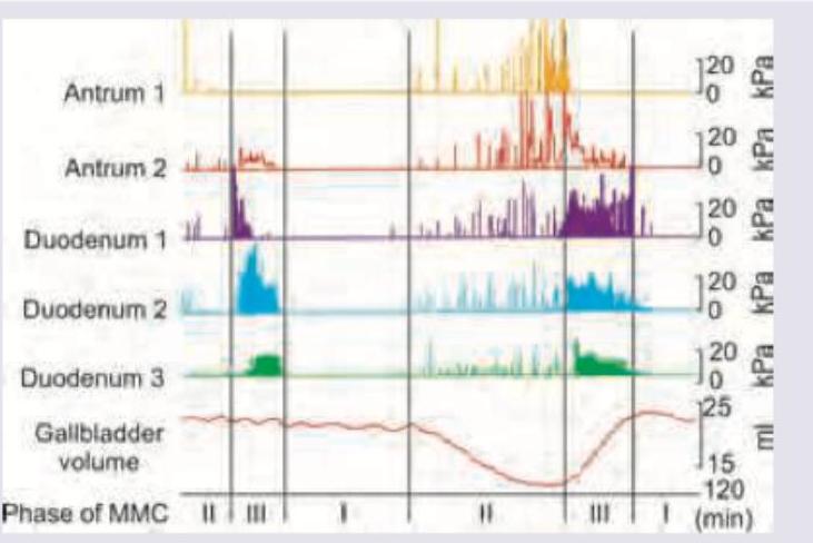
The image shows migrating motor complexes in various parts of gut. Identify the correct statement.

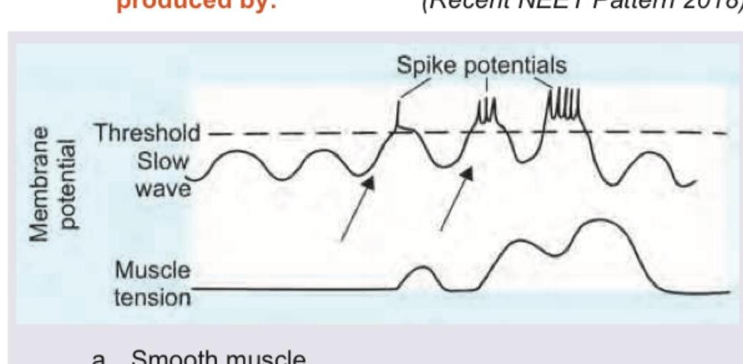
The following marked recordings in GIT are produced by:

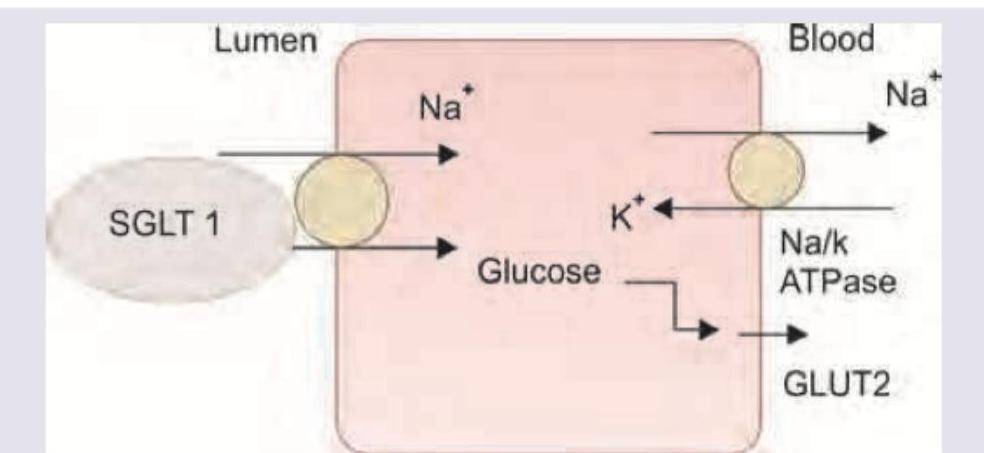
Which of these is true about the highlighted transporter?

Explanation: **Explanation:** **1. Understanding Normal Intra-Abdominal Pressure (IAP):** Intra-abdominal pressure is the steady-state pressure concealed within the abdominal cavity. In a healthy, resting adult who is breathing spontaneously, the normal IAP ranges from **0 to 8 mmHg** (often cited as approximately 5 mmHg). It is influenced by the abdominal wall compliance and the volume of the abdominal contents. It varies slightly with respiration—increasing during inspiration and decreasing during expiration. **2. Analysis of Incorrect Options:** * **10-15 mmHg (Option B):** This range is considered **elevated**. While it can occur transiently (e.g., during coughing or heavy lifting), a sustained IAP of 12 mmHg or higher is the diagnostic threshold for **Intra-abdominal Hypertension (IAH)**. * **15-20 mmHg (Option C):** This represents Grade II or III IAH. At these levels, venous return is compromised, and organ perfusion begins to decline. * **20-26 mmHg (Option D):** This is a critical range. An IAP >20 mmHg associated with new organ dysfunction (e.g., oliguria, respiratory failure) defines **Abdominal Compartment Syndrome (ACS)**, a surgical emergency. **3. High-Yield Clinical Pearls for NEET-PG:** * **Gold Standard Measurement:** IAP is most commonly measured indirectly via **intra-bladder pressure** (using a Foley catheter) with a maximum instillation volume of 25 mL of sterile saline. * **Abdominal Perfusion Pressure (APP):** Calculated as **MAP minus IAP**. A target APP of >60 mmHg is associated with better survival in critically ill patients. * **Grades of IAH:** * Grade I: 12–15 mmHg * Grade II: 16–20 mmHg * Grade III: 21–25 mmHg * Grade IV: >25 mmHg
Explanation: **Explanation:** Cholecystokinin (CCK) is a peptide hormone secreted by the **I cells** of the duodenum and jejunum in response to the presence of peptides, amino acids, and fatty acids. It acts as a master regulator of post-prandial digestion. 1. **Contraction of the Gallbladder:** CCK causes the gallbladder to contract while simultaneously relaxing the **Sphincter of Oddi**. This coordinated action ensures the delivery of bile into the duodenum for fat emulsification. 2. **Pancreatic Enzyme Secretion:** CCK acts on the pancreatic acinar cells to stimulate the secretion of a **large volume of pancreatic juice rich in enzymes** (lipase, amylase, and proteases). Note that Secretin, by contrast, stimulates a juice rich in bicarbonate. 3. **Increased Secretion of Enterokinase:** CCK also stimulates the release of **enterokinase** (enteropeptidase) from the duodenal mucosa. Enterokinase is the "master switch" that converts trypsinogen to active trypsin, initiating the protein digestion cascade. Since CCK performs all these functions to facilitate digestion, **Option D** is the correct answer. **High-Yield NEET-PG Pearls:** * **Stimulus:** The most potent stimulus for CCK release is the presence of **fatty acids** and amino acids in the duodenum. * **Satiety:** CCK acts on the hypothalamus to inhibit food intake (satiety signal). * **Gastric Emptying:** CCK **inhibits gastric emptying** (enterogastrone effect) to allow sufficient time for digestion in the small intestine. * **Trophic Effect:** It has a trophic (growth-promoting) effect on the exocrine pancreas.
Explanation: ### Explanation **Core Concept: Iron Absorption Dynamics** Dietary iron exists in two forms: **Heme iron** (from animal sources) and **Non-heme iron** (from plant sources). Non-heme iron is primarily found in the **ferric state (Fe³⁺)**, which is insoluble and cannot be absorbed by the apical transporters of the enterocytes. **Why Option C is Correct:** Ascorbic acid (Vitamin C) acts as a potent **reducing agent**. It converts ferric iron (Fe³⁺) into the **ferrous state (Fe²⁺)**. The ferrous form is more soluble and is the specific substrate for the **Divalent Metal Transporter 1 (DMT-1)** located on the brush border of the duodenum. Additionally, Vitamin C forms a soluble chelate with iron, preventing it from precipitating in the alkaline environment of the small intestine. **Analysis of Incorrect Options:** * **Option A:** Heme iron is absorbed via a distinct pathway (Heme Carrier Protein 1) and is relatively unaffected by dietary enhancers like Vitamin C. * **Option B:** Heme oxygenase is an intracellular enzyme that releases iron from the heme molecule *after* it has entered the enterocyte; Vitamin C does not modulate its activity. * **Option D:** Ferritin is the intracellular storage form of iron. Decreasing its production would not enhance absorption; rather, iron absorption is regulated by **Hepcidin**, which degrades ferroportin. **High-Yield Clinical Pearls for NEET-PG:** * **Site of Absorption:** Iron is predominantly absorbed in the **duodenum** and proximal jejunum. * **Inhibitors of Absorption:** Phytates (cereals), Oxalates, Tannins (tea), and Phosphates inhibit non-heme iron absorption. * **The "Acid" Connection:** Gastric HCl also aids absorption by maintaining an acidic pH, which keeps iron in the reduced Fe²⁺ state. * **Clinical Application:** Iron supplements are often prescribed with Vitamin C or orange juice to maximize bioavailability.
Explanation: **Explanation:** Secretin is a hormone produced by the **S-cells of the duodenum** in response to the entry of acidic chyme (pH < 4.5) from the stomach. **Why Option B is Correct:** The primary and most potent action of secretin is to stimulate the **pancreatic ductal cells** to secrete a large volume of watery fluid rich in **bicarbonate (HCO₃⁻)**. This bicarbonate is essential for neutralizing gastric acid in the duodenum, providing an optimal alkaline pH for the functioning of pancreatic digestive enzymes. **Analysis of Incorrect Options:** * **Option A:** While neutralizing acid is the *ultimate goal* or physiological outcome, it is not the direct "action" of the hormone itself. Secretin acts on the pancreas; the resulting bicarbonate then neutralizes the acid. * **Option C:** Secretin does inhibit gastric acid secretion (acting as an "enterogastrone"), but this is considered a secondary or minor effect compared to its stimulatory effect on the pancreas. * **Option D:** Secretin generally decreases gastric motility and increases the tone of the pyloric sphincter to prevent further acid entry into the duodenum, but this is not its "most important" regulatory function. **NEET-PG High-Yield Pearls:** * **Nature's Antacid:** Secretin is often referred to as "Nature's Antacid." * **Mechanism:** It acts via the **cAMP** second messenger system. * **Potentiation:** Secretin and CCK (Cholecystokinin) exhibit **potentiation**; secretin increases the bicarbonate response to CCK, and CCK increases the enzyme response to secretin. * **Clinical Use:** The **Secretin Stimulation Test** is the gold standard for diagnosing Exocrine Pancreatic Insufficiency (e.g., in Chronic Pancreatitis).
Explanation: ### Explanation **1. Why Glucose and Fructose are Correct:** The digestion of dietary carbohydrates occurs in two main phases: luminal digestion (via salivary and pancreatic amylase) and brush border digestion. Amylase breaks down starches into maltose, maltotriose, and α-limit dextrins. However, the **final products** of carbohydrate digestion—ready for absorption—are monosaccharides produced by brush border enzymes (disaccharidases) in the small intestine: * **Maltase** breaks down maltose into **Glucose + Glucose**. * **Sucrase** breaks down sucrose (table sugar) into **Glucose + Fructose**. * **Lactase** breaks down lactose (milk sugar) into **Glucose + Galactose**. Since Glucose and Fructose are the primary monosaccharides derived from common dietary sugars (starch and sucrose), they are the predominant final sugars found in the intestinal chyme. **2. Why the Other Options are Incorrect:** * **Option B (Ribose and Mannose):** Ribose is a pentose sugar found in nucleic acids (RNA), and Mannose is found in some polysaccharides, but they are not major products of dietary carbohydrate digestion. * **Option C & D (Xylulose and Xylose):** Xylulose is an intermediate in the pentose phosphate pathway and the uronic acid pathway. While small amounts of pentoses like xylose exist in fruits, they are not the "final sugars" of primary digestion. **3. NEET-PG High-Yield Clinical Pearls:** * **Absorption Mechanism:** Glucose and Galactose are absorbed via **SGLT-1** (Secondary active transport with Na+), while Fructose is absorbed via **GLUT-5** (Facilitated diffusion). * **Basolateral Exit:** All three monosaccharides (Glucose, Galactose, Fructose) exit the enterocyte into the blood via the **GLUT-2** transporter. * **Rate-Limiting Step:** In carbohydrate assimilation, the rate-limiting step is usually the **absorption** of sugars, except for lactose, where the **hydrolysis** (by lactase) is the rate-limiting step. * **Lactose Intolerance:** Caused by a deficiency of lactase, leading to osmotic diarrhea and flatulence due to bacterial fermentation of undigested lactose in the colon.
Explanation: **Explanation:** The correct answer is **D. High fecal levels of tryptophan and indole derivatives.** While the name "Maple Syrup Urine Disease" (MSUD) is classically associated with a specific urinary odor, this question tests a deeper understanding of metabolic transport defects. In MSUD, there is a deficiency of the **branched-chain alpha-keto acid dehydrogenase (BCKAD) complex**, leading to an accumulation of branched-chain amino acids (Leucine, Isoleucine, and Valine). A critical clinical observation in these patients is the **malabsorption of neutral amino acids**, particularly tryptophan, in the gut. When tryptophan is not absorbed, it is metabolized by intestinal bacteria into **indoles and skatoles**, which are then excreted in the feces. This biochemical marker is a high-yield diagnostic feature for MSUD in advanced physiology and biochemistry assessments. **Analysis of Incorrect Options:** * **Option A:** While a burnt sugar smell is characteristic of MSUD (due to isoleucine metabolites), it is a *physical* observation rather than a *laboratory* quantification of metabolites mentioned in the context of the other options. (Note: In many competitive exams, if a more specific biochemical marker like Option D is provided, it is prioritized). * **Option B:** High plasma phenylalanine is the hallmark of **Phenylketonuria (PKU)**, not MSUD. * **Option C:** High levels of citrulline in the urine are suggestive of **Citrullinemia**, a urea cycle disorder. **NEET-PG High-Yield Pearls:** * **Enzyme Defect:** BCKAD complex (requires Thiamine/B1 as a cofactor). * **Amino Acids Involved:** Leucine, Isoleucine, Valine (Mnemonic: **LIV**er). * **Odor:** The "burnt sugar" or "maple syrup" odor is specifically due to **S-alloisoleucine**. * **Hartnup Disease Connection:** Do not confuse this with Hartnup disease, which also involves tryptophan transport defects but primarily presents with pellagra-like symptoms and neutral aminoaciduria.
Explanation: This question tests the fundamental understanding of gastric physiology and the regulation of parietal cell function. **Explanation of Options:** * **Option A (Secretin):** Secretin is an enterogastrone released from S-cells in the duodenum in response to low pH. It acts as a physiological "brake" by inhibiting gastrin release and directly inhibiting parietal cells, thereby **decreasing gastric acid secretion**. * **Option B (H2 Blockers):** Histamine is a potent paracrine stimulator of acid secretion via H2 receptors on parietal cells. H2 blockers (e.g., Ranitidine) competitively inhibit these receptors, **preventing the release** of acid stimulated by histamine and reducing the potentiating effect of gastrin and acetylcholine. * **Option C (Total Acid Output):** The **Maximal Acid Output (MAO)**, measured after stimulation with pentagastrin, is directly proportional to the **total parietal cell mass**. This is a classic physiological marker used to assess the functional capacity of the gastric mucosa. Since all three statements are physiologically accurate, **Option D** is the correct answer. **High-Yield Clinical Pearls for NEET-PG:** * **Potentiation:** The combined effect of Histamine, Gastrin, and Acetylcholine is greater than the sum of their individual effects. Blocking one (e.g., with H2 blockers) significantly reduces the efficacy of the others. * **Proton Pump Inhibitors (PPIs):** These are the "final common pathway" inhibitors, acting on the H+/K+ ATPase pump. They are more potent than H2 blockers. * **Somatostatin:** Known as the "universal endocrine off-switch," it is the primary inhibitory paracrine mediator of gastric acid secretion. * **Zollinger-Ellison Syndrome:** Characterized by a gastrinoma leading to massive parietal cell hyperplasia and extremely high MAO.
Explanation: ***Occur every 100 minutes*** - The Migrating Motor Complex (MMC) is a pattern of electromechanical activity observed in the gastrointestinal tract during fasting, typically occurring in cycles of **approximately 90-120 minutes**, or about every 100 minutes. - This cyclical activity during the interdigestive period serves to **clear residual indigestible material and bacteria** from the small intestine into the colon. *Triggered by secretin* - The MMC is primarily triggered by **motilin**, a polypeptide hormone that increases gastrointestinal motility. - **Secretin** is known to inhibit gastric acid secretion and stimulate bicarbonate and water secretion from the pancreas and bile ducts, but it is not the primary trigger for MMC. *Phase 3 has least activity* - Phase 3 of the MMC is characterized by a **burst of intense, regular propagated contractions** that sweep from the stomach to the ileum, often referred to as the "housekeeping wave." - This phase exhibits the **most vigorous motor activity**, not the least, as seen by the high-amplitude spike bursts in the image. *Intake of meal exaggerates the migrating motor complex* - The MMC is a **fasting phenomenon** and is **abolished or inhibited by the intake of a meal**. - Feeding initiates a different motor pattern characterized by continuous, irregular contractions, which serve to mix and propel chyme through the digestive tract.
Explanation: ***Interstitial cells of Cajal*** - The image displays characteristic **slow waves** (also known as basal electrical rhythm) and superimposed **spike potentials** in the gastrointestinal tract, which are generated by the interstitial cells of Cajal (ICCs). - ICCs act as **pacemaker cells** in the GI tract, initiating the bioelectrical activity that dictates the rhythm of smooth muscle contractions. *Smooth muscle* - While GI smooth muscle cells contract in response to these electrical activities, they **do not generate the slow waves or spike potentials** themselves. - Smooth muscle cells are responsible for **muscle tension**, as shown in the lower tracing, which is directly triggered by the spike potentials. *Myenteric plexus* - The myenteric plexus primarily consists of **neurons** that regulate GI motility and secretions, influencing the activity of smooth muscle cells and ICCs. - It does not directly produce the characteristic slow waves or spike potentials observed as the intrinsic electrical rhythm. *Parasympathetic stimulation* - Parasympathetic stimulation, primarily via the **vagus nerve**, generally **increases the frequency and amplitude** of slow waves and spike potentials. - However, it modulates the activity rather than initiating the intrinsic electrical rhythm itself.
Explanation: ***One molecule goes in, other molecule goes out*** - The highlighted transporter is the **Na+/K+ ATPase**, which actively pumps 3 **Na+ ions out** of the cell and 2 **K+ ions into** the cell, maintaining an electrochemical gradient. - This counter-transport (one molecule type going in and another going out) is characteristic of an **antiporter** pump. *Both molecules go in* - This option would describe a **symporter** mechanism where two different molecules move in the **same direction** across the membrane. - The Na+/K+ ATPase explicitly shows Na+ moving out and K+ moving in, which contradicts simultaneous inward movement. *Both molecules go out* - This would mean two molecules are expelled from the cell. The Na+/K+ ATPase, however, has K+ entering the cell. - While Na+ is pumped out by this transporter, K+ is actively transported inward. *One molecule goes in and two exit* - The Na+/K+ ATPase transports three Na+ ions out of the cell and two K+ ions into the cell, which is a 3:2 ratio and not one in and two out. - This option incorrectly describes the stoichiometry and directionality of ions for the Na+/K+ ATPase.
Gastrointestinal Motility
Practice Questions
Gastrointestinal Secretions
Practice Questions
Digestion and Absorption
Practice Questions
Gastrointestinal Hormones
Practice Questions
Hepatobiliary Physiology
Practice Questions
Pancreatic Exocrine Function
Practice Questions
Gastrointestinal Circulation
Practice Questions
Intestinal Immune System
Practice Questions
Gut Microbiome
Practice Questions
Regulation of Food Intake
Practice Questions
Get full access to all questions, explanations, and performance tracking.
Start For Free