Which of the following is a FALSE statement regarding hemodynamic changes occurring during exercise?
Which of the following causes coronary vasodilation?
If the contractility of the heart is decreased, which of the following is seen ?
During heavy exercise the cardiac output (CO) increases up to five fold while pulmonary arterial pressure rises very little. This physiological ability of the pulmonary circulation is best explained by
Concentric hypertrophy of left ventricle is seen in -
Blood supply to the brain during moderate exercise:
During exercise in physiological limits, what is the effect on end systolic volume?
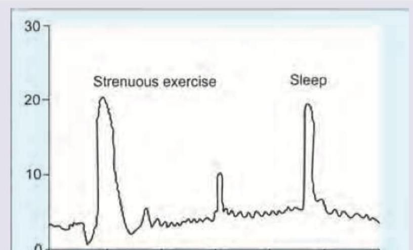
The blood levels of hormones are elevated during exercise and sleep as shown. Which hormone would exhibit this diurnal pattern?

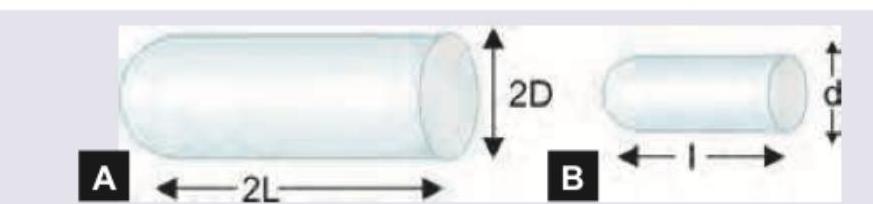
There are two blood vessels shown below. Assuming that pressure along both the vessels is same and both of them follow linear flow pattern, what will be the amount of blood flow in A compared to B?

Cushing reflex is associated with all except?
Explanation: ***Venoconstriction in exercising muscles as well as increased cardiac output leads to marked increase in systemic blood pressure.*** - This is the **FALSE statement**. During exercise, **vasodilation (not venoconstriction) occurs in exercising muscles** to increase blood flow to active tissues. Venoconstriction occurs in **non-exercising vascular beds** to redistribute blood. - While cardiac output increases significantly, **systemic vascular resistance (SVR) decreases** due to vasodilation in exercising muscles, which counteracts the rise in cardiac output. - The net effect is a **moderate increase in mean arterial pressure**, not a "marked increase." **Systolic BP rises** due to increased cardiac output, but **diastolic BP remains stable or slightly decreases** due to reduced SVR. - Therefore, this statement incorrectly describes both the vascular response in exercising muscles and the magnitude of systemic blood pressure change. *Venous return is augmented by the pumping action of skeletal muscles.* - **TRUE statement**. The **skeletal muscle pump** compresses veins during muscle contraction, pushing blood back toward the heart and increasing venous return. - This mechanism is crucial during exercise to maintain cardiac output and prevent blood pooling in lower extremities. *End-diastolic volume increases in the failing heart during exercise.* - **TRUE statement**. In a **failing heart**, the Frank-Starling mechanism operates on a flatter curve with reduced contractile reserve. - During exercise, increased venous return leads to **increased end-diastolic volume (preload)**, but the failing heart cannot adequately increase stroke volume proportionally, leading to volume accumulation and potential pulmonary congestion. *The increased adrenergic nerve impulses to the heart as well as an increased concentration of circulating catecholamines help to augment the contractile state of the myocardium.* - **TRUE statement**. During exercise, **sympathetic nervous system activation** increases, releasing **norepinephrine from adrenergic nerves** and **epinephrine from the adrenal medulla**. - These **catecholamines** bind to **beta-1 adrenergic receptors** on cardiomyocytes, increasing **heart rate (chronotropy)**, **contractility (inotropy)**, and **conduction velocity (dromotropy)**, thereby enhancing cardiac performance.
Explanation: ***Adenosine*** - **Adenosine** is a potent **endogenous vasodilator** in the coronary circulation, released in response to myocardial ischemia and hypoxia. - It acts on **A2a receptors** on smooth muscle cells, leading to increased cAMP production and subsequent relaxation. *Noradrenergic stimulation* - **Noradrenergic stimulation** primarily causes **vasoconstriction** in most vascular beds, including the coronary arteries, through activation of **alpha-1 adrenergic receptors**. - While beta-2 receptors can cause vasodilation, the overall effect in the coronaries under strong noradrenergic stimulation is often vasoconstrictive or has a complex interplay. *Hypocarbia* - **Hypocarbia** (decreased CO2) leads to **vasoconstriction** in many vascular beds, including the cerebral circulation and, to a lesser extent, the coronary arteries. - This effect is mediated by the pH change in the smooth muscle cells; reduced CO2 causes **alkalosis**, which generally promotes vasoconstriction. *None of the options* - This option is incorrect because **adenosine** is a well-established and potent coronary vasodilator.
Explanation: ***Decreased stroke volume*** - A decrease in the **contractility** of the heart directly reduces the force of myocardial contraction. - This weaker contraction results in less blood being ejected from the ventricle per beat, leading to a **decreased stroke volume**. *Increased ejection fraction* - **Ejection fraction** is the percentage of blood ejected from the ventricle with each beat, calculated as (stroke volume / end-diastolic volume) x 100. - When contractility decreases, **stroke volume** decreases, which would typically lead to a *decreased* ejection fraction, not an increased one. *Increased stroke work* - **Stroke work** is a measure of the work done by the ventricle to eject blood, and it depends on both stroke volume and aortic pressure. - With decreased contractility, **stroke volume** falls, which would *decrease* the stroke work, assuming afterload remains constant. *Increased cardiac output* - **Cardiac output** is the product of stroke volume and heart rate (CO = SV x HR). - Since decreased contractility leads to a **decreased stroke volume**, without a compensatory increase in heart rate, cardiac output would *decrease*, not increase.
Explanation: ***Increase in the number of open capillaries*** - During heavy exercise, the significant increase in cardiac output is accommodated by the **recruitment of previously closed pulmonary capillaries**. - This recruitment, along with **distension of existing capillaries**, reduces overall pulmonary vascular resistance, allowing blood flow to increase without a substantial rise in pulmonary arterial pressure. *Large amount of smooth muscle in pulmonary arterioles* - While pulmonary arterioles do contain smooth muscle, their primary role is in **regulating regional blood flow** and response to hypoxia, not facilitating large increases in overall blood flow during exercise. - The pulmonary circulation is characterized by **low resistance** and high capacitance compared to the systemic circulation, meaning it has less smooth muscle tone at baseline. *Sympathetically mediated greater distensibility of pulmonary vessels* - The pulmonary vasculature has **limited sympathetic innervation** compared to systemic vessels, and sympathetic activity plays a minor role in its distensibility during exercise. - Changes in pulmonary vascular resistance during exercise are primarily due to **mechanical factors** (recruitment and distension) rather than neurogenic control. *Smaller surface area of pulmonary circulation* - The pulmonary circulation actually has a **vast capillary surface area** crucial for efficient gas exchange. - A smaller surface area would lead to **higher resistance** and a greater pressure increase for a given flow, which contradicts the observation during exercise.
Explanation: ***Congenital aortic stenosis due to bicuspid aortic valve*** - **Aortic stenosis** creates a **pressure overload** on the left ventricle, leading to a compensatory increase in myocardial wall thickness without significant chamber dilation, which is the classic example of **concentric hypertrophy** [1]. - A **bicuspid aortic valve** is a common congenital anomaly that causes aortic stenosis and thus concentric left ventricular hypertrophy [2]. - This represents **acquired concentric hypertrophy** due to hemodynamic stress. *Mitral Stenosis* - **Mitral stenosis** primarily causes a pressure overload on the **left atrium**, leading to left atrial enlargement [3]. - While it can indirectly affect the left ventricle, it typically does not cause **concentric left ventricular hypertrophy** itself. *Aortic Regurgitation* - **Aortic regurgitation** leads to a **volume overload** on the left ventricle as blood flows back into the ventricle during diastole. - This typically results in **eccentric hypertrophy**, where both the ventricular wall thickness and chamber size increase significantly (dilated ventricle with increased mass) [1]. *Hypertrophic Obstructive Cardiomyopathy* - **Hypertrophic obstructive cardiomyopathy (HOCM)** is a **primary genetic myocardial disease** characterized by **asymmetric septal hypertrophy** rather than uniform concentric hypertrophy. - While HOCM involves significant myocardial hypertrophy, it represents a distinct pathophysiologic entity with **asymmetric distribution** (predominantly septal), not the classic concentric pattern seen with pressure overload states. - The hypertrophy in HOCM is **intrinsic (genetic)** rather than **adaptive (hemodynamic)** like in aortic stenosis. **References:** [1] Kumar V, Abbas AK, et al.. Robbins and Cotran Pathologic Basis of Disease. 9th ed. The Heart, p. 536. [2] Kumar V, Abbas AK, et al.. Robbins and Cotran Pathologic Basis of Disease. 9th ed. The Heart, pp. 562-563. [3] Kumar V, Abbas AK, et al.. Robbins and Cotran Pathologic Basis of Disease. 9th ed. Diseases of Infancy and Childhood, pp. 533-534.
Explanation: ***Correct: Remains constant*** - Cerebral blood flow is **autoregulated** to ensure a stable supply of oxygen and nutrients to the brain, regardless of changes in systemic blood pressure or metabolic demand during moderate exercise. - This autoregulation mechanism maintains a relatively constant blood flow (~750 mL/min or 50 mL/100g brain tissue/min) within a wide range of mean arterial pressures (60-150 mmHg). - The brain receives approximately **15% of cardiac output** at rest, and this proportion is maintained during moderate exercise. *Incorrect: Fluctuates unpredictably* - While there can be minor variations, the brain's **autoregulatory mechanisms** work to stabilize blood flow, preventing unpredictable fluctuations that would harm brain function. - Significant, unpredictable fluctuations would indicate a failure of these crucial physiological controls. *Incorrect: Increases* - Though overall cardiac output increases during exercise, the brain's demand for blood flow does **not significantly increase** in proportion to the body's other organs. - The brain prioritizes a constant, rather than an increased, supply to maintain stable function during moderate exercise. *Incorrect: Decreases* - A decrease in cerebral blood flow would lead to **cerebral hypoperfusion**, compromising brain function and potentially causing symptoms like dizziness or syncope. - The body's physiological responses during exercise are designed to prevent such a dangerous outcome.
Explanation: ***ESV decreases*** - During exercise, **sympathetic nervous system activity** increases, leading to enhanced cardiac contractility. - Improved contractility allows the heart to eject a greater percentage of its end-diastolic volume, resulting in a smaller **residual volume** in the ventricle after systole. *ESV increase* - An increase in ESV would indicate a **reduced ejection fraction** and poorer cardiac efficiency, which is contrary to the physiological adaptations during exercise. - This typically occurs in conditions of **heart failure** or myocardial dysfunction, not healthy exercise. *ESV first decrease and then increases* - While there are complex physiological responses during exercise, the primary and sustained effect on ESV within physiological limits is a **net decrease** due to increased contractility. - A subsequent increase would suggest a decline in cardiac function or the onset of fatigue beyond physiological limits. *ESV remain unchanged* - An unchanged ESV would imply no significant alteration in **cardiac contractility** or **ejection efficiency**, which is inconsistent with the cardiovascular demands and adaptations during exercise. - The body actively works to optimize cardiac output by increasing stroke volume, partly by reducing ESV during exercise.
Explanation: ***Growth hormone*** - **Growth hormone (GH)** secretion is known to increase significantly during both **strenuous exercise** and **sleep**, particularly during deep sleep stages. - The elevated levels during exercise promote **lipolysis** and **glucose production**, while during sleep, it facilitates **tissue repair** and **growth**. *Insulin* - **Insulin** levels typically **decrease during exercise** to promote the utilization of fat as fuel and increase during sleep in response to reduced metabolic demand and preparation for morning. - Its primary role is to regulate blood glucose, and its secretion is mainly stimulated by **high blood glucose** rather than exercise or sleep directly in this pattern. *Cortisol* - **Cortisol** secretion follows a **circadian rhythm**, peaking in the early morning and gradually decreasing throughout the day, reaching its lowest point at night. - While exercise can acutely increase cortisol, its **sleep-related pattern** is the opposite of what is shown, typically decreasing during early sleep. *Thyroid* - **Thyroid hormones (T3 and T4)** maintain a relatively **stable level** throughout the day and night, with minor diurnal fluctuations. - Their primary function is to regulate **metabolism** and they do not exhibit sharp, distinct peaks in response to exercise or sleep in the manner depicted.
Explanation: ***32 times*** - According to **Poiseuille-Hagen equation**: Q = (ΔP × π × r⁴) / (8 × η × L), where flow is directly proportional to the fourth power of radius and inversely proportional to vessel length. - From the diagram: Vessel A has diameter 2D and length 2L, while Vessel B has diameter d and length l. - **Key interpretation**: For the answer to be 32 times, the diameter of A must be twice that of B (radius_A = 2r), while the length of A is half that of B (length_A = L/2). - **Calculation**: - Q_A ∝ (2r)⁴ / (L/2) = 16r⁴ × 2/L = 32r⁴/L - Q_B ∝ r⁴ / L - **Q_A/Q_B = 32** - This demonstrates the **powerful effect of radius** (fourth power relationship) combined with **inverse length relationship** on blood flow. - **Clinical relevance**: Small changes in vessel diameter cause dramatic changes in blood flow, which is why vasoconstriction/vasodilation are potent mechanisms for regulating tissue perfusion. *Incorrect Option: 4 times* - Would require a different radius-to-length ratio than what's given in the problem. *Incorrect Option: 8 times* - This would result if diameter of A is 2× that of B AND length of A is also 2× that of B (not half). - Calculation: (2r)⁴/(2L) ÷ (r⁴/L) = 16r⁴/2L ÷ r⁴/L = 8 *Incorrect Option: 16 times* - This would occur if radius of A is 2× that of B but both vessels have the same length. - Calculation: (2r)⁴/L ÷ (r⁴/L) = 16
Explanation: ***Hypotension*** - The **Cushing reflex** is a compensatory response to increased intracranial pressure (ICP) aiming to maintain cerebral perfusion, which typically involves **hypertension**, not hypotension. - While prolonged or severe ICP can lead to decompensation and eventual hypotension, it is not a direct component of the reflex itself. *Increased intracranial pressure* - The **Cushing reflex** is triggered by an elevation in **intracranial pressure (ICP)**, as the body attempts to maintain blood flow to the brain. - This increased ICP reduces cerebral perfusion pressure, prompting a systemic response to raise mean arterial pressure. *Bradycardia* - **Bradycardia** is a classic component of the **Cushing reflex**, occurring as a compensatory response to the reflex hypertension. - The increased arterial blood pressure stimulates carotid and aortic baroreceptors, leading to a vagal response that slows the heart rate. *Irregular respiration* - **Irregular respiration** is another key component of the **Cushing reflex**, often manifesting as **Cheyne-Stokes breathing** or **ataxic breathing**. - This respiratory dysregulation is due to direct compression and dysfunction of the brainstem, specifically the medullary respiratory centers, caused by increased ICP.
Get full access to all questions, explanations, and performance tracking.
Start For Free