A woman with eclampsia is started on magnesium sulfate. What is the first sign of magnesium sulfate toxicity?
Which enzyme requires zinc as a cofactor?
Hypomagnesemia due to increased excretion by the kidney is caused by all except:
Hypermagnesemia may be observed in:
Normal anion gap is___ mmol/L?
In Bartter's syndrome there is a defect in
In renal failure, what is the primary cause of metabolic acidosis?
Which of the following statements is not true?
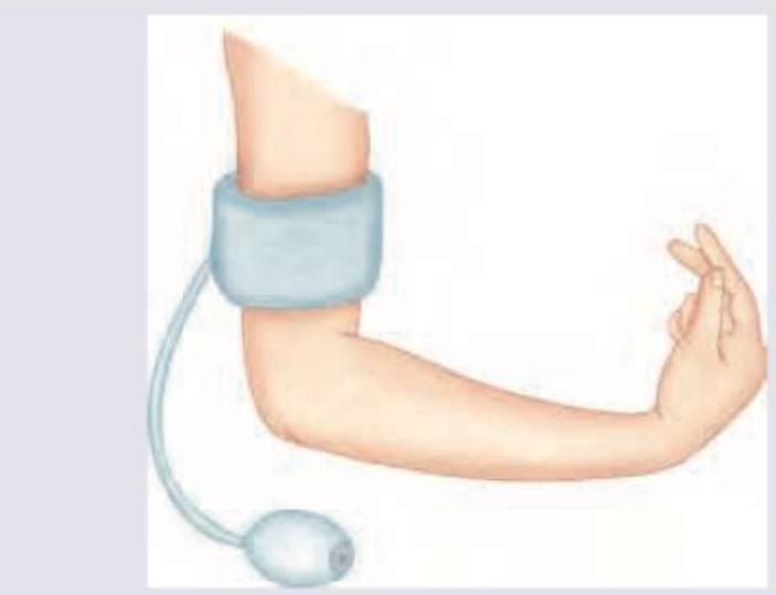
In a child with suspected tetany, the following test is performed. Identify the sign?

Intracellular water constitutes what percentage of total body water?
Explanation: ***Loss of knee jerk*** - **Diminished or absent deep tendon reflexes**, particularly the knee jerk, is the **earliest clinical sign** of magnesium sulfate toxicity. - This occurs at serum magnesium levels between **7-10 mEq/L** (8.5-12 mg/dL) due to magnesium's depressant effect on the nervous system and neuromuscular transmission. *Respiratory depression* - **Respiratory depression** is a more severe and later sign of magnesium toxicity, occurring at higher serum levels (typically >12 mEq/L). - It indicates significant central nervous system depression and potential for respiratory arrest, usually after reflexes are already lost. *Hypotension* - While magnesium sulfate can cause **vasodilation** and a subsequent drop in blood pressure, it is generally **not the first sign of toxicity** and often occurs concurrently with other mild to moderate signs. - Hypotension may be part of the therapeutic effect to reduce blood pressure in eclampsia, rather than an initial indicator of toxicity. *Reduced muscle tone* - **Reduced muscle tone** or **flaccidity** is also a consequence of magnesium's neuromuscular blocking effect but typically manifests **after the loss of deep tendon reflexes**. - It signifies more profound neuromuscular impairment, closer to the progression towards respiratory depression.
Explanation: ***Carbonic anhydrase*** - **Carbonic anhydrase** is a critical enzyme that rapidly interconverts carbon dioxide and water into carbonic acid, which then dissociates into a proton and a bicarbonate ion. - This enzyme contains a **zinc ion** in its active site, which is essential for its catalytic activity, particularly in binding and activating water for the hydration of CO2. *Lactate dehydrogenase* - **Lactate dehydrogenase (LDH)** catalyzes the reversible conversion of pyruvate to lactate, a key step in anaerobic glycolysis. - LDH primarily relies on **NAD+ or NADH** as cofactors and does not require zinc. *Glutathione peroxidase* - **Glutathione peroxidase (GPx)** is an antioxidant enzyme that catalyzes the reduction of hydrogen peroxide and organic hydroperoxides to water using glutathione. - Most mammalian glutathione peroxidases are **selenium-dependent enzymes**, incorporating selenocysteine at their active site, rather than zinc. *Hexokinase* - **Hexokinase** is an enzyme that phosphorylates hexoses, most notably glucose, to glucose-6-phosphate, the first step in glycolysis. - This enzyme requires **magnesium (Mg2+)** as a cofactor for its activity, as it forms a complex with ATP, facilitating the transfer of the phosphate group.
Explanation: ***Digitalis*** - **Digitalis** (digoxin) is a cardiac glycoside that inhibits the **Na+/K+-ATPase pump**; it is not directly associated with renal magnesium wasting. Instead, its toxicity can be exacerbated by hypomagnesemia, but it does not cause it. - While it affects electrolyte balance intracellularly, it does not primarily lead to increased **urinary excretion of magnesium**. *Aminoglycoside* - **Aminoglycosides** like gentamicin can cause **renal tubular damage**, particularly in the distal tubules. - This damage impairs the kidney's ability to reabsorb magnesium, leading to increased **urinary magnesium excretion** and hypomagnesemia. *Furosemide* - **Furosemide** is a **loop diuretic** that inhibits the Na-K-2Cl cotransporter in the thick ascending limb of the loop of Henle. - This inhibition reduces the **transepithelial potential difference**, which in turn reduces the passive reabsorption of magnesium and calcium, leading to increased urinary excretion. *Cisplatin* - **Cisplatin** is a chemotherapeutic agent known to cause **renal tubular toxicity**, particularly affecting the distal convoluted tubule. - This toxicity often results in impaired magnesium reabsorption and consequently increased **urinary magnesium loss**.
Explanation: ***Acute Kidney Injury*** - When the kidneys are unable to adequately excrete excess magnesium, it accumulates in the body, leading to **hypermagnesemia**. - This is a common cause of hypermagnesemia, especially in patients who are also receiving **magnesium-containing medications** (e.g., antacids, laxatives). *Hypothyroidism* - Hypothyroidism is typically associated with **hypo**magnesemia due to altered renal handling and increased urinary excretion. - It is also commonly linked with **hypo**calcemia. *Primary hypoparathyroidism* - Primary hypoparathyroidism is characterized by **decreased parathyroid hormone (PTH)**, leading to **hypocalcemia** [1] and often **hyperphosphatemia**. - Magnesium levels are typically normal or slightly reduced, as PTH plays a role in magnesium reabsorption in the tubule [2]. *Adrenal insufficiency* - Adrenal insufficiency (Addison's disease) is characterized by a deficiency in mineralocorticoids, leading to **hyponatremia** and **hyperkalemia**. - Magnesium levels are usually normal or can be slightly elevated due to hemoconcentration, but it is not a direct cause of significant hypermagnesemia.
Explanation: ***8-16*** - The normal range for the **anion gap** is generally considered to be 8-16 mmol/L, reflecting the unmeasured anions in the plasma. - This range can vary slightly between laboratories, but **8-16 mmol/L** is the most commonly accepted range in clinical practice. *30-34* - This range is significantly **higher than normal** and would indicate a **high anion gap metabolic acidosis**, rather than a normal anion gap. - A high anion gap suggests an accumulation of **unmeasured acids** in the body, such as in lactic acidosis or ketoacidosis. *20-24* - This value is also **elevated** compared to the normal range, suggesting a high anion gap. - An anion gap in this range would prompt investigation into causes of **metabolic acidosis** with an increased anion gap. *0-4* - This range is significantly **lower than normal** and could indicate a **low or negative anion gap**, which is a rare finding. - A low anion gap is often associated with conditions like **hypoalbuminemia**, multiple myeloma (due to paraproteins), or severe hypernatremia.
Explanation: ***Thick ascending limb of LOH*** - **Bartter's syndrome** is characterized by a genetic defect affecting the **Na-K-2Cl cotransporter (NKCC2)** located in the thick ascending limb of the loop of Henle. - This defect impairs the reabsorption of sodium, potassium, and chloride ions, leading to significant **electrolyte imbalances** such as hypokalemia, metabolic alkalosis, and hyperreninemia. *Descending limb of LOH* - The descending limb is primarily permeable to **water** due to aquaporin channels, and impermeable to solutes. - Defects in this segment are not typically associated with the electrolyte derangements seen in Bartter's syndrome. *DCT* - The **distal convoluted tubule (DCT)** is where fine-tuning of sodium and calcium reabsorption occurs, primarily through the Na-Cl cotransporter (NCC) and active calcium transport. - Defects in the DCT are characteristic of **Gitelman's syndrome**, which has similar but generally milder symptoms compared to Bartter's syndrome. *PCT* - The **proximal convoluted tubule (PCT)** is responsible for the bulk reabsorption of filtered substances, including glucose, amino acids, bicarbonate, and about 65-70% of filtered sodium. - While defects here can lead to various syndromes (e.g., Fanconi syndrome), they do not directly cause the specific electrolyte abnormalities seen in Bartter's syndrome.
Explanation: ***Decreased excretion of acids*** - In **renal failure**, the kidneys lose their ability to effectively excrete metabolic acid byproducts, leading to their accumulation in the body. - This accumulation of acids, such as **sulfates**, **phosphates**, and **urea**, consumes bicarbonate buffers, resulting in metabolic acidosis. *Increased H+ production* - While overproduction of **H+ ions** can cause acidosis, like in **ketoacidosis** or **lactic acidosis**, it's not the primary underlying mechanism in most cases of renal failure. - The problem in renal failure is primarily one of **impaired elimination**, not excessive generation, of acids. *Loss of HCO3-* - Loss of **bicarbonate (HCO3-)** can occur in conditions like severe diarrhea or renal tubular acidosis, but it's not the primary cause of metabolic acidosis in general renal failure. - In renal failure, decreased **ammoniagenesis** and impaired reabsorption of bicarbonate can contribute, but the main driver is reduced acid excretion. *Use of diuretics* - The use of **diuretics** (especially loop or thiazide diuretics) typically causes **metabolic alkalosis** due to increased potassium and hydrogen ion excretion, rather than acidosis. - Some diuretics, like **carbonic anhydrase inhibitors**, can cause a mild metabolic acidosis, but this is less common and not the primary cause of renal failure-associated acidosis.
Explanation: ***The unionized fraction of calcium in the plasma is an important determinant of PTH secretion*** - The **ionized (free)** fraction of calcium, not the unionized fraction, is the physiologically active form that is critical for regulating **PTH secretion** and other cellular processes. - The parathyroid glands respond to the level of ionized calcium in the extracellular fluid to maintain **calcium homeostasis**. - This is the **FALSE statement** because unionized calcium is not the active determinant. *Mg2+ deficiency impairs PTH secretion and can lead to hypocalcemia* - **Hypomagnesemia** impairs PTH secretion and also causes target tissue resistance to PTH. - This leads to **hypocalcemia** that is difficult to correct until magnesium levels are restored. - Magnesium is a necessary cofactor for normal parathyroid function. *Parathyroid hormone-related protein is responsible for causing hypercalcemia in cancer patients* - **Parathyroid hormone-related protein (PTHrP)** is indeed a common cause of **humoral hypercalcemia of malignancy (HHM)**, mimicking the actions of PTH and leading to high calcium levels in cancer patients. - Many tumors, particularly squamous cell carcinomas, produce PTHrP, which binds to **PTH receptors** in bone and kidneys, resulting in increased bone resorption and renal calcium reabsorption. *Ca2+ influences PTH secretion by acting on a calcium sensor G-protein coupled receptor located in the parathyroid gland* - **Calcium (Ca2+)** directly regulates PTH secretion via the **calcium-sensing receptor (CaSR)**, which is a G-protein coupled receptor located on the chief cells of the parathyroid glands. - When **extracellular ionized calcium levels** are high, CaSR is activated, leading to inhibition of PTH secretion; conversely, low calcium levels reduce CaSR activation, stimulating PTH release.
Explanation: ***Trousseau sign*** - The image depicts a blood pressure cuff inflated on the arm, leading to **carpopedal spasm** in the hand, which is characteristic of the **Trousseau sign**. - This sign is indicative of **latent tetany** and is often seen in conditions causing **hypocalcemia**. *Chvostek sign* - The Chvostek sign involves a **facial muscle twitch** elicited by tapping the facial nerve anterior to the ear. - This sign is also associated with hypocalcemia but differs clinically from the presentation in the image. *Allen sign* - The Allen test (not "sign") is performed to assess the **patency of the ulnar and radial arteries** before arterial puncture or cannulation. - It involves digitally compressing both arteries and observing the return of color to the hand after releasing one artery, which is unrelated to the image. *Turner sign* - The Turner sign refers to **flank ecchymosis** (bruising) and is a physical finding associated with **hemorrhagic pancreatitis**. - This sign indicates retroperitoneal bleeding, which is not represented by the image or related to tetany.
Explanation: ***60%*** - **Intracellular fluid (ICF)** makes up approximately **two-thirds (67%)** of the total body water. - Among the given options, **60% is the closest approximation** to the actual value. - ICF refers to the fluid contained within cells, crucial for mediating cellular reactions and maintaining cell volume. - ICF comprises about **40% of total body weight** (67% of 60% TBW). *40%* - This represents the approximate percentage of **total body weight** that is intracellular water, not the percentage of total body water. - As a proportion of total body water, ICF is much higher (approximately 67%). *25%* - This value is significantly lower than the actual proportion of intracellular water. - No major fluid compartment accounts for 25% of total body water. *80%* - This percentage is much higher than the actual proportion of intracellular water. - An 80% proportion would be physiologically inconsistent with normal fluid distribution between ICF and ECF compartments.
Get full access to all questions, explanations, and performance tracking.
Start For Free